1.Key Laboratory of Beam Technology of Ministry of Education, College of Nuclear Science and Technology, Beijing Normal University, Beijing 100875, China 2.Beijing Radiation Center, Beijing 100875, China
Fund Project:Project supported by the Young Scientists Fund of the National Natural Science Foundation of China (Grant No.11905010), the Fundamental Research Funds for the Central Universities, China(Grant No.2018NTST04), and the China Postdoctoral Science Foundation funded project(Grant No.2019M650526)
Received Date:06 January 2020
Accepted Date:13 March 2020
Published Online:20 May 2020
Abstract:The optical and electrical properties of ZnO related on the type and the concentration of defects in ZnO crystal. Ion implantation and annealing can change the type and the concentration of defects in ZnO. To understand the variation of defects in ZnO during ion implantation and after different temperature annealing, in situ luminescence measurements of ZnO crystal samples were carried out by ion beam induced luminescence (IBIL) during ion implantation of 2 MeV H+ and then after annealing at 473 K and 800 K in vacuum on the GIC4117 tandem accelerator in Beijing Normal University.IBIL spectra of ZnO showtwo emission peaks: UV emission, which is called near band emission (NBE), and visible emission, which is called deep band emission (DBE).The high-intensity of DBE and weak NBE of IBIL spectra of ZnOmay be due to the NBE is intrinsic to ZnO samples and therefore is just visibly observed from samples that are virtually defect-free. With the ion implantation, the destruction of the crystal structure and the arising of a mass of defects, inducing the weak intensity NBE and intense DBE.In addition, the overall IBIL spectra of ZnOreveal decrease intensity with the ion fluence,which indicates that the concentration of luminescence centersdecreases duringion implantation.With the H+ fluence, the concentration of the point defects increases. The point defects migrate and subsequently agglomerate into larger defect clusters. These defect clusters serve as traps for catching electrons and holes, which result in the quenching of luminescence centres. Annealing can help todecompose the defect clusters and repair the defects of crystal. However, amounts of defects and clusters still remain in the irradiated sample annealed at 473 K in vacuum, which acted as nonradiative center and suppress the luminescence induced weak intensity of IBIL. Annealing the sample at 800 K in vacuum may facilitate the decomposition of defect clusters during ion irradiation to point defects and the point defect return to the lattice position that can reduce the nonequilibrium defects inside the crystal and improve the crystallinity of the crystal, which increase the intensity of its IBIL. Keywords:ions beam induced luminescence/ annealing/ ZnO
3.实验结果与讨论本实验使用的样品为MTI合肥公司提供的大小为10 mm × 10 mm × 0.45 mm的双面抛光ZnO单晶, 晶向为(001). 实验过程中使用的是束流大小为15 nA, 束斑大小为1 mm, 能量为2 MeV的质子束. 首先对样品进行常温下的辐照, 测量其IBIL光谱, 然后选取两片在相同条件下辐照过的样品进行真空下的退火处理, 温度分别为473和800 K, 时间为3 h; 之后在常温下测量其IBIL光谱, 分析辐照和退火对缺陷的影响. 为了使结果更有说服力, 本实验对473 K真空退火3 h后的样品进行辐照, 然后在800 K的真空下进行二次退火处理3 h, 再测其IBIL光谱. 图2所示是常温下ZnO的IBIL光谱随注量的演变. 与光致发光[15]类似, ZnO的IBIL光谱显示出两个发光带, 分别是较宽的可见发光带和较窄的UV发射带. ZnO的可见发光带又可称为深能级发射(deep band emission, DBE), 主要受晶体内部的缺陷杂质的影响[5]; ZnO的UV发射也可叫做近带边发射(near band emission, NBE), 其发射和激子的复合有关, 一般来说晶体内的缺陷越少, 其NBE峰就越强, 对应的ZnO晶体也就越完美[16]. 通常来说, ZnO晶体的DBE会对其NBE有影响, 两个发射带在一定程度上有着竞争关系, 所以在光致发光中通常能见到较强的NBE峰和较弱的DBE峰[17]. 然而, 由于IBIL光谱反映的是在离子辐照过程中的ZnO的原位信息, 其内部会产生大量的缺陷, 所以在IBIL中的NBE相对较弱, 和缺陷相关的DBE较强. 随着离子的注入, ZnO的IBIL光谱呈单一下降的趋势, 这很可能是由于在辐照过程中产生了大量的点缺陷, 而这些点缺陷逐渐聚集成一个大的缺陷复合体, 并作为一个非辐射发光中心抑制发光造成的[18]. 除此之外, 注入的H+也可能和ZnO中的O的自由基形成O-H, 又或者和锌空位形成VZn-H. 这些都可能会抑制ZnO的发光[19]. 图 2 常温下ZnO的IBIL光谱随离子注量演变情况 Figure2. The Normalized IBIL spectra of ZnO at various fluences at room temperature.
ZnO晶体在经过退火处理后, 其内部原子的扩散系数将提高[20], 晶体内部应力减小, 结晶度提高. 这是由于退火时给原子内部的能量使得一些填隙减少, 错位移动到原晶格位置. 对于内部有大量团簇的被辐照的晶体, 退火处理还可以有效地分解团簇, 减少一些非平衡缺陷, 从而提高晶体的结晶度[11]. 图3和图4分别是辐照后的样品在473和800 K退火后的IBIL光谱. IBIL光谱中的DBE和晶体内部的缺陷浓度密切相关. 由图3和图4可以明显地看出, 473 K退火后的样品的DBE很弱, 而800 K退火后的样品的DBE峰显著地增强. ZnO晶体受到质子辐照时, 晶体内部产生大量缺陷, 这些缺陷快速移动、聚集, 形成团簇. 而将这些被辐照的样品进行退火处理后, 有一部分聚合的团簇会分解, 有些点缺陷可能会回到晶格位置. 退火对晶体结构的恢复有着较大的帮助, 但从实验中可以发现, 退火温度的高低对这些缺陷的影响有着较大的差别. 图 3 辐照后的样品在473 K退火后的IBIL光谱 Figure3. The Normalized IBIL of the sample with irradiation by 2 MeV H+ and annealing at 473 K.
图 4 辐照后的样品在800 K退火后的IBIL光谱 Figure4. The Normalized IBIL of the sample with irradiation by 2 MeV H+ and annealing at 800 K.
473 K退火后样品的IBIL的DBE光谱的强度很低, 说明473 K的退火很难分解辐照后形成的团簇, 晶体内部还是存在大量的缺陷和团簇, 且内部缺陷浓度大大超过原始晶体. 所以在进行IBIL测量时, 注入的质子和晶体内存在的缺陷再次聚集成团簇, 从而抑制发光. 而800 K退火后的样品则有着完全相反的情况. 其IBIL光谱强度反而高于原始晶体, 这是因为辐照后的样品在经过800 K的退火之后, 其晶体内的团簇在获得能量后分解, 且晶体内的填隙原子和一些错位恢复, 晶体的结晶度将接近原始晶体. 但在800 K退火后晶体内部还存在少量的缺陷, 而这些少量的缺陷将作为发光中心促进发光, 因此800 K退火后晶体的IBIL的DBE光谱比原始晶体的IBIL的DBE光谱还强. 图5为上述两个样品IBIL光谱NBE强度随离子注量的变化情况. 由图5可以明显看出, 473 K退火后样品的NBE峰非常弱, 再次说明其内部存在大量的团簇和缺陷, 晶体的结晶度很差. 而800 K退火后的晶体的NBE强度不仅远远超过473 K退火后晶体的NBE强度, 而且也比原始晶体的NBE稍强. 这是因为800 K退火后的晶体相比于473 K退火后的晶体有着更完整的晶体结构, 所以其NBE强度远大于473 K退火晶体的NBE强度. 800 K退火后的晶体内还存在少量缺陷, 且晶体结晶度稍低于原始晶体, 其NBE强度本应该稍低于原始晶体的NBE强度, 但由于NBE峰是激子峰和一些缺陷峰(Zni或者VZn)的重叠峰[5], 其发射强度并不完全取决于晶体结构的完整性(由激子峰的发射强度所体现), 与近带边发光相关的缺陷的浓度也可能会对其发光产生影响, 所以800 K退火后的晶体的NBE强度比原始晶体的NBE强度稍强. 图 5 473 K和800 K退火后的样品的NBE强度随离子注量的演变情况 Figure5. Evolutions of the luminescence peak intensitiesof NBE with the irradiation fluence at annealingtemperatures of 473 K and 800 K for irradiated samples.
从上文的介绍中可知, 473和800 K对缺陷的恢复和团簇的分解效果是不同的, 800 K退火对辐照后产生的缺陷的恢复以及团簇的分解效果都比较显著, 而473 K退火则无明显成效. 为了再次验证800 K下退火对样品的恢复能力, 取相同批次的ZnO在473 K真空退火3 h后, 对其进行IBIL分析; 之后再将辐照过的样品进行退火处理(800 K真空退火3 h), 接着进行IBIL分析. 473 K真空退火3 h后样品的IBIL光谱如图6所示, 473 K退火后的ZnO受到辐照后发光非常弱, 这很大程度上是由于473 K退火后样品内产生了大量的点缺陷所导致的. Gruzintsev和Yakimov[20]提到, 在退火温度超过473 K时, 晶体内的氧原子获得能量后很容易逃离其晶格位置, 从而在原位处形成一个氧空位, 而游离的氧原子也可能在间隙处形成氧填隙, 所以473 K退火后晶体内部是存在大量的点缺陷的. 473 K退火后的ZnO在受到质子辐照时, 晶体内部游离的氧原子会迅速和注入的质子形成O-H, 在注入的过程中形成的缺陷也可能会和退火后产生的缺陷(例如氧空位和氧填隙)快速聚集形成团簇, 这一系列的过程都会抑制ZnO发光, 所以其473 K退火后的IBIL光谱强度很弱. 让此样品再在800 K真空退火3 h, 然后测其IBIL光谱, 如图7所示, 其IBIL光谱明显增强. 这就可以再次说明800 K退火对晶体内缺陷的恢复和团簇的分解作用. 473 K退火再受到质子辐照后, 晶体内部存在大量的团簇和缺陷, 而800 K退火之后, 这些团簇、缺陷明显减少, 其IBIL的发射强度也就有了明显增强. 从图8显示出的两样品的NBE峰强度可以明显看出, 473 K退火后的样品在离子注入, 再在800 K下退火后, 其NBE强度显著提高, 说明800 K退火后晶体的结构更完整, 也进一步说明800 K退火对晶体内缺陷的恢复、团簇的分解以及晶体结晶度的提高都是有帮助的. 图 6 473 K真空退火3 h后的样品的IBIL光谱 Figure6. The Normalized IBIL of the sample with annealing at 473 K in vacuum for 3 h.
图 7 473 K真空退火3 h后再辐照的样品, 在800 K真空退火3 h后的IBIL光谱 Figure7. The Normalized IBIL of the sample annealed at 473 K in vacuum for 3 h was irradiated by 2 MeV H+, and then was annealed at 800 K in vacuum in vacuum for 3 h.
图 8 473 K退火和473 K退火后辐照, 再800 K退火的样品的NBE强度随离子注量的演变情况 Figure8. Evolutions of the luminescence peak intensities of NBE with the irradiation fluence at annealing temperatures of 473 K for virgin samples and annealing temperature of 800 K for irradiated samples which has been annealed at 473 K.
 图 1 高低温IBIL 装置简图
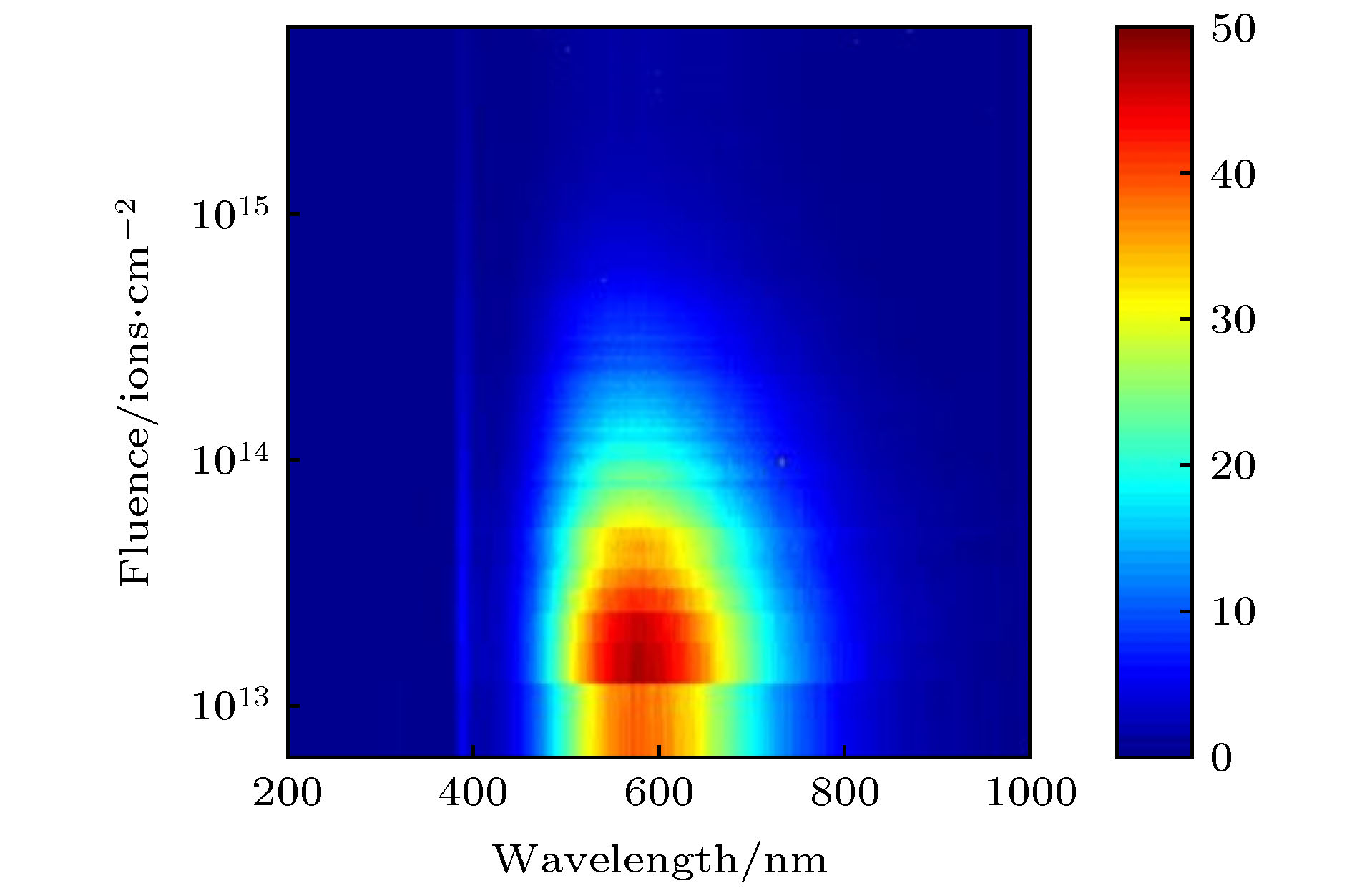 图 2 常温下ZnO的IBIL光谱随离子注量演变情况
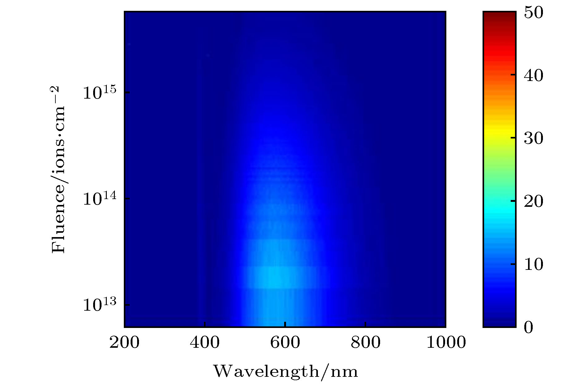 图 3 辐照后的样品在473 K退火后的IBIL光谱
 图 4 辐照后的样品在800 K退火后的IBIL光谱
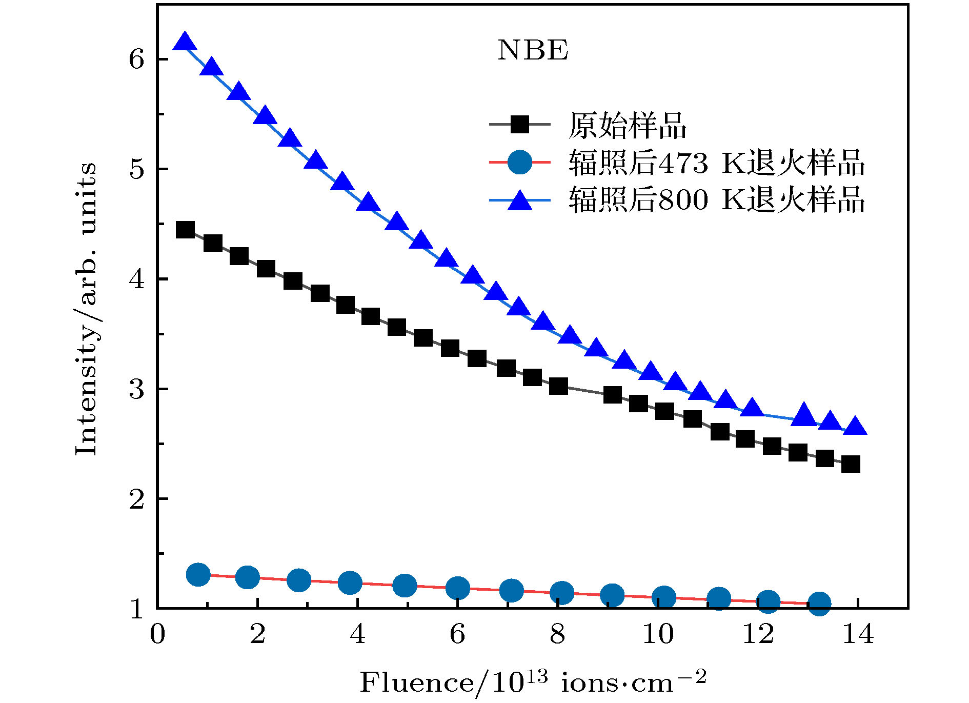 图 5 473 K和800 K退火后的样品的NBE强度随离子注量的演变情况
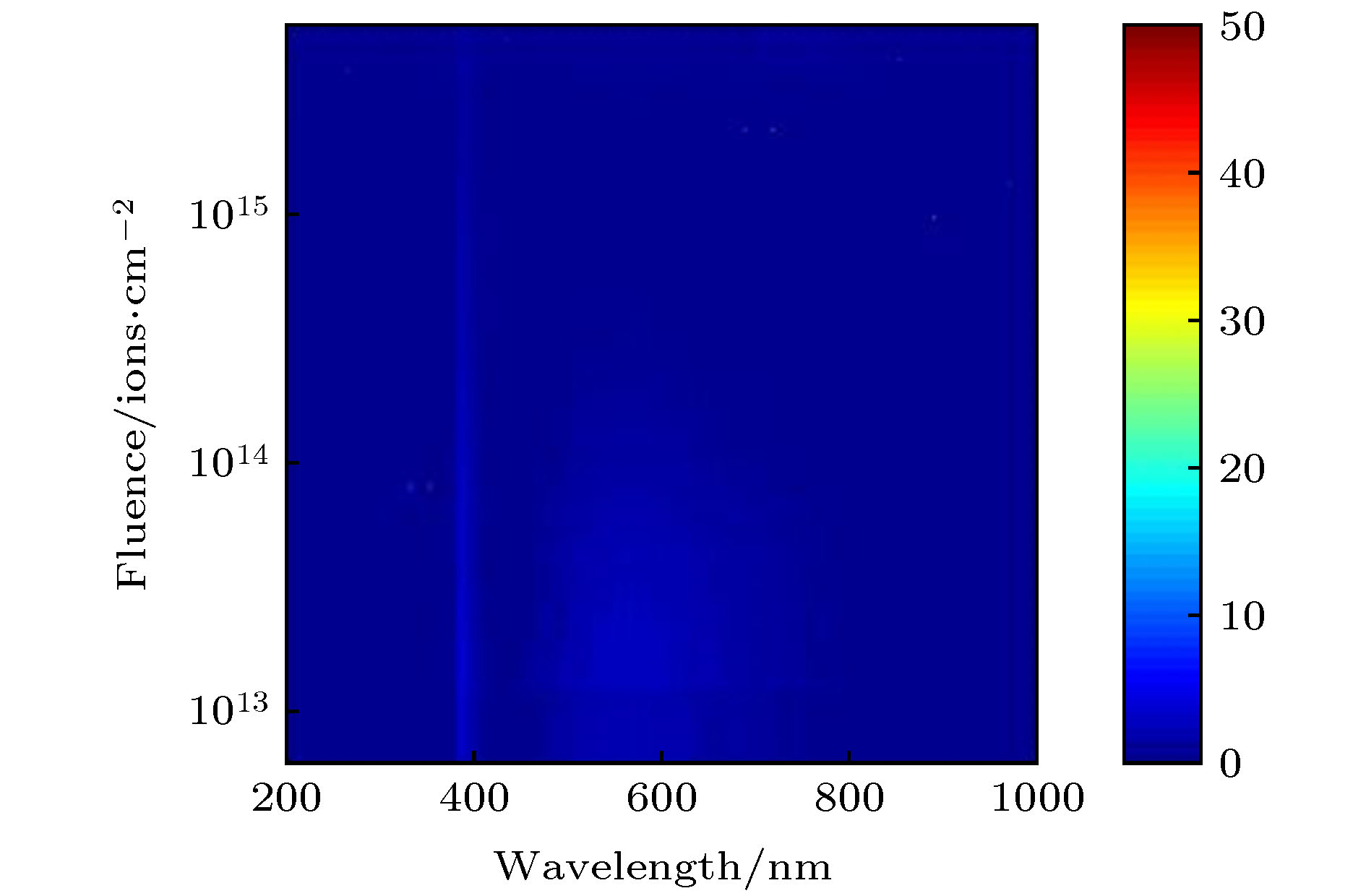 图 6 473 K真空退火3 h后的样品的IBIL光谱
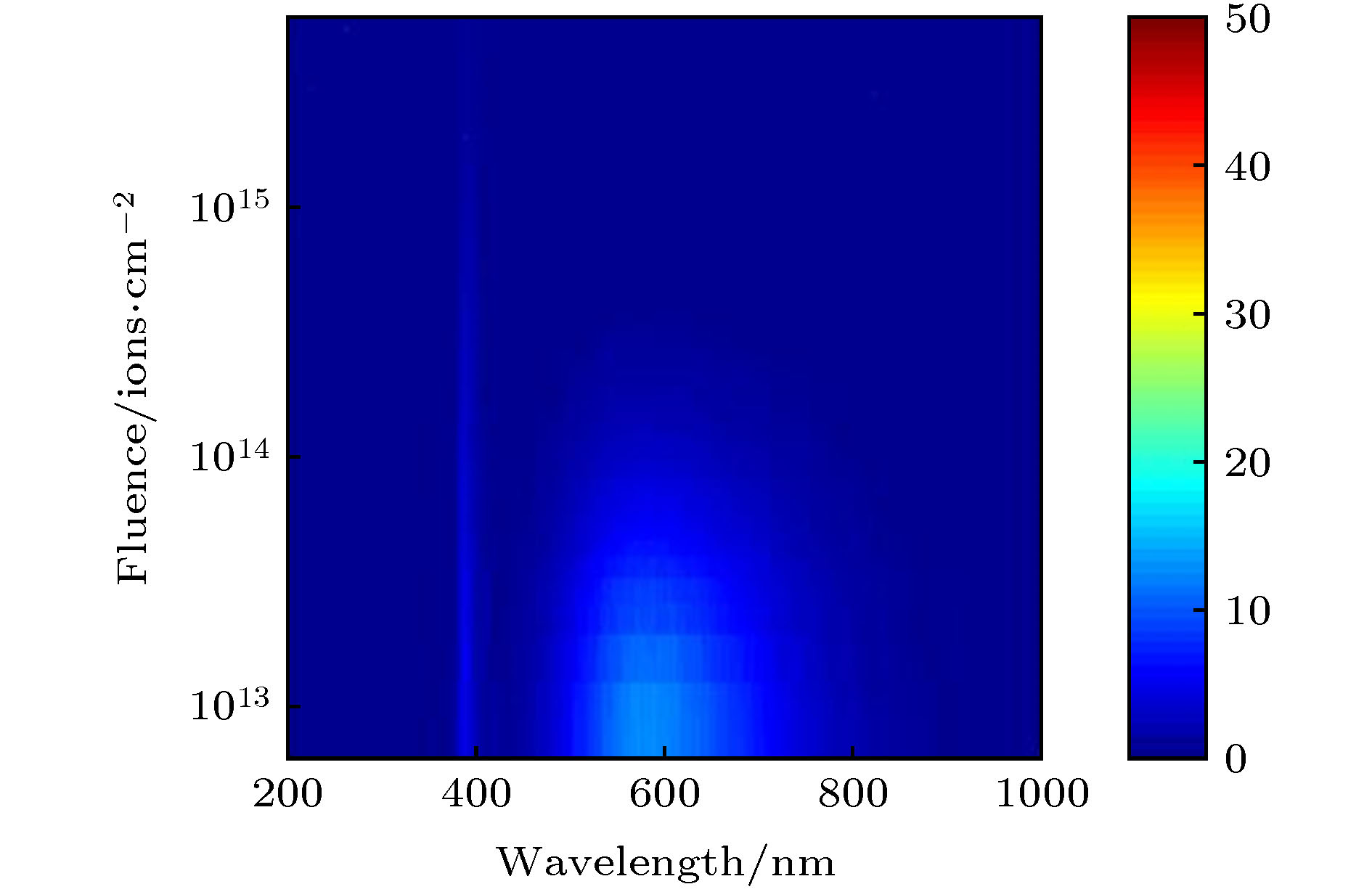 图 7 473 K真空退火3 h后再辐照的样品, 在800 K真空退火3 h后的IBIL光谱
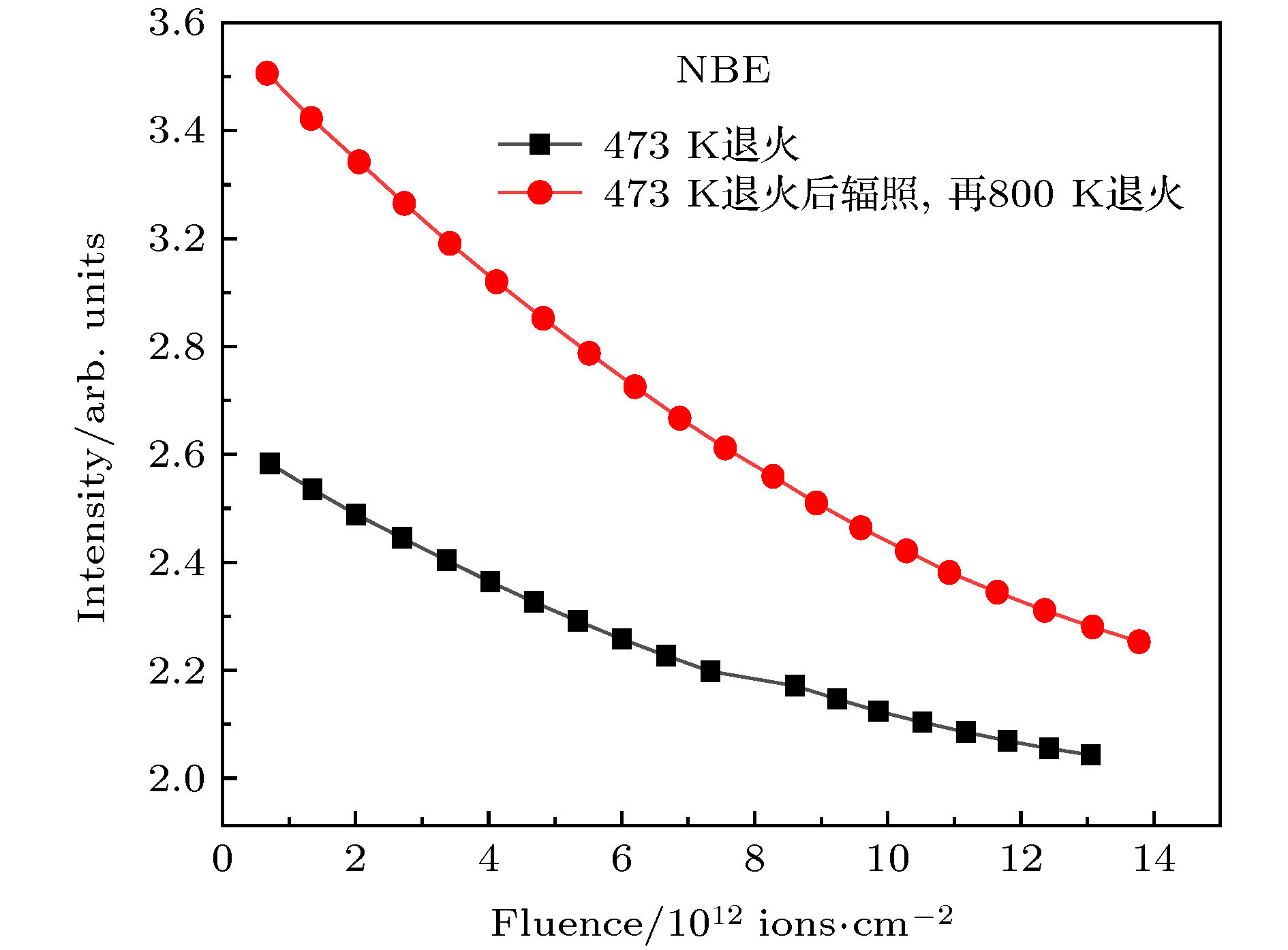 图 8 473 K退火和473 K退火后辐照, 再800 K退火的样品的NBE强度随离子注量的演变情况