 ,�й�ũҵ��ѧԺ����������ҽ�о���,���� 100193
Progress on genetic mapping and genetic mechanism of cattle and sheep horns
Xiaohong He, Lin Jiang, Yabin Pu, Qianjun Zhao, Yuehui Ma,Institute of Animal Science, Chinese Academy of Agricultural Science, Beijing 100193, ChinaͨѶ����: ���»ԣ���ʿ���о�Ա���о����������Ŵ���Դ�о���E-mail:yuehui.ma@263.net
��ί: ����
�ո�����:2020-07-21������:2020-11-19�����������:2021-01-20
| ��������: |
Received:2020-07-21Revised:2020-11-19Online:2021-01-20
| Fund supported: |
����� About authors
������,��ʿ,���о�Ա,�о����������Ŵ���Դ�о���E-mail:

ժҪ
�����ڶ����Ǹ�����,Ϊ��ۻ���������С�ţ(Bos taurus)������(Ovis aries)�ǵı��Ͱ���Ұ�������DZ��͡��˹�ѱ�����DZ��͡����νǵȶ��֡�ţ�������Dz����ǵ�������״��������״֮��Ĺ�ϵ�Լ�������״�Ķ������ػ��Ƶȷ�������붯��ģ�͡�������,�Խ���״�о���������,�ڲ�����������Դ��������Ȼѡ���Ա�ѡ����˹�ѡ��ԽDZ��͵�Ӱ��ȷ���ȡ����һϵ�н�չ��������ϸ�����˽ǵ��о��ſ�����DZ����Ŵ���λ����λ������Ŵ���λ�ͻ��νǵ�,����Ŀǰţ������ǵ��Ŵ����Ƽ����ڵ���������˷���,����Ϊ��ۻ�������״��������������״�Ŵ������о��ṩ�ο���
�ؼ��ʣ�
Abstract
Horns are cranial appendages, which are unique in ruminants. Cattle (Bos taurus) and sheep (Ovis aries) cranial appendages exhibit various forms of morphology, including wild-type two-horn phenotype, polled phenotype and scur phenotype. These animals provide an ideal model for studies on the underlying relationship between quality and quantitative traits of cattle and sheep horn and the molecular mechanisms of horn phenotype as a polygenic regulation for quality traits. In recent years, some research progresses of cattle and sheep horns are successively reported, which helps us better understand the evolutionary origin of new organ, the effects of natural selection, sex selection and artificial selection on horn phenotypes. In this review, we introduce in details the recent advances on the research of horn traits in cattle and sheep, and summarize the genetic mapping of multi-horned phenotypes, the genetic mapping of polled locus, and studies on scur phenotype. Moreover, we discuss potential problems in such research, thereby providing a reference for investigation on the genetic mechanisms of horn traits in ruminants.
Keywords��
PDF (629KB)Ԫ������ά�����������������EndNote|Ris|Bibtex�ղر���
�������ø�ʽ
������, ����, ���DZ�, ��ٻ��, ���»�. ţ������ǵ��Ŵ���λ���Ŵ������о���չ. �Ŵ�[J], 2021, 43(1): 40-51 doi:10.16288/j.yczz.20-229
Xiaohong He.
����������Ǹ�����(cranial appendages),�ֳƹ��ʽ�(headgear),��Ҫ�����ڷ�ۻ�����С��Ǹ�������Ҫ����4��,�ֱ�Ϊţ��(Bovidae)����Ķ���[1,2]������¹��(Giraffidae)�����ǡ�������(Antilocapridae)�IJ�Ǻ�¹��(Cervidae)��ʵ�ǡ�������ţ�ƶ������еĸ�����,����ţ(Bos taurus)������(Ovis aries)��ɽ��(Capra hircus)��,������Ҫ�ɹ��ʽ��ĺ��ⲿ�����ļ�Ӳ���������,����������[1,3]��ţ�������ܹ��ڰ�����ԭ��ɳĮ���ڵĹ㷺��̬���������沢��Ӧ���ֻ���[4,5],��FAOSTAT (Food and Agriculture Organization of the United Nations)���ݿ�ͳ��,������2018��,ȫ����ţ����������ֱ�Ϊ14.90��12.09��ֻ��ţ������DZ��;��зḻ�Ķ�̬��,����Ұ���͵����DZ��͡��˹�ѡ���γɵ��DZ����Լ��ڹ���Ʒ���д��ڵĶ�DZ���(��������),��������������ȫ�Ļ��νDZ���,��Щ����Ϊ����״�ṩ�˷ḻ���о��زġ�����ţ������Ƕ������ı�������,������һֱ����Ҫ���о��ȵ�֮һ,Ҳ�Ƕ�������Ŵ������о�����Ҫģ�͡�
1 ��DZ����о���չ
1.1 �������Ʒ�ּ��ֲ�
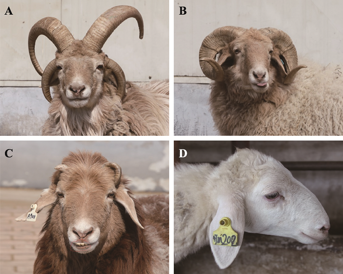
������Ψһ���ж�DZ��͵ļ�����ۻ����������״���зḻ�Ķ�����,���սǵ���������Ϊ�ǡ����ǡ���DZ���,���ж���Ǽ�������Ʒ���д��ڵĹ��϶���ϡ�ı���[6,7],���������ٶ�����ǰ���Ѿ�����[6]���������(multi-horned sheep)��ͷ�ϴ���2ֻ���Ͻǵ�����,һ����3~6ֻ��,�ݱ������ǿ��Դﵽ9ֻ,һ����4ֻ�DZ������,�����ֳ�Ϊ�Ľ���(ͼ1A)����������ֲַ�������[8]��ŷ�ޡ���������½[9,10,11],�ǿ�չ������Ŵ����ص����붯��ģ�͡�ͼ1
 �´��ڴ�|����ԭͼZIP|����PPT
ͼ1����ǵı���
A����DZ���;B�����DZ���;C�����ν�;D���DZ��͡�
Fig. 1The phenotype of sheep
���������ʷ�����������ޡ�ŷ�㷺�ֲ�[12],����ǿ�ҵ��˹�ѡ��,�ñ���Ŀǰ����ʮ����Ʒ����������,��Ⱥ����������С���ִ�Ķ������Ʒ����Ҫ����Jacob[13]��Manx Loaghtan��Hebridean��Navajo-Churro[10]��Icelandic[9]��Damara[14]������̩�����ɹ���[15]����ʲ����[16,17]����ˮ��Ƥ��[18]�ȡ���ЩƷ����һ���ⶼ�ǵ��صĹ�������Ʒ�֡����,���ҹ���ظ�ԭ����5200�ĵ�������ʱ,�·����˲�����Ʒ���еĶ��Ⱥ�塪����Dz�����,Ҳ��ĿǰΨһ�ڸߺ��ε������ֵĶ������Ⱥ��[19]������,������Ȼ�粢�����ڶ�������Ұ������,�ɴ��Ʋ�������������ڽ���ʱ����ɵ�ѱ��[20]��
1.2 ��DZ����Ŵ���λ
����1913��,Nature��־�����˹��ڶ��������о�[7];Alderson[21]�Ʋ��������״��2������������,��������������Ϊ���Դ��ϻ�����ʱ,�������Ϊ��DZ���;Dyrmundsson[9]��Ϊ��DZ��Ͷ����DZ��Ͷ���Ϊ�����Ŵ�,���DZ���Ϊ�����Ŵ���He��[22]����̩������ɹŶ������ˮ��Ƥ��Ϊ�о�����,��34ֻ���Ǻ�32ֻ��������ȫ�������������(genome wide association study, GWAS),�ɹ�����ǻ���λ�㶨λ������2��Ⱦɫ���132.6~132.7 Mb ����,�����ֻ��νDZ��Ͳ���Ӱ���DZ��͵��Ŵ���Ren��[23]����700K Illumina���ܶ�оƬ��24ֻ���Ǻ�22ֻ�Ľ���ˮ��Ƥ�����GWAS����,������2��Ⱦɫ��132.0~133.1 Mb����ɸѡ��4��������SNP (single nucleotide polymorphisms)λ�㡣Kijas��[24]����оƬ�Զ��Jacobs��Navajo-Churro�����GWAS����,������2��Ⱦɫ��131.9~132.6 Mb����ɸѡ��10��������SNPλ��,��������λ����132.568 Mb��,�ҷ���Navajo-Churro�������λ�㶨λ��10��Ⱦɫ��29.3~29.5 Mb�䡣Greyvenstein��[25]��26ֻ��Ǻ�16ֻ���ǵ�Damara�������GWAS����,���λ�㶨λ������2��Ⱦɫ��128~135 Mb����,û�з��ֶ�DZ��͵�CNV(copy number variations),�ҷ���������SNP�ڶ�Ǹ����϶����Ӻϻ����͡���������,�Էֲ����й������ޡ�����ŷ��6���������Ⱥ����ж��λ���Ŵ���λ�о�(��1),�������λ�㶨λ������2��Ⱦɫ��,�״γɹ��������϶�λ����ǻ������λ�㡣2020��Li��[26]���������о���չ,���ø�ͨ���ز������ݶ���ˮ��Ƥ��(��DZ���)��Сβ����(���DZ���)���н���״SNP����������Ʒ�ּ��ѡ������ɨ����,��2��Ⱦɫ��HOXD�����(homeobox D cluster)��HOXD1��HOXD3��HOXD8��HOXD9��HOXD10�Ȼ�����ɸѡ���������ź�,������ȷ�������ǻ�����Ŵ���λ��He��[19]�Զ�Dz������������鷢��,�������Ⱥ���д��ڽ�����3~6�����ȵ��Ŵ�����,����4���DZ�����������,������Ƿ�Ϊ���Ͷ�DZ���(4����)�ͷǵ��Ͷ�DZ���(3��5����6����)����,���ֱ��Ͷ�λ��Ⱦɫ�����ͬλ��,��λ������2��Ⱦɫ��132.8 Mb��,�ý��������DZ��͵���������(���ͺͷǵ��Ͷ��)���ܾ�����ͬ���Ŵ�λ��(��1)��
Table 1
��1
��1�������״����Ŵ����䡢SNPs�ͺ�ѡ����
Table 1
| ���ͻ��� | �Ŵ����䡢��ѡ�����SNPs | �������� | �������� | ���ֻ�Ʒ�� | Ұ������� | �ο����� |
|---|---|---|---|---|---|---|
| �� | Chr.10: OarHH41��AGLA226 | ���DZ�� | �������� | (����ū��×��ķ����)×����ū�� | ���� | [40] |
| Chr.10: 7.4 cM���� | ���Ǻ� ��λø��� | �������� | ������ | Ұ�� | [44,48] | |
| Chr.10: 200 kb���� | SNPs | �������� | ����ū��×��ķ���� | ���� | [41] | |
| Chr.10:OAR10_29546872,RXFP2 | DNAоƬ�� �ز������� | ѡ���źŷ���������������ѡ������ɨ���� | ������������ū��;Сβ������ | ���� | [26,37] | |
| Chr.10:29.3~29.5 Mb,RXFP2��EEF1DP3 | DNAоƬ�� ���� | ������ƽ�������оƬGWAS��������̬�Լ�� | ��������ū��Navajo-Churro����̲���������� | ���� | [24,42,52] | |
| RXFP2 3′UTR 1.8 kbƬ�β��� | ������ | ���з��� | 7����ʿ����Ʒ�� | ���� | [51] | |
| �ǵĶ�̬�� | Chr.10:RXFP2 | DNAоƬ;RXFP2������ | GWAS������ѡ��ϵ����ƽ��Ƶ�ʷ�����������֮�����Ӧ�Ȳ������ | ������ | Ұ�� | [49,50] |
| �ǵij��ȡ� �ǵķ��� | Chr.10:CSRD87��OarSEJ09 Chr.10:RXFP2 | ���DZ�� DNAоƬ�� �ز��� | ����ͼ��QTL��λ��GWAS������ѡ������ɨ���� | ��������� ������ | Ұ�������� | [45,46,48~50,55] |
| �����ڷ��� | (1) SOX9��HOXD; (2) SOX10��SNAI1��SNAI2��TFAP2A��NGFR��COL11A2 | ��������� ת¼������ | �����顢ת¼����� | �нǷ�ۻ���� | Ұ���ͼ��� | [7] |
| ���ν� | COL6A2��COL6A1��PARVA��TNN��TNC | ��������ѧ ���� | ������� | ����̩�� | ���� | [62] |
| Chr.10��RXFP2 | DNAоƬ | GWAS���� | ������ | Ұ�� | [49] | |
| ��DZ��� | Chr.2: 128~135 Mb��HOXD����ء�MTX2��EVX2��KIAA1715 | DNAоƬ�� �ز������� | GWAS����������������Ʒ�ּ��ѡ������ɨ���� | ����̩���ɹ�����ˮ��Ƥ��Jacobs��Navajo-Churro���Damara���� | ���� | [9,22~24,26] |
| ������� | Chr.2:132.8 Mb,MTX2��HOXD����� | DNAоƬ | оƬGWAS���� | ��Dz����� | ���� | [19] |
�´��ڴ�|����CSV
2 �DZ����о���չ
2.1 ţ�DZ���
2.1.1 ţ��λ��(POLLED)���Ŵ���λ1993��,Georges��[27]��ţ����λ�㶨λ��1��Ⱦɫ����,����һ������Χ��С��1��Ⱦɫ�����˿������[28]���о���Ա½���ڸ����䷢��4����ͻ��(��2)��Medugorac��[29]������ŷ�����ص���ţƷ���з���Celtic POLLED (PC)ͻ��,��ͻ��λ��IFNAR2��OLIG1����֮��,��һ��202 bp�IJ���-ȱʧ������,�о���Աͨ������༭�����Ƴɺ���ͻ��ĺ�˹̹ţ����άϸ��,ͨ����¡�õ����DZ��͵Ķ�ţ[30]��Medugorac��[29]��Rothammer��[31]ͨ���Ժ�˹̹ţ����,�����˵ڶ���ͻ��,�о�������260 kb�ĵ������ϴ���5�����˹̹ţ�DZ�����صĺ�ѡͻ��,��ͻ����PCͻ��˴˲�����Ҳ���Ӱ��,��ΪFriesian POLLED(PF)ͻ��(��2),��ͻ��λ��PCλ������200 kb��,��һ��80 kb���ظ�����,���Ǻ�˹̹ţ����Я��PCͻ��[32]��Utsunomiya��[33]���������ţ�з����˵�3��ͻ�䡪��Guarani POLLED(PG)ͻ��,��ͻ����һ��110 kb���ظ�Ƭ��,�������ָ��ǻ�����������ͨţ�����һ����Mongolian POLLED(PM)ͻ��[34],�������ɹ���ţ���ɹ�Turanoţ,�ɹ���ţ����ͻ�䶨λ��POLLEDλ��800 kb��������,����2�������ͣ�һ������ԭ��������61 bp������219 bp�ظ�-����Ƭ��;�ڶ���ͻ������ԭ��������621 bp��6 bpȱʧ��7 bp�IJ��롣PMͻ���219 bp�ظ�Ƭ������һ��11 bp�Ļ���,����ţ�ƶ�������ȫ����,PMͻ��ͬʱҲλ��PF��PG�����ڡ������ͷ�������,PM��������Turanoţ���뵽�ɹ���ţ��[34]��
Table 2
��2
��2ţ����״����Ŵ�ͻ��ͺ�ѡ����
Table 2
| ���� | ͻ��λ�㡢��ѡ���� | �������� | �������� | Ʒ�� | �ο����� |
|---|---|---|---|---|---|
| �� | PCͻ��,IFNAR2��OLIG1 | DNAоƬ,ת¼�� | �����鴿���ӷ����� ���������� | ���ŷ��ţƷ�� | [29,30] |
| PFͻ�� | DNAоƬ,ȫ��������� | �����ͷ��� | ��˹̹ţ | [29,31,32] | |
| PGͻ�� | ȫ��������� | �����ͷ��� | �������ţ | [33] | |
| PMͻ�� | DNAоƬ,ȫ��������� | ����������� | �ɹ���ţ���ɹ�Turanoţ | [34] | |
| RXPF2��FOXL2 | ת¼�����, | ���������� | ��˹̹ţ | [32] | |
| OTOP3��OLIG1 | ��������� | ��ѡ����� | ţ�ƶ��� | [70] | |
| ���� | TWIST1 | DNAоƬ | ȫ������������� | ������ţ | [59,61] |
�´��ڴ�|����CSV
2.1.2 ţ�DZ���ת¼���о�
������Աͨ���Խ�ѿ�ͽǽ���ת¼�鼰�����������,��һ����չ�˽Ƿ������DZ��͵��ź�ͨ·�о���Allais-Bonnet��[35]��̥����90��PC��������Ļ�������lincRNA (long intergenic non- coding RNA)���з���,�����нǺ��DZ��͵Ľ�ѿ��֯��RXFP2�����LincRNA#1�����������졣�DZ���ţ̥����ѿ��λRXFP2���������������нDZ���(P < 0.05),LincRNA#1�ı�������нDZ���(P = 0.052)��Wiedemar��[32]��ţ̥��150��Ľ���֯���DZ��ͽ�ѿ��λRNA����,����OLIG1��OLIG2��C1H21/f62��RXFP2��FOXL2�����������,ͬʱLincRNA#2������Ҳ��������;��̥����70~175���ѿ�ͶƤ������,�����нDZ��͵�RXFP2��FOXL2�� LincRNA#2�������������DZ���,��δ�ﵽ�������졣�������о�����,RXFP2�������о��Ĺ�ͬ�������Ļ���,�����������ijһʱ���ڴ��ڱ�����졣Li��[36]��PMͻ������ţ80~90��̥����ѿ��֯��λ���е�����ѧ����,ȷ����29�������ϵ�����71�������µ�����,�����ϵ������漰��л�,�����µ������漰ϸ�����ӡ�ϸ���Ǽ��γɺ�ϸ���ɷ���֯���нDZ��ͺ��DZ����ڽ�ѿ��֯�ṹ�ϵ�������ܵ�����ɸѡ�����쵰����ϸ���ṹ��ء�
2.2 �����DZ���
2.2.1 ������(Ho)λ���Ŵ���λ������ѱ������ʱ,ͬʱ��ëɫ����ë���͡����͵���״������ѡ��[37]����Ҳ���������翪ʼ�о�����״֮һ��Lundrigan[38]����Ұ������͵ط�����Ʒ�ֹ���һ���н�,����ѧ�Ҵ�16���Ϳ�ʼѡ��������,��������Ĺ���ʼ�����DZ���,�о���Ա�ԡ�����-�ǡ���Ա���Ҳ��չ�˴����о���
�ڼ��������о���������,�����о���Ϊλ�ڳ�Ⱦɫ��Ho�������ϵ�3����λ������������нǶ��DZ���,�ֱ�ΪHoP��Ho+��Hoh1[39]��Montgomery[40]������ū�����ķ������ӽ�Ⱥ����,������ġ���λ�㡱��λ��10��Ⱦɫ���ϡ�Pickering[41]��������ū�����ķ������ӽ�Ⱥ��,����λ�㶨λ������С��50 kb��������,17����ǹ��ɵĵ������ܹ�ʹ����DZ���Ԥ�����ȷ�ʴ�97%��Kijas��[37]����ȫ�������ź�ѡ������������غ�����ū��,������10��Ⱦɫ���Ϸ����DZ��͵�������SNPλ��(OAR10_29546872),��λ���ٽ�RXFP2����Navajo-Churro����ĸ��ܶ�оƬȫ�����������������,��λ�㶨λ������10��Ⱦɫ��29.3~29.5 Mb����[24](��1)��Dominik��[42]Ҳ�ڰ�������ū����Ⱥ�巢�ֿ��Լ����DZ��͵ĵ������̬�ԡ�������ū��Ʒ����,����OAR10_ 29546872.1��OAR10_29458450����SNPλ��,��ĸ����Ԥ��ȷ�ʴﵽ32.3%~71.3%,������Ԥ��ȷ�ʴﵽ62%~72.5% [43],����������SNPs��������ͻ��,���Բ���100%��Ԥ��DZ��͡�ͬʱ,Ұ��������Ҳ������λ��,����251�����Ǻ͵�λø���,Dario��[44]��������(Soay)���DZ��Ͷ�λ��10��Ⱦɫ�塣
2.2.2 ��������Ա�ѡ��������о�
���ڹ����ͻ�ý���Ȩ�����ƻ����з�������Ҫ����,�ǵijߴ�Խ��,����ķ�ֳ�ɹ��ʾ�Խ�ߡ��Ա�ѡ����Ұ������ǿ��������ѡ���Դ�������Դ�����ϵ�о�����,�����dzߴ��QTLλ��10��Ⱦɫ��[45],�о���Ա�Դ������л����ز�����ź�ѡ�����,���ֹ����Ľ���RXFP2�ܵ�ǿ�ҵ���ѡ����γɵ�(��1)[46],ͬʱ���Ծ�����Խ��,�������Ա�ѡ��ǿ�Ⱦ�Խ��[47]������Ұ��������Ⱥ����,��Ȼ���д�ǵĹ�����ͬ�Ծ����о�������,������Ⱥ���Ա����˽DZ��͵Ķ�̬��,Ⱥ���ڴ���������Ǻͻ��ν����ֱ���,��ĸ��Ⱥ���д��������ǡ����νǺ���3�ֱ���[48,49]�����ֶ�̬����ͨ��RXFP2��������Ȼѡ����Ա�ѡ�������Э���γɵ�[50],����������RXFP2��������λ����,��ǵ�λ����Ho+��߷�ֳ�����,С�ǵ�λ����HoP���������,�����γ��Ӻ�������,Ⱥ���д���Ho+Ho+��Ho+HoP��HoPHoP���ֻ�������,Ho+Ho+��Ho+HoP��������ı�����������,HoPHoP�������д�Լ50%����Ϊ���νDZ��͡�
2.2.3 �����DZ����Ŵ��ĸ�����
Wiedemar��[51]ͨ����5��ŷ������Ʒ�ֽ�һ������,����RXFP2����3��UTR����һ��1.8 kb����Ƭ�����DZ������,���Ƕ����DZ���Ϊ���ԡ�Wang��[52]���й�̲����Ҳ����RXFP2������һ��ͬ��ͻ�����DZ����������,���Թ���34������Ʒ��489������Ĵ�������ⷢ��,λ��RXFP2����3��UTR����IJ���Ƭ��ֻ�ڲ�������Ʒ�ֵĽDZ����г��ַ���[53],�����ҹ��ط�����Ʒ�ְ���̩�����GWAS������δ����������λ��,ͬʱ,��Ⱒ��̩��RXFP2����3��UTR����1.8 kb����Ƭ��,������ָò���Ƭ�����DZ��Ͳ����������[22]��2020��,Li��[26]���ҹ�Сβ����ͺ�����л����ز���Ľ���״����������Ʒ�ּ�ѡ������ɨ����,�������CNV��SNP���������Լ�ѡ������ɨ����������λ��10��Ⱦɫ��RXFP2�������ź�(��1)����������,�������ڱ�����ֻ����һ������,��Ȼ�Ѿ��ڻ������ϳɹ���λ���ǵ��Ŵ�����,����ͬ������Ⱥ���г��ֽ�Ȼ��ͬ�Ľ��,��ЩƷ�ֽ�����RXFP2��������������,��һЩƷ�����ڹ���,ͬʱ�DZ����ڲ�ͬƷ���еõ����Ӷ��Ľ�������DZ��ͻ����Ŵ����и����ԡ�
3 �������DZ��͵�������״
���,����״����Ϊ�ǵ��͵�������״,����������(ͼ1B)����(ͼ1D)��������������о������µķ��֡�Ұ���������������״��Ⱥ���о��зḻ�ı���,��Ϊ�о����Ŵ����ص�����ģ�͡�Dario��[44]ͨ������ͼ��Ұ��������Ľ���״��λ��10��Ⱦɫ����,Johnston��[48]��������һ������С��7.4 cM��Χ��,���ҽ�ȷ���dz��Ⱥͽǻ���λ�ܳ��ĵ��ػ���Ҳ��λ����һ����Johnston��[49]��һ������оƬ����ȷ����������ǹؼ���ѡ����RXFP2,�û����ܽ��;߱������ǹ���Ľdz�76%������QTL,����RXFP2��������DZ��ͺ�ѡ����(������״),Ҳ�ǽdz��Ⱥʹ�ϸ��������״����Ч����������Ұ����Ⱥ���ϲ�δ�õ����ƵĽ��,Miller��[54]��76�������(Ұ������)���ڸ��ܶ�оƬ��GWAS����,��û�з���Ӱ��dz��Ⱥͽǻ���λ�ܳ���QTL������,Pan��[55]��89���й�������ز����������RXFP����������İ�Ұ�����,����dz��ͽ���������(������ˮƽ����)���(��1)����֮,�����DZ���(������״,���нǻ�����)�������dz��Ⱥʹ�ϸ��������״QTL����λ��ͬһ�Ŵ������ͬһ��ѡ����RXFP2����4 ���ν��о���չ
����״����������ͬ��,�䷢���̶�Ҳ�кܴ����,���պ��߿ɷ�Ϊ�����ǡ����ν�[56]����[12]�����ν���С�ġ��������,�Ҳ����ι̸������ǵĽ�(ͼ1C)�����ν������ڹ�Ԫǰ3800~3500����Ѿ��ڼ����д���[57]���ںܶ�����ɽ��ţ��Ʒ���ж����ڻ��ν�����[52],���ֽǵı��ͽ����˼���ļ�ֵ[58]������ѧ�ϻ��ν�����������2����Ҫ������һ�ǻ��νDz���������,���������ͻ;���ǻ��νǵĹ��ʽ��ĸ�������[59]��4.1 ţ���ν�
ţ�ϴ���2�����͵Ļ��ν��Ŵ�,White��[60]��Ϊţ�ϴ��ڵ���ţ�ǻ��α��͵�SCURSλ��(Scλ��),�����䶨λ��19��Ⱦɫ��[58],II�ͻ��νDZ������ɷ���������ţTWIST1�����ͻ�䵼��(��2)[59,61],�����нǶ����Ŵ�λ�㶨λ������ͬ��4.2 ������ν�
�����о�����,�������Ұ�������Ļ��νDZ��Ͷ�λ������10��Ⱦɫ��RXFP2����,����������нǶ���λ�㶨λ��ͬ���Ի��νǵ�iTRAQ����������PARVA��TNN��TNC��COL6A1��COL6A2��һϵ�������Բ��쵰��(��1),������(ECM)- receptor interactions��focal adhesion��PI3K-Akt��Ӱ������Ƿ���(����)����Ҫ�ź�ͨ·[62]�������о�����,ǰ�����ź�ͨ·����ϸ��ճ������[63,64],�����ܲ���ϸ������ϸ������[65]����Ȥ����,Mariasegaram��[66]��ţ���о��з���(ECM)-receptor interactions�ź�ͨ·Ҳ�ǻ��νǶ��ǵ��������ź�ͨ·��PI3K-Akt�ź�ͨ·�ܵ�����Ƥϸ����ϸ������ʵı���[67],focal adhesion�ź�ͨ·�����ڻ��������Թ��γ�[68]��5 �ǵ��Ŵ�����
5.1 ţ���Ŵ�����
ţ���нDZ���ΪҰ����,����λ��(POLLED)�������Ŵ�,I�ͻ��νǶ�������λ�Ŵ�,ͬʱ���Ա�Ӱ��[32,60];II�ͻ��ζ������DZ��������Ե�,�������Ӻ���,Ŀǰû�з��ִ��ϵ�TWIST1ͻ��,�Ʋ��ͻ������̥����;����ţ��ţ���нDZ��Ͷ��DZ���Ϊ��λ�Ŵ�[69],�ڷ�����ţ�Ͱ���˹ţ���ӽ�Ⱥ����,���ĸţȫ�����DZ���,�������ţΪ3�ֱ���,�ֱ�Ϊ�нǡ����νǺ���,�Ʋ���ܴ�������һ��Ha�������ǵ��Ŵ���Ŀǰ�Ѿ�����4��ţ���DZ����Ŵ�λ��,����λ��ţ1��Ⱦɫ��һ�αȽϼ��е�����,�ֱ�ΪPC��PF��PM��PGͻ��(��2),��4������û�ж�λ���κ���֪����lncRNA(long non-coding RNA)����miRNAs��,�Ʋ���ͨ��Ӱ��DNA��������,����ǿ�������ػ���ı����һ�������23���������ͷDZ����������˽ṹ��,����OLIG1��OLIG2��IFNAR2��PAXBP1�ȡ�Wang��[70]�о�����OLIG1���нǷ�ۻ�ද�����������ѡ�����,�������ֻ�ͨ·���[71],��OLIG1�������65 kb�����212 bp���ظ�Ƭ����ţ�DZ��͵�����ͻ��[29,30]��OLIG1��������ڹ��ʽǵķ����Ϸ�����Ҫ����[70]��ͬʱ,Tetens��[72]����OLIG2��FOXL2�� RXFP2�Ȼ���Ϊ��ѿ�ֻ���ػ���
����֯�������ܵļ����ϸ���γ�[73,74],������Ƥϸ�����Ǩ�Ƽ����ϸ������Ϊ��Ƥ-����ת��(epithelial-to-mesenchymal transition, EMT),�ù��̵���ϸ�����Ͷ�Ԫ�����γ����ٵ���֯����[75]��Betancur��[71]����TWIST1��SNAI2��SOX9��HOXD����صȲ�����ϸ��Ǩ�㡣ͬʱ,�о�����TWIST������سɹǹ���,��ͻ�������������[76,77],��TWIST��ţII�ͻ��νǵ�ͻ�����ͬʱWang��[70]�о�������SOX10��SNAI1��SNAI2��TFAP2A��NGFR��COL11A2��6����ϸ��Ǩ����ػ����ڽ���֯�������Ա���,�ü����COL1A1��COL1A2��COL5A1��COL5A2��COL6A6�Ȼ�����Ӱ��ţ���νǵIJ������[66]���о�����TWIST1��TWIST2��ZEB2��FOXC2��ת¼���ӿ���ֱ������E-��ճ���ı���[78],��E-��ճ������EMT�ı�ǵ���,����ͨ��Ӱ����ϸ��Ǩ��Ӱ��ţ�ǵķ����������о�������Щ��ѡ�������ͨ��Ӱ����ϸ��Ǩ��Ӱ��ǵ��γɺͷ���,�Ӷ�Ӱ��ţ�ǵı��͡�
5.2 ����ǵ��Ŵ�����
�������״������2��λ�����,һ����λ��10��Ⱦɫ��ġ���λ�㡱,��һ����λ��2��Ⱦɫ��ġ����λ�㡱,��DZ��Ͷ����DZ���Ϊ�����Ŵ���Ŀǰ����ǵ��Ŵ����Ʋ������,����λ���DZ��ͺͶ�DZ��͵��Ŵ�����ͺ�ѡ���������DZ��͵ĺ�ѡ����ΪRXFP2 (�ɳ���/���ȵ���������������2,relaxin/insulin like family peptide receptor 2),λ������10��Ⱦɫ�塣Wang��[70]�о�����RXFP2����Ϊ����֯�߱������,RXFP2ΪG����ż�����嵰��,��ͻ����Ե��¹�������[79]��RXFP2�����RLN����ͨ�������Ĥ�ڻ���������,����ALP��RUNX2��BMP2�յ��ɹǷֻ�[80],����RXFP2�ԽǷ�������Ҫ����,���RXFP2����,����ͨ������������ɳ��صĽ��,���ƳɹǷֻ�,�Ӷ����ƹ��ʽ��ĵ��γɡ����λ����Ŵ�����λ��2��Ⱦɫ��128~135 Mb,���������MTX2��HNRNPA3��NFE2L2��HOXD����ص�,����HOXD����HOXת¼���Ӽ����Ա,��һ��ʮ����Ҫ���ڽ����ϱ��ص�ת¼����,����Ҫ������Э��֫��ԳƷ���,�Ӷ�Ӱ��֫�����̬���о�����HOXD����س���Լ100 kb,����13������,����HOXD 4����2.7 kb��ȱʧ���Ե�����(Equus caballus)�ļ�����ȱ��[81],HOXD������岿�ֳ�Ա��ͻ����Ե����˵Ķ�ֺ����[82],��HOXD10��HOXD11��HOXD12��HOXD13����ָ�ͽ�ֺ�ķ������,��ͻ���Ӱ����ָ�ͽ�ֺ������[83],HOXD13������֫��ĩ�˱���,���ж����ٽ�������غͿ���ĩ�˷������ص�ת������[84],ͨ����һ����ĵ��شӶ�ʵ��֫�����������ĩ�����������ص�ת��[85]��Jerkovi?��[86]��һ�����о�������,HOXD9~HOXD13������ͨ����������DNA����,����HOXDת¼����ֱ����DNA���ϡ��������Ҫ���ⲿ��Ӳ�Ľ��ʻ����ʺ��ڲ����ʻ����������ֹ���[87],�������м仹������Ĥ��Ƥ�½����֯����Ƥ�ͱ�Ƥ[3]�����ʽ�����Ҫ���ɹ���֯����,��Դ�����߲�(�������߲�Ͳ����߲�)�������������߲��γ��������,���߹ǡ��Ǻ��ǵĶ��ǡ������߲��γɸ�������,����֫[73,88]����ϸ��Ǩ���γɶ�Ǻ����[74],��ЩӰ�����߲��γɺ���ϸ��Ǩ�ƵĻ�������ǽ��γɵĺ�ѡ����Betancur��[71]����HOXD������Լ�SNAI2��TWIST1��SOX9 �Ȼ��������ϸ��Ǩ��,Wang��[70]��HOXD��������η���һ��3.6 kb�нǷ�ۻ��������ת�����Ӳ���,��һ���������ָò������һ��25 bp�������Ա���Ԫ��;ͬʱ����COL11A2����ϸ��Ǩ����ػ����ڽ��������Ա���,��COL11A2������������ν��������쵰��COL6A2��COL6A3��COL6A1��COL1A2ͬ���ڽ�ԭ�������Ա,��ЩԪ���ͻ�����ܶԽǷ�������Ҫ���á�
6 ������չ��
���Ƿ�ۻ����ı�־������(����Ұ����ۻ�����������),���ݻ���Ϊ�ɹ�������֮һ,�ǵķ������������Ŵ�����һֱ���Ŵ�ѧ�о����ȵ�֮һ,Ҳ�������������Ŵ���״�о��IJο�ģ��֮һ��2019��,����ũ�ֿƼ���ѧ�����ŶӺ�������ҵ��ѧ�����Ŷ�[70,89]��Science��������������ƪ��ۻ����ǵ�����о�����,���ַ�ۻ����ĽǾ��й�ͬ�Ļ���ϸ������֯��Դ;ѱ¹CCND1�������ε�ͻ�丳���ۼ����������Ĺ����Խ�ϻ���,���ܵ��´���¹��������ǰ�˶�ţ������ǵ��Ŵ����ؿ�չ�˶�����о�,�ڶ�DZ��͡��DZ��ͺͻ��νǵ��Ŵ�λ�㶨λ���Ŵ����Ƶȷ���ȡ������Ҫ��չ,Ŀǰ���о�����,ţ�DZ�����4��ͻ��,��λ��1��Ⱦɫ��,���ν���2��ͻ��λ��;�������״������2��λ�����,�ֱ���λ��10��Ⱦɫ��ġ���λ�㡱��λ��2��Ⱦɫ��ġ����λ�㡱�����Ƕ�ţ���������״�γɵķ��ӻ������д����������о�,����ȷ�����γɵ����ͻ����������ڷ����ؼ�����,�����Dz�ͬ���͵��Ŵ����ػ��ơ�ţ������Ϊ�����ṩ�⡢�̡�ë(Ƥ)��������������,������ũҵ��չ�а�������Ҫ��ɫ,ͬʱҲ������ũ��������������Ҫ��ɲ��֡�ţ���������״���зḻ�ı���,��չ���γɺ��Ŵ����Ƶ��о����������ƽ�������������״�Ķ������ػ��ƻ�����������������Դ�����Ȼ����о��Ľ�չ,Ϊ������������״���غ��Ŵ��ֻ��е������ṩ�ο���
�ο����� ԭ��˳��
������ȵ���
������������
�����ڿ�Ӱ������
DOI:10.1002/(ISSN)1097-010XURL [��������: 2]
[��������: 1]
[��������: 1]
DOI:10.1098/rspb.2011.0938URLPMID:21733893 [��������: 2]

The horns, ossicones and antlers of ruminants are familiar and diverse examples of cranial appendages. We collectively term ruminant cranial appendages 'headgear'; this includes four extant forms: antlers (in cervids), horns (in bovids), pronghorns (in pronghorn antelope) and ossicones (in giraffids). Headgear evolution remains an open and intriguing question because phylogenies (molecular and morphological), adult headgear structure and headgear development (where data are available) all suggest different pictures of ruminant evolution. We discuss what is known about the evolution of headgear, including the evidence motivating previous hypotheses of single versus multiple origins, and the implications of recent phylogenetic revisions for these hypotheses. Inclusion of developmental data is critical for progress on the question of headgear evolution, and we synthesize the scattered literature on this front. The areas most in need of attention are early development in general; pronghorn and ossicone development in particular; and histological study of fossil forms of headgear. An integrative study of headgear development and evolution may have ramifications beyond the fields of systematics and evolution. Researchers in organismal biology, as well as those in biomedical fields investigating skin, bone and regenerative medicine, may all benefit from insights produced by this line of research.
DOI:10.1093/molbev/msu264URL [��������: 1]
Following domestication, sheep (Ovis aries) have become essential farmed animals across the world through adaptation to a diverse range of environments and varied production systems. Climate-mediated selective pressure has shaped phenotypic variation and has left genetic "footprints" in the genome of breeds raised in different agroecological zones. Unlike numerous studies that have searched for evidence of selection using only population genetics data, here, we conducted an integrated coanalysis of environmental data with single nucleotide polymorphism (SNP) variation. By examining 49,034 SNPs from 32 old, autochthonous sheep breeds that are adapted to a spectrum of different regional climates, we identified 230 SNPs with evidence for selection that is likely due to climate-mediated pressure. Among them, 189 (82%) showed significant correlation (P a parts per thousand currency sign 0.05) between allele frequency and climatic variables in a larger set of native populations from a worldwide range of geographic areas and climates. Gene ontology analysis of genes colocated with significant SNPs identified 17 candidates related to GTPase regulator and peptide receptor activities in the biological processes of energy metabolism and endocrine and autoimmune regulation. We also observed high linkage disequilibrium and significant extended haplotype homozygosity for the core haplotype TBC1D12-CH1 of TBC1D12. The global frequency distribution of the core haplotype and allele OAR22_18929579-A showed an apparent geographic pattern and significant (P a parts per thousand currency sign 0.05) correlations with climatic variation. Our results imply that adaptations to local climates have shaped the spatial distribution of some variants that are candidates to underpin adaptive variation in sheep.
DOI:10.1038/s41467-018-04737-0URLPMID:29904051 [��������: 1]
Cattle domestication and the complex histories of East Asian cattle breeds warrant further investigation. Through analysing the genomes of 49 modern breeds and eight East Asian ancient samples, worldwide cattle are consistently classified into five continental groups based on Y-chromosome haplotypes and autosomal variants. We find that East Asian cattle populations are mainly composed of three distinct ancestries, including an earlier East Asian taurine ancestry that reached China at least ~3.9 kya, a later introduced Eurasian taurine ancestry, and a novel Chinese indicine ancestry that diverged from Indian indicine approximately 36.6-49.6 kya. We also report historic introgression events that helped domestic cattle from southern China and the Tibetan Plateau achieve rapid adaptation by acquiring ~2.93% and ~1.22% of their genomes from banteng and yak, respectively. Our findings provide new insights into the evolutionary history of cattle and the importance of introgression in adaptation of cattle to new environmental challenges in East Asia.
[��������: 2]
[��������: 2]
[��������: 1]
[��������: 1]
[��������: 4]
DOI:10.2527/2004.82102900xURLPMID:15484940 [��������: 2]
The objectives of this study were to 1) evaluate the genetic diversity of Navajo-Churro sheep using pedigree information; 2) examine the distribution of the Navajo-Churro population; and 3) evaluate the effect of breeder dynamics on genetic conservation of the breed. Pedigree data and breeder information (city and state) were obtained from the Navajo-Churro Sheep Breed Association. Inbreeding coefficients were calculated for each individual animal using pedigree information. A geographic information system program was used to divide the United States into four regions and overlay breeder locations, flock size, and flock inbreeding level. The small correlation between level of inbreeding and flock size (r = -0.07, P = 0.07) indicated that inbreeding levels are not different across flock sizes. The mean flock inbreeding levels ranged from 0 to 11% across regions. The level of inbreeding did not differ among regions (P = 0.15), except for Region 4 (Kansas and Missouri; P = 0.001). The number of breeders registering sheep averaged 34 per year. Most of the breeders were transient, with only eight breeders maintaining ownership for more than 7 yr. Average inbreeding level for 2000 was found to be 1.2%, with a linear increase in inbreeding of 0.1%/yr over the period studied, suggesting a minimal loss of genetic diversity for the Navajo-Churro. However, given the relatively small effective population size (92) and the transient nature of the breeders, development of an ex situ cryo-preserved germplasm bank may be the best long-term strategy for maintaining this breed's genetic diversity.
[��������: 1]
[��������: 2]
[��������: 1]
[��������: 1]
[��������: 1]
[��������: 1]
[��������: 1]
[��������: 1]
[��������: 1]
URL [��������: 1]
��ˮ��Ƥ��ԭ����ɽ��ʡ��ˮ�ص���������������ͷ��2~6ֻ�ǣ���״���죬��Ŀǰ�ҹ�����Ψһ�Ķ������Ʒ�֡���ˮ��Ƥ��������أ�����56.12��5.63 kg,ĸ��40.67��4.85 kg��ĸ��ֳ��121.05%������ˮ��Ƥ��������ë��Ƥ�������ۣ�Ƥ�����ᣬ�������ߵ�Ƥ�µ��ϵ�ԭ�ϡ���չ����ˮ��Ƥ����о������������ش�Ŀǰ������������2000ֻ��ʵʩ��Ҫ��Ʒ�ֱ�����ʩ���ڱ��С���
DOI:10.1111/age.12644URLPMID:29441598 [��������: 3]
[��������: 1]
[��������: 1]
DOI:10.1111/age.12464URLPMID:27427781 [��������: 3]
Four-horned sheep are an ideal animal model for illuminating the genetic basis of horn development. The objective of this study was to locate the genetic region responsible for the four-horned phenotype and to verify a previously reported polled locus in three Chinese breeds. A genome-wide association study (GWAS) was performed using 34 two-horned and 32 four-horned sheep from three Chinese indigenous breeds: Altay, Mongolian and Sishui Fur sheep. The top two significant single nucleotide polymorphisms (SNPs) associated with the four-horned phenotype were both located in a region spanning positions 132.6 to 132.7 Mb on sheep chromosome 2. Similar locations for the four-horned trait were previously identified in Jacob, Navajo-Churro, Damara and Sishui Fur sheep, suggesting a common genetic component underlying the four-horned phenotype. The two identified SNPs were both downstream of the metaxin 2 (MTX2) gene and the HOXD gene cluster. For the top SNP-OAR2:g.132619300G>A-the strong associations of the AA and AG genotypes with the four-horned phenotype and the GG genotype with the two-horned phenotype indicated the dominant inheritance of the four-horned trait. No significant SNPs for the polled phenotype were identified in the GWAS analysis, and a PCR analysis for the detection of the 1.8-kb insertion associated with polled sheep in other breeds failed to verify the association with polledness in the three Chinese breeds. This study supports the hypothesis that two different loci are responsible for horn existence and number. This study contributes to the understanding of the molecular regulation of horn development and enriches the knowledge of qualitative traits in domestic animals.
DOI:10.1038/srep21111URLPMID:26883901 [��������: 1]
Horns are a cranial appendage found exclusively in Bovidae, and play important roles in accessing resources and mates. In sheep (Ovies aries), horns vary from polled to six-horned, and human have been selecting polled animals in farming and breeding. Here, we conducted a genome-wide association study on 24 two-horned versus 22 four-horned phenotypes in a native Chinese breed of Sishui Fur sheep. Together with linkage disequilibrium (LD) analyses and haplotype-based association tests, we identified a genomic region comprising 132.0-133.1 Mb on chromosome 2 that contained the top 10 SNPs (including 4 significant SNPs) and 5 most significant haplotypes associated with the polycerate phenotype. In humans and mice, this genomic region contains the HOXD gene cluster and adjacent functional genes EVX2 and KIAA1715, which have a close association with the formation of limbs and genital buds. Our results provide new insights into the genetic basis underlying variable numbers of horns and represent a new resource for use in sheep genetics and breeding.
DOI:10.1111/age.12409URLPMID:26767438 [��������: 4]
Phenotypic variability in horn characteristics, such as their size, number and shape, offers the opportunity to elucidate the molecular basis of horn development. The objective of this study was to map the genetic determinant controlling the production of four horns in two breeds, Jacob sheep and Navajo-Churro, and examine whether an eyelid abnormality occurring in the same populations is related. Genome-wide association mapping was performed using 125 animals from the two breeds that contain two- and four-horned individuals. A case-control design analysis of 570 712 SNPs genotyped with the ovine HD SNP Beadchip revealed a strong association signal on sheep chromosome 2. The 10 most strongly associated SNPs were all located in a region spanning Mb positions 131.9-132.6, indicating the genetic architecture underpinning the production of four horns is likely to involve a single gene. The closest genes to the most strongly associated marker (OAR2_132568092) were MTX2 and the HOXD cluster, located approximately 93 Kb and 251 Kb upstream respectively. The occurrence of an eyelid malformation across both breeds was restricted to polled animals and those carrying more than two horns. This suggests the eyelid abnormality may be associated with departures from the normal developmental production of two-horned animals and that the two conditions are developmentally linked. This study demonstrated the presence of separate loci responsible for the polled and four-horned phenotypes in sheep and advanced our understanding of the complexity that underpins horn morphology in ruminants.
DOI:10.1111/age.12411URLPMID:26767563 [��������: 1]
Polyceraty (presence of multiple horns) is rare in modern day ungulates. Although not found in wild sheep, polyceraty does occur in a small number of domestic sheep breeds covering a wide geographical region. Damara are fat-tailed hair sheep, from the south-western region of Africa, which display polyceraty, with horn number ranging from zero to four. We conducted a genome-wide association study for horn number with 43 Damara genotyped with 606 006 SNP markers. The analysis revealed a region with multiple significant SNPs on ovine chromosome 2, in a location different from the mutation for polled in sheep on chromosome 10. The causal mutation for polyceraty was not identified; however, the region associated with polyceraty spans nine HOXD genes, which are critical in embryonic development of appendages. Mutations in HOXD genes are implicated in polydactly phenotypes in mice and humans. There was no evidence for epistatic interactions contributing to polyceraty. This is the first report on the genetic mechanisms underlying polyceraty in the under-studied Damara.
DOI:10.1038/s41467-020-16485-1URLPMID:32499537 [��������: 4]
Understanding the genetic changes underlying phenotypic variation in sheep (Ovis aries) may facilitate our efforts towards further improvement. Here, we report the deep resequencing of 248 sheep including the wild ancestor (O. orientalis), landraces, and improved breeds. We explored the sheep variome and selection signatures. We detected genomic regions harboring genes associated with distinct morphological and agronomic traits, which may be past and potential future targets of domestication, breeding, and selection. Furthermore, we found non-synonymous mutations in a set of plausible candidate genes and significant differences in their allele frequency distributions across breeds. We identified PDGFD as a likely causal gene for fat deposition in the tails of sheep through transcriptome, RT-PCR, qPCR, and Western blot analyses. Our results provide insights into the demographic history of sheep and a valuable genomic resource for future genetic studies and improved genome-assisted breeding of sheep and other domestic animals.
DOI:10.1038/ng0693-206URLPMID:8348158 [��������: 1]
The presence or absence of horns in Bos taurus is thought to be under the genetic control of the autosomal polled locus, characterized by two alleles: P dominant over p, and causing the polled or hornless phenotype. We have demonstrated genetic linkage between the polled locus and two microsatellite markers, GMPOLL-1 and GMPOLL-2, and have assigned the corresponding linkage group to bovine chromosome 1. This confirms the existence of the postulated polled locus and the hypothesized inheritance pattern. It will allow marker assisted selection for the polledness trait in breeding programs and is a first step towards positional cloning and molecular study of a gene that has been subjected to both natural and artificial selection.
DOI:10.1007/BF00354293URLPMID:8563169 [��������: 1]
Five Charolais families known to segregate for both horned and polled were selected and tested for linkage analysis by use of microsatellites and karyotyping for Robertsonian translocation 1;29. No recombinants were found between any of these markers and the polled phenotype or each other. When statistical analysis was performed, the logarithm of the odds (LOD) indicated that there was 100% linkage occurring between the markers and the phenotype (p < 0.001). These microsatellite markers, TGLA49 and BM6438, can be assumed to be very close to the actual gene that determines the polled phenotype. Another linked marker, SOD1, was physically mapped, which places all of these markers within 1q12-14, very near the centromere of Chromosome (Chr) 1. A homozygous polled cow was identified in this study by following the alleles at both markers and the phenotypes in her family.
DOI:10.1371/journal.pone.0039477URLPMID:22737241 [��������: 5]
The persistent horns are an important trait of speciation for the family Bovidae with complex morphogenesis taking place briefly after birth. The polledness is highly favourable in modern cattle breeding systems but serious animal welfare issues urge for a solution in the production of hornless cattle other than dehorning. Although the dominant inhibition of horn morphogenesis was discovered more than 70 years ago, and the causative mutation was mapped almost 20 years ago, its molecular nature remained unknown. Here, we report allelic heterogeneity of the POLLED locus. First, we mapped the POLLED locus to a approximately 381-kb interval in a multi-breed case-control design. Targeted re-sequencing of an enlarged candidate interval (547 kb) in 16 sires with known POLLED genotype did not detect a common allele associated with polled status. In eight sires of Alpine and Scottish origin (four polled versus four horned), we identified a single candidate mutation, a complex 202 bp insertion-deletion event that showed perfect association to the polled phenotype in various European cattle breeds, except Holstein-Friesian. The analysis of the same candidate interval in eight Holsteins identified five candidate variants which segregate as a 260 kb haplotype also perfectly associated with the POLLED gene without recombination or interference with the 202 bp insertion-deletion. We further identified bulls which are progeny tested as homozygous polled but bearing both, 202 bp insertion-deletion and Friesian haplotype. The distribution of genotypes of the two putative POLLED alleles in large semi-random sample (1,261 animals) supports the hypothesis of two independent mutations.
DOI:10.1038/nbt.3560URL [��������: 3]
DOI:10.1186/1297-9686-46-44URL [��������: 2]
URLPMID:24671182 [��������: 5]
The molecular regulation of horn growth in ruminants is still poorly understood. To investigate this process, we collected 1019 hornless (polled) animals from different cattle breeds. High-density SNP genotyping confirmed the presence of two different polled associated haplotypes in Simmental and Holstein cattle co-localized on BTA 1. We refined the critical region of the Simmental polled mutation to 212 kb and identified an overlapping region of 932 kb containing the Holstein polled mutation. Subsequently, whole genome sequencing of polled Simmental and Holstein cows was used to determine polled associated genomic variants. By genotyping larger cohorts of animals with known horn status we found a single perfectly associated insertion/deletion variant in Simmental and other beef cattle confirming the recently published possible Celtic polled mutation. We identified a total of 182 sequence variants as candidate mutations for polledness in Holstein cattle, including an 80 kb genomic duplication and three SNPs reported before. For the first time we showed that hornless cattle with scurs are obligate heterozygous for one of the polled mutations. This is in contrast to published complex inheritance models for the bovine scurs phenotype. Studying differential expression of the annotated genes and loci within the mapped region on BTA 1 revealed a locus (LOC100848215), known in cow and buffalo only, which is higher expressed in fetal tissue of wildtype horn buds compared to tissue of polled fetuses. This implicates that the presence of this long noncoding RNA is a prerequisite for horn bud formation. In addition, both transcripts associated with polledness in goat and sheep (FOXL2 and RXFP2), show an overexpression in horn buds confirming their importance during horn development in cattle.
DOI:10.1111/age.12764URLPMID:30644114 [��������: 2]
DOI:10.1038/ng.3775URLPMID:28135247 [��������: 3]
The yak is remarkable for its adaptation to high altitude and occupies a central place in the economies of the mountainous regions of Asia. At lower elevations, it is common to hybridize yaks with cattle to combine the yak's hardiness with the productivity of cattle. Hybrid males are sterile, however, preventing the establishment of stable hybrid populations, but not a limited introgression after backcrossing several generations of female hybrids to male yaks. Here we inferred bovine haplotypes in the genomes of 76 Mongolian yaks using high-density SNP genotyping and whole-genome sequencing. These yaks inherited approximately 1.3% of their genome from bovine ancestors after nearly continuous admixture over at least the last 1,500 years. The introgressed regions are enriched in genes involved in nervous system development and function, and particularly in glutamate metabolism and neurotransmission. We also identified a novel mutation associated with a polled (hornless) phenotype originating from Mongolian Turano cattle. Our results suggest that introgressive hybridization contributed to the improvement of yak management and breeding.
DOI:10.1371/journal.pone.0063512URLPMID:23717440 [��������: 1]
Despite massive research efforts, the molecular etiology of bovine polledness and the developmental pathways involved in horn ontogenesis are still poorly understood. In a recent article, we provided evidence for the existence of at least two different alleles at the Polled locus and identified candidate mutations for each of them. None of these mutations was located in known coding or regulatory regions, thus adding to the complexity of understanding the molecular basis of polledness. We confirm previous results here and exhaustively identify the causative mutation for the Celtic allele (PC) and four candidate mutations for the Friesian allele (PF). We describe a previously unreported eyelash-and-eyelid phenotype associated with regular polledness, and present unique histological and gene expression data on bovine horn bud differentiation in fetuses affected by three different horn defect syndromes, as well as in wild-type controls. We propose the ectopic expression of a lincRNA in PC/p horn buds as a probable cause of horn bud agenesis. In addition, we provide evidence for an involvement of OLIG2, FOXL2 and RXFP2 in horn bud differentiation, and draw a first link between bovine, ovine and caprine Polled loci. Our results represent a first and important step in understanding the genetic pathways and key process involved in horn bud differentiation in Bovidae.
DOI:10.1186/s12953-018-0141-9URL [��������: 1]
DOI:10.1371/journal.pbio.1001258URLPMID:22346734 [��������: 3]
Through their domestication and subsequent selection, sheep have been adapted to thrive in a diverse range of environments. To characterise the genetic consequence of both domestication and selection, we genotyped 49,034 SNP in 2,819 animals from a diverse collection of 74 sheep breeds. We find the majority of sheep populations contain high SNP diversity and have retained an effective population size much higher than most cattle or dog breeds, suggesting domestication occurred from a broad genetic base. Extensive haplotype sharing and generally low divergence time between breeds reveal frequent genetic exchange has occurred during the development of modern breeds. A scan of the genome for selection signals revealed 31 regions containing genes for coat pigmentation, skeletal morphology, body size, growth, and reproduction. We demonstrate the strongest selection signal has occurred in response to breeding for the absence of horns. The high density map of genetic variability provides an in-depth view of the genetic history for this important livestock species.
DOI:10.2307/1382822URL [��������: 1]
[��������: 1]
DOI:10.1093/oxfordjournals.jhered.a023014URLPMID:8904835 [��������: 2]
The presence or absence of horns in Merino sheep is under the genetic control of the autosomal Horns (Ho) locus. Sheep chromosome OOV1 is a candidate region for the Ho locus because it shows conserved synteny with cattle chromosome BBO1 where the cattle polled locus has been located. We demonstrate that the Ho locus in sheep is excluded from sheep chromosome OOV1 and we identified linkage between the Ho locus and markers from sheep chromosome OOV10. These data suggest that there are at least two loci affecting the presence or absence of horns in sheep and cattle. The orthologous regions to OOV10 are likely to be on cattle, human, and mouse chromosomes BBO12, HSA13, and MMU14.
[��������: 2]
DOI:10.1111/j.1365-2052.2011.02271.xURL [��������: 2]
The aim of this study was to fine map the genomic location of the Horns locus in the Australian Merino sheep population and to identify markers that can be used to predict the horn phenotype. A linkage disequilibrium analysis of horn data from Australian Merino sheep mapped the Horns locus to a small region on chromosome 10. A single nucleotide polymorphism in the region was found to be highly predictive for the polled phenotype in an experimental population of Merino sheep. This was owing to a dominance effect of one of the alleles when inherited maternally. It was suggested that a genetic test would provide a good predictor of the polled phenotype. Finally, an evaluation of industry data showed that the SNP is at very different frequencies in Poll Merino sheep that have been bred for polledness (based on phenotype alone) compared with the Merino sheep breed.
DOI:10.1186/s12711-018-0398-6URLPMID:29788905 [��������: 1]
BACKGROUND: In horned sheep breeds, breeding for polledness has been of interest for decades. The objective of this study was to improve prediction of the horned and polled phenotypes using horn scores classified as polled, scurs, knobs or horns. Derived phenotypes polled/non-polled (P/NP) and horned/non-horned (H/NH) were used to test four different strategies for prediction in 4001 purebred Merino sheep. These strategies include the use of single 'single nucleotide polymorphism' (SNP) genotypes, multiple-SNP haplotypes, genome-wide and chromosome-wide genomic best linear unbiased prediction and information from imputed sequence variants from the region including the RXFP2 gene. Low-density genotypes of these animals were imputed to the Illumina Ovine high-density (600k) chip and the 1.78-kb insertion polymorphism in RXFP2 was included in the imputation process to whole-genome sequence. We evaluated the mode of inheritance and validated models by a fivefold cross-validation and across- and between-family prediction. RESULTS: The most significant SNPs for prediction of P/NP and H/NH were OAR10_29546872.1 and OAR10_29458450, respectively, located on chromosome 10 close to the 1.78-kb insertion at 29.5 Mb. The mode of inheritance included an additive effect and a sex-dependent effect for dominance for P/NP and a sex-dependent additive and dominance effect for H/NH. Models with the highest prediction accuracies for H/NH used either single SNPs or 3-SNP haplotypes and included a polygenic effect estimated based on traditional pedigree relationships. Prediction accuracies for H/NH were 0.323 for females and 0.725 for males. For predicting P/NP, the best models were the same as for H/NH but included a genomic relationship matrix with accuracies of 0.713 for females and 0.620 for males. CONCLUSIONS: Our results show that prediction accuracy is high using a single SNP, but does not reach 1 since the causative mutation is not genotyped. Incomplete penetrance or allelic heterogeneity, which can influence expression of the phenotype, may explain why prediction accuracy did not approach 1 with any of the genetic models tested here. Nevertheless, a breeding program to eradicate horns from Merino sheep can be effective by selecting genotypes GG of SNP OAR10_29458450 or TT of SNP OAR10_29546872.1 since all sheep with these genotypes will be non-horned.
DOI:10.1534/genetics.106.057141URLPMID:16868121 [��������: 3]
An understanding of the determinants of trait variation and the selective forces acting on it in natural populations would give insights into the process of evolution. The combination of long-term studies of individuals living in the wild and better genomic resources for nonmodel organisms makes achieving this goal feasible. This article reports the development of a complete linkage map in a pedigree of free-living Soay sheep on St. Kilda and its application to mapping the loci responsible for three morphological polymorphisms for which the maintenance of variation demands explanation. The map was derived from 251 microsatellite and four allozyme markers and covers 3350 cM (approximately 90% of the sheep genome) at approximately 15-cM intervals. Marker order was consistent with the published sheep map with the exception of one region on chromosome 1 and one on chromosome 12. Coat color maps to chromosome 2 where a strong candidate gene, tyrosinase-related protein 1 (TYRP1), has also been mapped. Coat pattern maps to chromosome 13, close to the candidate locus Agouti. Horn type maps to chromosome 10, a location similar to that previously identified in domestic sheep. These findings represent an advance in the dissection of the genetic diversity in the wild and provide the foundation for QTL analyses in the study population.
DOI:10.1038/hdy.2011.69URLPMID:21847139 [��������: 2]
Dissecting the genetic architecture of fitness-related traits in wild populations is key to understanding evolution and the mechanisms maintaining adaptive genetic variation. We took advantage of a recently developed genetic linkage map and phenotypic information from wild pedigreed individuals from Ram Mountain, Alberta, Canada, to study the genetic architecture of ecologically important traits (horn volume, length, base circumference and body mass) in bighorn sheep. In addition to estimating sex-specific and cross-sex quantitative genetic parameters, we tested for the presence of quantitative trait loci (QTLs), colocalization of QTLs between bighorn sheep and domestic sheep, and sex x QTL interactions. All traits showed significant additive genetic variance and genetic correlations tended to be positive. Linkage analysis based on 241 microsatellite loci typed in 310 pedigreed animals resulted in no significant and five suggestive QTLs (four for horn dimension on chromosomes 1, 18 and 23, and one for body mass on chromosome 26) using genome-wide significance thresholds (Logarithm of odds (LOD) >3.31 and >1.88, respectively). We also confirmed the presence of a horn dimension QTL in bighorn sheep at the only position known to contain a similar QTL in domestic sheep (on chromosome 10 near the horns locus; nominal P<0.01) and highlighted a number of regions potentially containing weight-related QTLs in both species. As expected for sexually dimorphic traits involved in male-male combat, loci with sex-specific effects were detected. This study lays the foundation for future work on adaptive genetic variation and the evolutionary dynamics of sexually dimorphic traits in bighorn sheep.
DOI:10.1111/mec.13415URLPMID:26454263 [��������: 2]
The identification of genes influencing fitness is central to our understanding of the genetic basis of adaptation and how it shapes phenotypic variation in wild populations. Here, we used whole-genome resequencing of wild Rocky Mountain bighorn sheep (Ovis canadensis) to >50-fold coverage to identify 2.8 million single nucleotide polymorphisms (SNPs) and genomic regions bearing signatures of directional selection (i.e. selective sweeps). A comparison of SNP diversity between the X chromosome and the autosomes indicated that bighorn males had a dramatically reduced long-term effective population size compared to females. This probably reflects a long history of intense sexual selection mediated by male-male competition for mates. Selective sweep scans based on heterozygosity and nucleotide diversity revealed evidence for a selective sweep shared across multiple populations at RXFP2, a gene that strongly affects horn size in domestic ungulates. The massive horns carried by bighorn rams appear to have evolved in part via strong positive selection at RXFP2. We identified evidence for selection within individual populations at genes affecting early body growth and cellular response to hypoxia; however, these must be interpreted more cautiously as genetic drift is strong within local populations and may have caused false positives. These results represent a rare example of strong genomic signatures of selection identified at genes with known function in wild populations of a nonmodel species. Our results also showcase the value of reference genome assemblies from agricultural or model species for studies of the genomic basis of adaptation in closely related wild taxa.
DOI:10.1111/jeb.12883URLPMID:27090379 [��������: 1]
Sexual selection has a critical role in evolution, and it is fundamental to identify what ecological factors drive its variation. Disentangling the ecological correlates of sexual selection over the long term, however, is challenging and has rarely been done in nature. We sought to assess how demographic changes influenced the intensity, direction and form of sexual selection and whether selective pressures varied with age. We tested whether breeder sex ratio, number of competitors and age structure influenced selection differentials on horn length of wild bighorn rams (Ovis canadensis) of different age classes on Ram Mountain, Alberta. We used 21 years of data including a detailed pedigree, demographic parameters and repeated morphological measurements. Sexual selection on horn length of males of all ages was directional and positive. Selection intensity increased with the number of competitors, reflecting male-male encounter rate during the rut, but was independent of breeder sex ratio or age structure. This result can also be linked to changes in population size because the number of competitors was highly correlated to total number of sheep. This demographic effect likely arises from age-dependent mating tactics. Males aged 2-4 years are weakly competitive and experienced stronger sexual selection as they accounted for a greater proportion of all males. Selection experienced by mature males appeared independent of demography. Our study provides a rare description of the demographic determinants of sexual selection in nature.
DOI:10.1038/hdy.2009.109URLPMID:19690581 [��������: 4]
The evolution of male weaponry in animals is driven by sexual selection, which is predicted to reduce the genetic variability underlying such traits. Soay sheep have an inherited polymorphism for horn type in both sexes, with males presenting with either large, normal horns or small, deformed horns (scurs). In addition, there is additive genetic variation in horn length among males with normal horns. Given that scurred males cannot win conflicts with normal-horned males, it is unusual that genes conferring scurs should persist in the population. Identifying the genetic basis of these traits should help us in understanding their evolution. We developed microsatellite markers in a targeted region of the Soay sheep genome and refined the location of the Horns locus (Ho) to a approximately 7.4 cM interval on chromosome 10 (LOD=8.78). We then located quantitative trait loci spanning a 34 cM interval with a peak centred close to Ho, which explained the majority of the genetic variation for horn length and base circumference in normal-horned males (LOD=2.51 and LOD=1.04, respectively). Therefore, the genetic variation in both horn type and horn length is attributable to the same chromosomal region. Understanding the maintenance of horn type and length variation will require an investigation of selection on genotypes that (co)determine both traits.
DOI:10.1111/j.1365-294X.2011.05076.xURL [��������: 4]
Understanding the genetic architecture of phenotypic variation in natural populations is a fundamental goal of evolutionary genetics. Wild Soay sheep (Ovis aries) have an inherited polymorphism for horn morphology in both sexes, controlled by a single autosomal locus, Horns. The majority of males have large normal horns, but a small number have vestigial, deformed horns, known as scurs; females have either normal horns, scurs or no horns (polled). Given that scurred males and polled females have reduced fitness within each sex, it is counterintuitive that the polymorphism persists within the population. Therefore, identifying the genetic basis of horn type will provide a vital foundation for understanding why the different morphs are maintained in the face of natural selection. We conducted a genome-wide association study using similar to 36 000 single nucleotide polymorphisms (SNPs) and determined the main candidate for Horns as RXFP2, an autosomal gene with a known involvement in determining primary sex characters in humans and mice. Evidence from additional SNPs in and around RXFP2 supports a new model of horn-type inheritance in Soay sheep, and for the first time, sheep with the same horn phenotype but different underlying genotypes can be identified. In addition, RXFP2 was shown to be an additive quantitative trait locus (QTL) for horn size in normal-horned males, accounting for up to 76% of additive genetic variation in this trait. This finding contrasts markedly from genome-wide association studies of quantitative traits in humans and some model species, where it is often observed that mapped loci only explain a modest proportion of the overall genetic variation.
DOI:10.1038/nature12489URL [��������: 3]
Sexual selection, through intra-male competition or female choice, is assumed to be a source of strong and sustained directional selection in the wild(1,2). In the presence of such strong directional selection, alleles enhancing a particular trait are predicted to become fixed within a population, leading to a decrease in the underlying genetic variation(3). However, there is often considerable genetic variation underlying sexually selected traits in wild populations, and consequently, this phenomenon has become a long-discussed issue in the field of evolutionary biology(1,4,5). In wild Soay sheep, large horns confer an advantage in strong intra-sexual competition, yet males show an inherited polymorphism for horn type and have substantial genetic variation in their horn size(6). Here we show that most genetic variation in this trait is maintained by a trade-off between natural and sexual selection at a single gene, relaxin-like receptor 2 (RXFP2). We found that an allele conferring larger horns, Ho+, is associated with higher reproductive success, whereas a smaller horn allele, Ho-P, confers increased survival, resulting in a net effect of overdominance (that is, heterozygote advantage) for fitness at RXFP2. The nature of this trade-off is simple relative to commonly proposed explanations for the maintenance of sexually selected traits, such as genic capture(7,8) ('good genes') and sexually antagonistic selection(5,9). Our results demonstrate that by identifying the genetic architecture of trait variation, we can determine the principal mechanisms maintaining genetic variation in traits under strong selection and explain apparently counter-evolutionary observations.
DOI:10.1111/age.12309URLPMID:26103004 [��������: 2]
Sheep breeds show a broad spectrum of different horn phenotypes. In most modern production breeds, sheep are polled (absence of horns), whereas horns occur mainly in indigenous breeds. Previous studies mapped the responsible locus to the region of the RXFP2 gene on ovine chromosome 10. A 4-kb region of the 3'-end of RXFP2 was amplified in horned and polled animals from seven Swiss sheep breeds. Sequence analysis identified a 1833-bp genomic insertion located in the 3'-UTR region of RXFP2 present in polled animals only. An efficient PCR-based genotyping method to determine the polled genotype of individual sheep is presented. Comparative sequence analyses revealed evidence that the polled-associated insertion adds a potential antisense RNA sequence of EEF1A1 to the 3'-end of RXFP2 transcripts.
DOI:10.1016/j.smallrumres.2013.10.022URL [��������: 3]
DOI:10.1186/s12711-016-0256-3URLPMID:27760516 [��������: 1]
BACKGROUND: The mode of inheritance of horn status in sheep is far more complex than a superficial analysis might suggest. Observations, which were mostly based on crossbreeding experiments, indicated that the allele that results in horns is dominant in males and recessive in females, and some authors even speculated about the involvement of more than two alleles. However, all recent genome-wide association analyses point towards a very strong effect of a single autosomal locus on ovine chromosome 10, which was narrowed down to a putatively causal insertion polymorphism in the 3'-untranslated region of the relaxin/insulin-like family peptide receptor 2 gene (RXFP2). The main objective of this study was to test this insertion polymorphism as the causal mutation in diverse sheep breeds, including breeds with a variable and/or sex-dependent horn status. RESULTS: After re-sequencing a region of about 246 kb that covered the RFXP2 gene and its flanking regions for 24 sheep from six completely horned and six completely polled breeds, we identified the same insertion polymorphism that was previously published as segregating with horn status in these breeds. Multiplex PCR genotyping of 489 sheep from 34 breeds and some crosses between sheep breeds showed a nearly perfect segregation of the insertion polymorphism with horn status in sheep breeds of Central and Western European origin. In these breeds and their crossings, heterozygous males were horned and heterozygous females were polled. However, this segregation pattern was not, or at least not completely, reproducible in breeds with sex-dependent and/or variable horn status, especially in sheep that originated from even more southern European regions and from Africa. In such breeds, we observed almost all possible combinations of genotype, sex and horn status phenotype. CONCLUSIONS: The 1.78-kb insertion polymorphism in the 3'-untranslated region of RXFP2 and SNPs in the 3'-UTR, exon 14 and intron 11 of this gene that we analyzed in this study cannot be considered as the only cause of polledness in sheep and are not useful as a universal marker to define the genetic horn status in sheep.
DOI:10.7717/peerj.4364URL [��������: 1]
DOI:10.1093/gigascience/gix134URLPMID:29300887 [��������: 2]
Background: Characterization of genetic variations in maize has been challenging, mainly due to deterioration of collinearity between individual genomes in the species. An international consortium of maize research groups combined resources to develop the maize haplotype version 3 (HapMap 3), built from whole-genome sequencing data from 1218 maize lines, covering predomestication and domesticated Zea mays varieties across the world. Results: A new computational pipeline was set up to process more than 12 trillion bp of sequencing data, and a set of population genetics filters was applied to identify more than 83 million variant sites. Conclusions: We identified polymorphisms in regions where collinearity is largely preserved in the maize species. However, the fact that the B73 genome used as the reference only represents a fraction of all haplotypes is still an important limiting factor.
DOI:10.1086/281224URL [��������: 1]
DOI:10.1016/j.jas.2009.12.024URL [��������: 1]
Abstract
Archaeological finds of a ritual character from Hostivice–Litovice are classed as Eneolithic cattle burials (depositions). One pit from the Funnel Beaker Period (Baalberge group; ca. 3800–3500 BC) contained a subadult bovine skeleton, whose skull bore loose horns (scurs) while still alive. This type of horn, which is movable or possibly hanging, is known from recent breeds and is caused by a special combination of alleles on two locuses and its phenotypic expression is sexually specific. However, this can also be simply a pathological state (teratology, atrophy, dysplasia or fractures), possibly caused by deliberate deformative manipulation on the horns. Such manipulations are known from recent breeding as well as from the prehistory. Both possible causations of this unique find from Hostivice–Litovice and related finds of hornlessness are discussed.DOI:10.1111/j.1365-2052.2003.01079.xURLPMID:14731227 [��������: 2]
Polled, or the absence of horns, is a desirable trait for many cattle breeders. However, the presence of scurs, which are small horn-like structures that are not attached to the skull, can lower the value of an animal. The scurs trait has been reported as sex influenced. Using a genome scan with 162 autosomal microsatellite markers genotyped across three full-sib families, the scurs locus was mapped near BMS2142 on cattle chromosome 19 (LOD = 4.21). To more precisely map scurs, the families from the initial analysis and three additional families were genotyped for 16 microsatellite markers and SNPs in three genes on chromosome 19. In this subsequent analysis, the scurs locus was mapped 4 cM distal of BMS2142 (LOD = 4.46) and 6 cM proximal to IDVGA46 (LOD = 2.56). ALOX12 and MFAP4 were the closest genes proximal and distal, respectively, to the scurs locus. Three microsatellite markers on the X chromosome were genotyped across these six families but were not linked to scurs, further demonstrating that this trait was not sex linked. Because the polled locus has been mapped to the centromeric end of chromosome 1 and scurs has now been mapped to chromosome 19, these two traits are not linked in Bos taurus.
DOI:10.1371/journal.pone.0022242URLPMID:21814570 [��������: 3]
The developmental pathways involved in horn development are complex and still poorly understood. Here we report the description of a new dominant inherited syndrome in the bovine Charolais breed that we have named type 2 scurs. Clinical examination revealed that, despite a strong phenotypic variability, all affected individuals show both horn abnormalities similar to classical scurs phenotype and skull interfrontal suture synostosis. Based on a genome-wide linkage analysis using Illumina BovineSNP50 BeadChip genotyping data from 57 half-sib and full-sib progeny, this locus was mapped to a 1.7 Mb interval on bovine chromosome 4. Within this region, the TWIST1 gene encoding a transcription factor was considered as a strong candidate gene since its haploinsufficiency is responsible for the human Saethre-Chotzen syndrome, characterized by skull coronal suture synostosis. Sequencing of the TWIST1 gene identified a c.148_157dup (p.A56RfsX87) frame-shift mutation predicted to completely inactivate this gene. Genotyping 17 scurred and 20 horned founders of our pedigree as well as 48 unrelated horned controls revealed a perfect association between this mutation and the type 2 scurs phenotype. Subsequent genotyping of 32 individuals born from heterozygous parents showed that homozygous mutated progeny are completely absent, which is consistent with the embryonic lethality reported in Drosophila and mouse suffering from TWIST1 complete insufficiency. Finally, data from previous studies on model species and a fine description of type 2 scurs symptoms allowed us to propose different mechanisms to explain the features of this syndrome. In conclusion, this first report on the identification of a potential causal mutation affecting horn development in cattle offers a unique opportunity to better understand horn ontogenesis.
DOI:10.1007/BF02982500URL [��������: 2]
DOI:10.1186/1471-2156-10-33URLPMID:19575823 [��������: 2]
BACKGROUND: Polled animals are valued in cattle industry because the absence of horns has a significant economic impact. However, some cattle are neither polled nor horned but have so-called scurs on their heads, which are corneous growths loosely attached to the skull. A better understanding of the genetic determinism of the scurs phenotype would help to fine map the polled locus. To date, only one study has attempted to map the scurs locus in cattle. Here, we have investigated the inheritance of the scurs phenotype in the French Charolais breed and examined whether the previously proposed localisation of the scurs locus on bovine chromosome 19 could be confirmed or not. RESULTS: Our results indicate that the inheritance pattern of the scurs phenotype in the French Charolais breed is autosomal recessive with complete penetrance in both sexes, which is different from what is reported for other breeds. The frequency of the scurs allele (Sc) reaches 69.9% in the French Charolais population. Eleven microsatellite markers on bovine chromosome 19 were genotyped in 267 offspring (33 half-sib and full-sib families). Both non-parametric and parametric linkage analyses suggest that in the French Charolais population the scurs locus may not map to the previously identified region. A new analysis of an Angus-Hereford and Hereford-Hereford pedigree published in 1978 enabled us to calculate the frequency of the Sc allele in the Hereford breed (89.4%) and to study the penetrance of this allele in males heterozygous for both polled and scurs loci (40%). This led us to revise the inheritance pattern of the scurs phenotype proposed for the Hereford breed and to suggest that allele Sc is not fully but partially dominant in double heterozygous males while it is always recessive in females. Crossbreeding involving the Charolais breed and other breeds gave results similar to those reported in the Hereford breed. CONCLUSION: Our results suggest the existence of unknown genetics factors modifying the expression of the scurs locus in double heterozygous Hereford and Angus males. The specific inheritance pattern of the scurs locus in the French Charolais breed represents an opportunity to map this gene and to identify the molecular mechanisms regulating the growth of horns in cattle.
DOI:10.1016/S2095-3119(17)61894-XURL [��������: 2]
URLPMID:2199285 [��������: 1]
Cell-cell and cell-substratum interactions are mediated through several different families of receptors. In addition to targeting cell adhesion to specific extracellular matrix proteins and ligands on adjacent cells, these receptors influence many diverse processes including cellular growth, differentiation, junction formation, and polarity. Several families of adhesion receptors have been identified. These include: 1) the integrins, heterodimeric molecules that function both as cell-substratum and cell-cell adhesion receptors; 2) the adhesion molecules of the immunoglobulin superfamily, which are involved in cell-cell adhesion and especially important during embryo-genesis, wound healing, and the inflammatory response; 3) the cadherins, developmentally regulated, calcium-dependent homophilic cell-cell adhesion proteins; 4) the LEC-CAMs, cell adhesion molecules with lectin-like domains that mediate white blood cell/endothelial cell adhesion; and 5) homing receptors that target lymphocytes to specific lymphoid tissue. In this review we summarize recent data describing the structure and function of some of these cell adhesion molecules (with special emphasis on the integrin family) and discuss the possible role of these molecules in development, inflammation, wound healing, coagulation, and tumor metastasis.
DOI:10.1186/1471-2350-12-174URLPMID:22208904 [��������: 1]
BACKGROUND: Spontaneous preterm delivery (PTD) has a multifactorial etiology with evidence of a genetic contribution to its pathogenesis. A number of candidate gene case-control studies have been performed on spontaneous PTD, but the results have been inconsistent, and do not fully assess the role of how two genotypes can impact outcome. To elucidate this latter point we re-analyzed data from a previously published case-control candidate gene study, using a case-parent triad design and a hybrid design combining case-parent triads and control-mother dyads. These methods offer a robust approach to genetic association studies for PTD compared to traditional case-control designs. METHODS: The study participants were obtained from the Norwegian Mother and Child Cohort Study (MoBa). A total of 196 case triads and 211 control dyads were selected for the analysis. A case-parent triad design as well as a hybrid design was used to analyze 1,326 SNPs from 159 candidate genes. We compared our results to those from a previous case-control study on the same samples. Haplotypes were analyzed using a sliding window of three SNPs and a pathway analysis was performed to gain biological insight into the pathophysiology of preterm delivery. RESULTS: The most consistent significant fetal gene across all analyses was COL5A2. The functionally similar COL5A1 was significant when combining fetal and maternal genotypes. PON1 was significant with analytical approaches for single locus association of fetal genes alone, but was possibly confounded by maternal effects. Focal adhesion (hsa04510), Cell Communication (hsa01430) and ECM receptor interaction (hsa04512) were the most constant significant pathways. CONCLUSION: This study suggests a fetal association of COL5A2 and a combined fetal-maternal association of COL5A1 with spontaneous PTD. In addition, the pathway analysis implied interactions of genes affecting cell communication and extracellular matrix.
DOI:10.4137/BMI.S25132URLPMID:25922570 [��������: 1]
BACKGROUND: Alzheimer's disease (AD) is the most common cause of dementia with no curative therapy currently available. Establishment of sensitive and non-invasive biomarkers that promote an early diagnosis of AD is crucial for the effective administration of disease-modifying drugs. MicroRNAs (miRNAs) mediate posttranscriptional repression of numerous target genes. Aberrant regulation of miRNA expression is implicated in AD pathogenesis, and circulating miRNAs serve as potential biomarkers for AD. However, data analysis of numerous AD-specific miRNAs derived from small RNA-sequencing (RNA-Seq) is most often laborious. METHODS: To identify circulating miRNA biomarkers for AD, we reanalyzed a publicly available small RNA-Seq dataset, composed of blood samples derived from 48 AD patients and 22 normal control (NC) subjects, by a simple web-based miRNA data analysis pipeline that combines omiRas and DIANA miRPath. RESULTS: By using omiRas, we identified 27 miRNAs expressed differentially between both groups, including upregulation in AD of miR-26b-3p, miR-28-3p, miR-30c-5p, miR-30d-5p, miR-148b-5p, miR-151a-3p, miR-186-5p, miR-425-5p, miR-550a-5p, miR-1468, miR-4781-3p, miR-5001-3p, and miR-6513-3p and downregulation in AD of let-7a-5p, let-7e-5p, let-7f-5p, let-7g-5p, miR-15a-5p, miR-17-3p, miR-29b-3p, miR-98-5p, miR-144-5p, miR-148a-3p, miR-502-3p, miR-660-5p, miR-1294, and miR-3200-3p. DIANA miRPath indicated that miRNA-regulated pathways potentially downregulated in AD are linked with neuronal synaptic functions, while those upregulated in AD are implicated in cell survival and cellular communication. CONCLUSIONS: The simple web-based miRNA data analysis pipeline helps us to effortlessly identify candidates for miRNA biomarkers and pathways of AD from the complex small RNA-Seq data.
DOI:10.1186/1471-2164-11-370URL [��������: 2]
DOI:10.1155/2015/920280URLPMID:25695094 [��������: 1]
Prolonged hyperglycemia is an important risk factor of the pathogenesis of diabetic retinopathy (DR). Extracellular matrix molecules, such as fibronectin, collagen IV, and laminin, are associated with fibrotic membranes. In this study, we investigated the expression of fibronectin, collagen IV, and laminin in RPE cells under high glucose conditions. Furthermore, we also detected the phosphorylation of protein kinase B (Akt) under high glucose conditions in RPE cells. Our results showed that high glucose upregulated fibronectin, collagen IV, and laminin expression, and activated Akt in RPE cells. We also found that pretreatment with LY294002 (an inhibitor of phosphatidylinositol 3-kinase) abolished high glucose-induced expression of fibronectin, collagen IV, and laminin in RPE cells. Thus, high glucose induced the expression of fibronectin, collagen IV, and laminin through PI3K/Akt signaling pathway in RPE cells, and the PI3K/Akt signaling pathway may contribute to the formation of fibrotic membrane during the development of DR.
[��������: 1]
DOI:10.1007/BF02983113URL [��������: 1]
[��������: 8]
DOI:10.1146/annurev.cellbio.042308.113245URLPMID:19575671 [��������: 3]
The neural crest is a multipotent stem cell–like population that gives rise to a wide range of derivatives in the vertebrate embryo including elements of the craniofacial skeleton and peripheral nervous system as well as melanocytes. The neural crest forms in a series of regulatory steps that include induction and specification of the prospective neural crest territory–neural plate border, specification of bona fide neural crest progenitors, and differentiation into diverse derivatives. These individual processes during neural crest ontogeny are controlled by regulatory circuits that can be assembled into a hierarchical gene regulatory network (GRN). Here we present an overview of the GRN that orchestrates the formation of cranial neural crest cells. Formulation of this network relies on information largely inferred from gene perturbation studies performed in several vertebrate model organisms. Our representation of the cranial neural crest GRN also includes information about direct regulatory interactions obtained from the cis-regulatory analyses performed to date, which increases the resolution of the architectural circuitry within the network.
DOI:10.1111/age.12237URLPMID:25645725 [��������: 1]
DOI:10.3340/jkns.2016.59.3.192URLPMID:27226848 [��������: 2]
Understanding the development of a skull deformity requires an understanding of the normal morphogenesis of the cranium. Craniosynostosis is the premature, pathologic ossification of one or more cranial sutures leading to skull deformities. A review of the English medical literature using textbooks and standard search engines was performed to gather information about the prenatal development and growth of the cranial vault of the neurocranium. A process of morphogenic sequencing begins during prenatal development and growth, continues postnatally, and contributes to the basis for the differential manner of growth of cranial vault bones. This improved knowledge might facilitate comprehension of the pathophysiology of craniosynostosis.
DOI:10.1387/ijdb.170051gcURLPMID:29139535 [��������: 2]
The mammalian skull vault is a highly regulated structure that evolutionally protects brain growth during vertebrate development. It consists of several membrane bones with different tissue origins (e.g. neural crest-derived frontal bone and mesoderm-derived parietal bone). Although membrane bones are formed through intramembranous ossification, the neural crest-derived frontal bone has superior capabilities for osteoblast activities and bone regeneration via TGF, BMP, Wnt, and FGF signaling pathways. Neural crest (NC) cells are multipotent, and once induced, will follow specific paths to migrate to different locations of the body where they give rise to a diverse array of cell types and tissues. Recent studies using genetic mouse models have greatly advanced our knowledge of NC cell induction, proliferation, migration and differentiation. Perturbations or disruptions of neural crest patterning lead to severe developmental defects or diseases. This review summarizes recent discoveries including novel functions of genes or signaling molecules that are capable of governing developmental processes of neural crest patterning, which may function as a gene regulatory network in controlling skull development. The proposed regulatory network will be important to understand how the signaling pathways and genes converge to regulate osteoblast activities and bone formation, which will be beneficial for the potential identification of molecular targets to prevent or alleviate human diseases or disorders involving defective neural crest development.
DOI:10.1172/JCI39104URLPMID:19487818 [��������: 1]
The origins of the mesenchymal cells participating in tissue repair and pathological processes, notably tissue fibrosis, tumor invasiveness, and metastasis, are poorly understood. However, emerging evidence suggests that epithelial-mesenchymal transitions (EMTs) represent one important source of these cells. As we discuss here, processes similar to the EMTs associated with embryo implantation, embryogenesis, and organ development are appropriated and subverted by chronically inflamed tissues and neoplasias. The identification of the signaling pathways that lead to activation of EMT programs during these disease processes is providing new insights into the plasticity of cellular phenotypes and possible therapeutic interventions.
DOI:10.1371/journal.pone.0099331URLPMID:24971743 [��������: 1]
BACKGROUND: Twist1 and Twist2 are highly homologous bHLH transcription factors that exhibit extensive highly overlapping expression profiles during development. While both proteins have been shown to inhibit osteogenesis, only Twist1 haploinsufficiency is associated with the premature synostosis of cranial sutures in mice and humans. On the other hand, biallelic Twist2 deficiency causes only a focal facial dermal dysplasia syndrome or additional cachexia and perinatal lethality in certain mouse strains. It is unclear how these proteins cooperate to synergistically regulate bone formation. METHODS: Twist1 floxed mice (Twist1(f/f)) were bred with Twist2-Cre knock-in mice (Twist2(Cre/+)) to generate Twist1 and Twist2 haploinsufficient mice (Twist1(f/+); Twist2(Cre/+)). X-radiography, micro-CT scans, alcian blue/alizarin red staining, trap staining, BrdU labeling, immunohistochemistry, in situ hybridizations, real-time PCR and dual luciferase assay were employed to investigate the overall skeletal defects and the bone-associated molecular and cellular changes of Twist1(f/+);Twist2(Cre/+) mice. RESULTS: Twist1 and Twist2 haploinsufficient mice did not present with premature ossification and craniosynostosis; instead they displayed reduced bone formation, impaired proliferation and differentiation of osteoprogenitors. These mice exhibited decreased expressions of Fgf2 and Fgfr1-4 in bone, resulting in a down-regulation of FGF signaling. Furthermore, in vitro studies indicated that both Twist1 and Twist2 stimulated 4.9 kb Fgfr2 promoter activity in the presence of E12, a Twist binding partner. CONCLUSION: These data demonstrated that Twist1- and Twist2-haploinsufficiency caused reduced bone formation due to compromised FGF signaling.
DOI:10.1242/jcs.000067URLPMID:17374642 [��������: 1]
Basic helix-loop-helix (bHLH) transcription factors are known as key regulators for mesenchymal differentiation. The present study showed that overexpression of Twist-1, a bHLH transcription factor, suppresses bone morphogenetic protein (BMP)-induced osteoblast differentiation, and downregulation of endogenous Twist-1 enhances BMP signaling. Maximal inhibition of BMP signaling was observed when Twist-1 was bound to E47, which markedly enhanced the stability of Twist-1. Co-immunoprecipitation assays revealed that Twist-1 formed a complex with Smad4 and histone deacetylase (HDAC) 1 in MC3T3-E1 cells stably expressing Twist-1. With trichostatin, an HDAC inhibitor, osteogenic factors such as alkaline phosphatase, Runx2 and osteopontin increased. Those results suggested that Twist-1 inhibited BMP signaling by recruiting HDAC1 to Smad4. Furthermore, the inhibitory effects of Twist-1 on BMP signaling were overcome by Id1 through induction of Twist-1 degradation. These findings suggest that Twist-1 can act as an inhibitor of BMP signaling, and Id1 can regulate BMP signaling through a positive feedback loop repressing Twist-1 function. These two molecules may therefore regulate differentiation of mesenchymal cells into progeny such as osteoblasts by controlling BMP signaling.
DOI:10.1002/jcp.25797URLPMID:28079253 [��������: 1]
The lineage transition between epithelium and mesenchyme is a process known as epithelial-mesenchymal transition (EMT), by which polarized epithelial cells lose their adhesion property and obtain mesenchymal cell phenotypes. EMT is a biological process that is often involved in embryogenesis and diseases, such as cancer invasion and metastasis. The EMT and the reverse process, mesenchymal-epithelial transition (MET), also play important roles in stem cell differentiation and de-differentiation (or reprogramming). In this review, we will discuss current research progress of EMT in embryonic development, cellular differentiation and reprogramming, and cancer progression, all of which are representative models for researches of stem cell biology in normal and in diseases. Understanding of EMT and MET may help to identify specific markers to distinguish normal stem cells from cancer stem cells in future.
DOI:10.1359/jbmr.080204URLPMID:18433302 [��������: 1]
INTRODUCTION: Insulin-like factor 3 (INSL3) is produced primarily by testicular Leydig cells. It acts by binding to its specific G protein-coupled receptor RXFP2 (relaxin family peptide 2) and is involved in testicular descent during fetal development. The physiological role of INSL3 in adults is not known, although substantial INSL3 circulating levels are present. The aim of this study was to verify whether reduced INSL3 activity could cause or contribute to some signs of hypogonadism, such as reduced BMD, currently attributed to testosterone deficiency. MATERIALS AND METHODS: Extensive clinical, biochemical, and hormonal study, including bone densitometry by DXA, was performed on 25 young men (age, 27-41 yr) with the well-characterized T222P mutation in the RXFP2 gene. Expression analysis of INSL3 and RXFP2 on human bone biopsy and human and mouse osteoblast cell cultures was performed by RT-PCR, quantitative RT-PCR, and immunohistochemistry. Real-time cAMP imaging analysis and proliferation assay under the stimulus of INSL3 was performed on these cells. Lumbar spine and femoral bone of Rxfp2-deficient mice were studied by static and dynamic histomorphometry and muCT, respectively. RESULTS: Sixteen of 25 (64%) young men with RXFP2 mutations had significantly reduced BMD. No other apparent cause of osteoporosis was evident in these subjects, whose testosterone levels and gonadal function were normal. Expression analyses showed the presence of RXFP2 in human and mouse osteoblasts. Stimulation of these cells with INSL3 produced a dose- and time-dependent increase in cAMP and cell proliferation, confirming the functionality of the RXFP2/INSL3 receptor-ligand complex. Consistent with the human phenotype, bone histomorphometric and muCT analyses of Rxfp2(-/-) mice showed decreased bone mass, mineralizing surface, bone formation, and osteoclast surface compared with wildtype littermates. CONCLUSIONS: This study suggests for the first time a role for INSL3/RXFP2 signaling in bone metabolism and links RXFP2 gene mutations with human osteoporosis.
[��������: 1]
DOI:10.1111/age.12531URLPMID:28111759 [��������: 1]
In the horse, the term occipitoatlantoaxial malformation (OAAM) is used to describe a developmental defect in which the first cervical vertebra (atlas) resembles the base of the skull (occiput) and the second cervical vertebra (axis) resembles the atlas. Affected individuals demonstrate an abnormal posture and varying degrees of ataxia. The homeobox (HOX) gene cluster is involved in the development of both the axial and appendicular skeleton. Hoxd3-null mice demonstrate a strikingly similar phenotype to Arabian foals with OAAM. Whole-genome sequencing was performed in an OAAM-affected horse (OAAM1) and seven unaffected Arabian horses. Visual inspection of the raw reads within the region of HOXD3 identified a 2.7-kb deletion located 4.4 kb downstream of the end of HOXD4 and 8.2 kb upstream of the start of HOXD3. A genotyping assay revealed that both parents of OAAM1 were heterozygous for the deletion. Additional genotyping identified two of 162 heterozygote Arabians, and the deletion was not present in 371 horses of other breeds. Comparative genomics studies have revealed that this region is highly conserved across species and that the entire genomic region between Hoxd4 and Hoxd3 is transcribed in mice. Two additional Arabian foals diagnosed with OAAM (OAAM 2 and 3) were genotyped and did not have the 2.7-kb deletion. Closer examination of the phenotype in these cases revealed notable variation. OAAM3 also had facial malformations and a patent ductus arteriosus, and the actual malformation at the craniocervical junction differed. Genetic heterogeneity may exist across the HOXD locus in Arabian foals with OAAM.
DOI:10.1086/302070URLPMID:9758628 [��������: 1]
Synpolydactyly (SPD) is a dominantly inherited congenital limb malformation consisting of 3/4 syndactyly in the hands and 4/5 syndactyly in the feet, with digit duplication in the syndactylous web. The condition recently has been found to result from different-sized expansions of an amino-terminal polyalanine tract in HOXD13. We report a novel type of mutation in HOXD13, associated in some cases with features of classic SPD and in all cases with a novel foot phenotype. In two unrelated families, each with a different intragenic deletion in HOXD13, all mutation carriers have a rudimentary extra digit between the first and second metatarsals and often between the fourth and fifth metatarsals as well. This phenotype has not been reported in any mice with genetic modifications of the HoxD gene cluster. The two different deletions affect the first exon and the homeobox, respectively, in each case producing frameshifts followed by a long stretch of novel sequence and a premature stop codon. Although the affected genes may encode proteins that exert a dominant negative or novel effect, they are most likely to act as null alleles. Either possibility has interesting implications for the role of HOXD13 in human autopod development.
DOI:10.1002/dvdy.23756URLPMID:22374744 [��������: 1]
BACKGROUND: Four posterior Hoxd genes, from Hoxd13 to Hoxd10, are collectively regulated during the development of tetrapod digits. Besides the well-documented role of Hoxd13, the function of the neighboring genes has been difficult to evaluate due to the close genetic linkage and potential regulatory interferences. We used a combination of five small nested deletions in cis, involving from two to four consecutive genes of the Hoxd13 to Hoxd9 loci, in mice, to evaluate their combined functional importance. RESULTS: We show that deletions leading to a gain of function of Hoxd13, via regulatory re-allocation, generate abnormal phenotypes, in agreement with the dominant negative role of this gene. We also show that Hoxd10, Hoxd11, and Hoxd12 all seem to play a genuine role in digit development, though less compelling than that of Hoxd13. In contrast, the nearby Hoxd9 contributed no measurable function in digits. CONCLUSIONS: We conclude that a slight and transient deregulation of Hoxd13 expression can readily affect the relative lengths of limb segments and that all posterior Hoxd genes likely contribute to the final limb morphology. We discuss the difficulty to clearly assess the functional share of individual genes within such a gene family, where closely located neighbors, coding for homologous proteins, are regulated by a unique circuitry and all contribute to shape the distal parts of our appendages.
DOI:10.1101/gad.281055.116URLPMID:27198226 [��������: 1]
During vertebrate limb development, Hoxd genes are regulated following a bimodal strategy involving two topologically associating domains (TADs) located on either side of the gene cluster. These regulatory landscapes alternatively control different subsets of Hoxd targets, first into the arm and subsequently into the digits. We studied the transition between these two global regulations, a switch that correlates with the positioning of the wrist, which articulates these two main limb segments. We show that the HOX13 proteins themselves help switch off the telomeric TAD, likely through a global repressive mechanism. At the same time, they directly interact with distal enhancers to sustain the activity of the centromeric TAD, thus explaining both the sequential and exclusive operating processes of these two regulatory domains. We propose a model in which the activation of Hox13 gene expression in distal limb cells both interrupts the proximal Hox gene regulation and re-enforces the distal regulation. In the absence of HOX13 proteins, a proximal limb structure grows without any sign of wrist articulation, likely related to an ancestral fish-like condition.
DOI:10.1101/gad.283598.116URLPMID:27222515 [��������: 1]
The striking correlation between the genomic arrangement of Hox genes and their temporal and spatial pattern of expression during embryonic development has been a source of fascination since its discovery. This correspondence has been used as a privileged example in the investigation of the connection between genomic architecture and function. In this issue of Genes & Development, Beccari and colleagues (pp. 1172-1186) make a big step forward in understanding Hox gene regulation during limb development by showing the pivotal role of HOXA13 and HOXD13 proteins in the transition from a proximal to a distal type of Hoxd transcriptional regulation.
DOI:10.1371/journal.pgen.1006567URLPMID:28103242 [��������: 1]
Homeotic genes code for key transcription factors (HOX-TFs) that pattern the animal body plan. During embryonic development, Hox genes are expressed in overlapping patterns and function in a partially redundant manner. In vitro biochemical screens probing the HOX-TF sequence specificity revealed largely overlapping sequence preferences, indicating that co-factors might modulate the biological function of HOX-TFs. However, due to their overlapping expression pattern, high protein homology, and insufficiently specific antibodies, little is known about their genome-wide binding preferences. In order to overcome this problem, we virally expressed tagged versions of limb-expressed posterior HOX genes (HOXA9-13, and HOXD9-13) in primary chicken mesenchymal limb progenitor cells (micromass). We determined the effect of each HOX-TF on cellular differentiation (chondrogenesis) and gene expression and found that groups of HOX-TFs induce distinct regulatory programs. We used ChIP-seq to determine their individual genome-wide binding profiles and identified between 12,721 and 28,572 binding sites for each of the nine HOX-TFs. Principal Component Analysis (PCA) of binding profiles revealed that the HOX-TFs are clustered in two subgroups (Group 1: HOXA/D9, HOXA/D10, HOXD12, and HOXA13 and Group 2: HOXA/D11 and HOXD13), which are characterized by differences in their sequence specificity and by the presence of cofactor motifs. Specifically, we identified CTCF binding sites in Group 1, indicating that this subgroup of HOX-proteins cooperates with CTCF. We confirmed this interaction by an independent biological assay (Proximity Ligation Assay) and demonstrated that CTCF is a novel HOX cofactor that specifically associates with Group 1 HOX-TFs, pointing towards a possible interplay between HOX-TFs and chromatin architecture.
DOI:10.1002/jemt.22681URLPMID:27184115 [��������: 1]
The sheep horn presents outstanding mechanical properties of impact resistance and energy absorption, which suits the need of the vehicle bumper design, but the mechanism behind this phenomenon is less investigated. The microstructure and mechanical properties of the sheep horn of Small Tailed Han Sheep (Ovis aries) living in northeast China were investigated in this article. The effect of sampling position and orientation of the sheep horn sheath on mechanical properties were researched by tensile and compression tests. Meanwhile, the surface morphology and microstructure of the sheep horn were observed using scanning electron microscopy (SEM). The formation mechanism of the mechanical properties of the sheep horn was investigated by biological coupling analysis. The analytical results indicated that the outstanding mechanical properties of the sheep horn are determined by configuration, structure, surface morphology and material coupling elements. These biological coupling elements make the sheep horn possess super characteristics of crashworthiness and energy absorption through the internal coupling mechanism. We suppose that these findings would make a difference in vehicle bumper design. Microsc. Res. Tech. 79:664-674, 2016. (c) 2016 Wiley Periodicals, Inc.
DOI:10.1016/j.semcdb.2016.01.010URLPMID:26805722 [��������: 1]
Various ultradian rhythms ensure proper temporal regulations during embryo development. The embryo molecular clock, which was first identified in the presomitic mesoderm (PSM) underlying periodic somite formation, is one among them. Somites are the earliest manifestation of the segmented vertebrate body and they are formed with strict temporal precision. The tetrapod limb is also a segmented structure and the formation of limb bone elements have also been associated with a molecular clock, operating in the distal limb mesenchyme. In both the PSM and the distal limb mesenchyme, the molecular clock (MC) is influenced by FGF, SHH and RA, which are also the key regulators of the development of these tissues. While somitogenesis has been continuously scrutinized to understand the mechanisms of the MC, the limb bud has served as an outstanding paradigm to study how a cohort of undifferentiated cells is organized into functional 3D structures. The fact that both the trunk and limb development are shaped by the MC and by common signaling molecules has prompted the exciting possibility of establishing parallelisms between somitogenesis and limb development. Systematically correlating various parameters during trunk and limb development will help us to appreciate the common principles underlying segmented structure formation and allow the rise of new questions in order to fill the gaps in our present understanding. In this review we have established the parallelisms between somitogenesis and limb development at the level of gene expression patterns and their regulation. Finally, we have also discussed the most evident new avenues this exercise could open to the scientific community.
DOI:10.1126/science.aax8961URLPMID:31221842 [��������: 1]
