 ,, 石田培, 赵志达, 胡文萍, 尚明玉, 张莉,中国农业科学院北京畜牧兽医研究所,北京 100193
The Analysis of PI3K-AKT Signal Pathway Based on the Proteomic Results of Sheep Embryonic Skeletal Muscle
WANG XinYue,, SHI TianPei, ZHAO ZhiDa, HU WenPing, SHANG MingYu, ZHANG Li,Institute of Animal Sciences, Chinese Academy of Agriculture Sciences, Beijing 100193通讯作者:
责任编辑: 林鉴非
收稿日期:2019-08-29接受日期:2020-03-30网络出版日期:2020-07-16
| 基金资助: |
Received:2019-08-29Accepted:2020-03-30Online:2020-07-16
作者简介 About authors
王欣悦,E-mail:

摘要
关键词:
Abstract
Keywords:
PDF (1015KB)元数据多维度评价相关文章导出EndNote|Ris|Bibtex收藏本文
本文引用格式
王欣悦, 石田培, 赵志达, 胡文萍, 尚明玉, 张莉. 基于绵羊胚胎骨骼肌蛋白质组学的PI3K-AKT信号通路分析[J]. 中国农业科学, 2020, 53(14): 2956-5963 doi:10.3864/j.issn.0578-1752.2020.14.018
WANG XinYue, SHI TianPei, ZHAO ZhiDa, HU WenPing, SHANG MingYu, ZHANG Li.
0 引言
【研究意义】胚胎期是绵羊骨骼肌生长发育的重要时期。胚胎骨骼肌纤维在该时期发生增殖、分化、融合、增粗及成熟等生物过程,直接影响出生后骨骼肌的生长[1]。因此,分析绵羊胚胎骨骼肌蛋白质组学数据对阐明其生长发育机制、筛选重要调控蛋白具有重要意义。【前人研究进展】骨骼肌生长发育研究一直备受关注,早期研究较多的MSTN,又称GDF8,是一种肌肉生长抑制素,对家畜肌肉生长发育具有重要作用,其活性的丧失或降低会促进动物肌肉的发育。随后,发现 Pax3、Pax7、CLPG和肌源性调节因子(myogenic regulatory factors, MRFs)调控肌源性祖细胞、成肌细胞和肌纤维的生长[2,3,4]。肌源性调节因子4(myogenic regulatory factor 4, Mrf4)、肌源因子5(myogenic factor 5, Myf5)、肌源性分化因子(myogenic differentiation 1, MyoD)和肌细胞生成素(Myogenin)是决定肌纤维最终分化的调控因子,Six家族蛋白质是参与肌肉早期发育的转录因子,并在胚胎骨骼肌发育过程中发挥重要作用[5,6]。研究发现PI3K-AKT等信号通路与骨骼肌生长发育密切相关,可以诱导肌肉的生成、调控基因的表达和成肌分化[7,8]。绵羊骨骼肌结构特征研究表明,绵羊胚胎期第50天至第100天是肌纤维生长发育的关键阶段,此阶段以后肌纤维的种类、数量和状态不再发生变化[9,10]。【本研究切入点】蛋白组学研究技术为揭示家畜骨骼肌生长发育提供了有效的技术手段。目前,蛋白质组学技术广泛应用于猪、鸡、牛和羊等动物的骨骼肌生长发育研究。通过该技术,研究人员已挖掘出一批调控骨骼肌生长发育的关键蛋白[11,12,13,14]。但现阶段,对绵羊胚胎骨骼肌蛋白质组学的研究非常少。本团队前期利用TMT技术[15]对胚胎期第85天(D85N)、第105天(D105N)和第135天(D135N)的绵羊胚胎背最长肌进行蛋白质定量研究,并鉴定到1316种差异丰度蛋白。本研究在此基础上利用生物信息学技术对差异丰度蛋白质进行分析与筛选[16]。【拟解决的关键问题】通过进一步分析差异丰度蛋白,揭示绵羊胚胎骨骼肌重要发育时间节点、挖掘发育相关调控蛋白,分析预测候选调控蛋白功能与结构,为提高绵羊产肉性能、阐明绵羊胚胎骨骼肌生长发育蛋白质调控机制提供新思路。1 材料与方法
试验于2018年7月在中国农业科学院北京畜牧兽医研究所完成。1.1 前期绵羊胚胎骨骼肌蛋白质组学分析
选择体况良好、体重相近的中国美利奴绵羊成年母羊进行同期发情与人工输精。通过手术法采集妊娠D85N、D105N和D135N母羊的胚胎相同部位的背最长肌(每阶段3个生物学重复)为样品进行TMT蛋白质组学定量。通过对二级质谱数据进行Maxquant (v1.5.2.8)检索(数据库为NCBI Ovis aries Oar_v4.0 https://www.ncbi.nlm.nih.gov/genome/?term=Ovis+aries),设置D105N vs D85N、D135N vs D105N和D135N vs D85N 3个比较组进行分析,共鉴定到1316种差异丰度蛋白质。本试验将利用GO、KEGG和R等生物信息学数据分析软件和平台对这些差异丰度蛋白质进行分析和筛选。1.2 差异丰度蛋白质聚类分析
为进一步分析差异丰度蛋白质功能,筛选调控绵羊胚胎骨骼肌生长发育候选蛋白,利用R中Mfuzz算法对前期定量到的1316种差异丰度蛋白进行表达模式聚类分析[17,18]。1.3 cluster 5蛋白GO和KEGG分析
利用InterProScan v.5.14-53.0(http://www.ebi.ac. uk/interpro/)、KAAS v.2.0(http://www. genome.jp/ kaas-bin/kaas_main)、KEGG mapper V2.5(http://www. kegg.jp/kegg/mapper.html)和Perl module(v.1.31 https://metacpan.org/pod/Text::NSP::Measures::2D:: Fisher)等软件对cluster 5蛋白进行功能注释及富集分析。1.4 AKT2蛋白生物信息学分析
使用ExPASy网站的ProtParam(http://web.expasy. org/protparam/)预测和分析蛋白质的分子量、等电点等物理参数[19]; TMHMM软件(http://www.cbs.dtu.dk/ services/TMHMM-2.0/)对蛋白进行跨膜区域预测[20];使用 Expasy(http://www.expasy.org/proteomics)软件分析蛋白质潜在的磷酸化和糖基化等位点[21,22,23];利用Protein Homology/analogY Recognition Engine V 2.0(Phyre2,http://www.sbg.bio.ic.ac.uk/phyre2/html/page. cgi?id=index)预测蛋白质的三级结构。2 结果
2.1 差异丰度蛋白质表达模式聚类分析
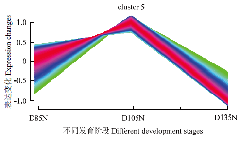
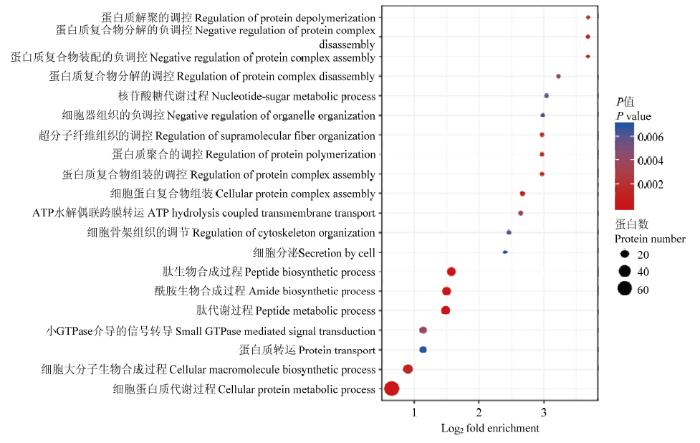
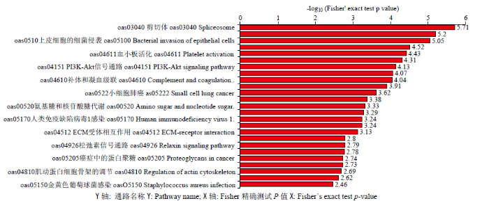
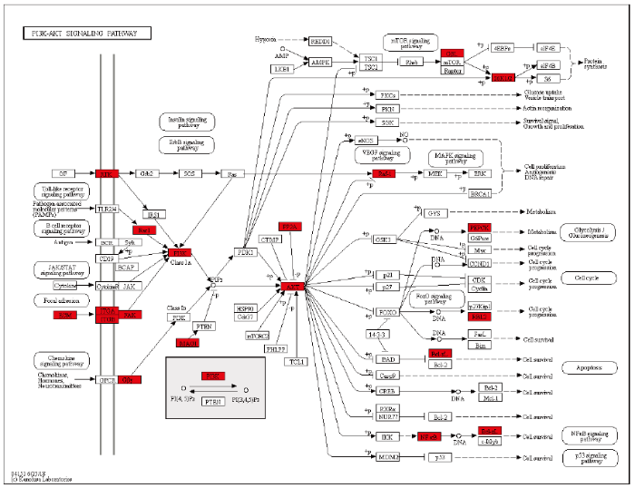
R语言表达模式聚类分析表明,cluster 5蛋白在D105N时具有较高表达趋势(图1)。通过GO和KEGG分析发现,cluster 5蛋白质参与胞内蛋白质代谢过程,并显著富集于PI3K-AKT信号通路。同时,ATK2蛋白在PI3K-AKT信号通路中显著上调(图2-4)。图1
 新窗口打开|下载原图ZIP|生成PPT
图1差异丰度蛋白质表达模式聚类分析
Fig. 1Expression patterns cluster analysis of differential abundance proteins
图2
 新窗口打开|下载原图ZIP|生成PPT
图2cluster 5差异丰度蛋白质GO生物学过程分析
Y轴:生物学过程 ; X轴:富集指数
Fig. 2GO biology process analysis of cluster 5 differential abundance proteins
Y: Enrichment index; X: Biology process
图3
 新窗口打开|下载原图ZIP|生成PPT
图3cluster 5差异丰度蛋白质KEGG富集
Y轴: 通路名称; X轴: Fisher精确测试P值
Fig. 3KEGG enrichment of cluster 5 differential abundance proteins
Y: Pathway name;X: Fisher’s exact test p-value
图4
 新窗口打开|下载原图ZIP|生成PPT
图4PI3K-AKT信号通路
红色:显著富集的上调基因
Fig. 4PI3K-AKT signaling pathway
Red: Significant enrichment up-regulation gene
2.2 AKT2蛋白生物信息学分析
2.2.1 AKT2蛋白的理化性质AKT2蛋白由481个氨基酸构成。使用ProtParam在线软件分析AKT2蛋白的理化性质,推测其分子式为C2490H3865N673O724S24,分子量为55.58kD,理论等电点(pI)为6.08,半衰期均是30 h,不稳定系数32.36,属于稳定蛋白。脂肪系数为76.61,亲水性平均系数(GRAVY)是-0.454,属于亲水性蛋白。负电荷(Asp + Glu)氨基酸残基72个,正电荷(Arg + Lys)氨基酸残基66个。
2.2.2 AKT2蛋白跨膜结构分析及其潜在N-糖基化、磷酸化位点预测
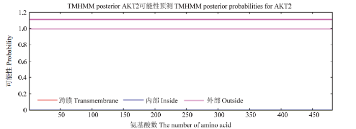
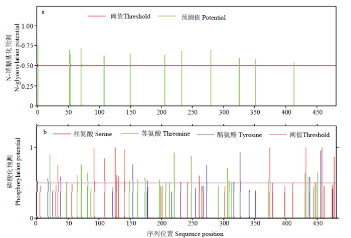
TMHMM在线预测表明,AKT2蛋白的481个氨基酸没有位于细胞膜上和膜内,全部位于膜外,属膜受体蛋白(图5)。PSORT II Prediction分析结果表明AKT2蛋白主要在65.2% 胞质、4.3% 线粒体、17.4%细胞核、4.3%分泌包囊、4.3%细胞支架。使用NetNGlyc 1.0 Server和NetPhos 3.1 Server分别预测AKT2蛋白N-端糖基化和磷酸化情况,结果显示:AKT2蛋白有12个N-糖基化位点,71个磷酸化位点,其中26个丝氨酸(Ser)磷酸化位点、26个苏氨酸(Thr)磷酸化位点、19个酪氨酸(Tyr)磷酸化位点(图6)。
图5
 新窗口打开|下载原图ZIP|生成PPT
图5AKT2蛋白跨膜结构分析
Fig. 5Transmembrane structure analysis of AKT2 protein
图6
 新窗口打开|下载原图ZIP|生成PPT
图6AKT2蛋白的糖基化和磷酸化位点预测
(a)AKT2 12个N-糖基化位点;(b)AKT2 71个磷酸化位点
Fig. 6Prediction on glycosylation and phosphorylation sites of AKT2 protein
(a)12 N-glycosylation sites in AKT2;(b)71 phosphorylation sites in AKT2
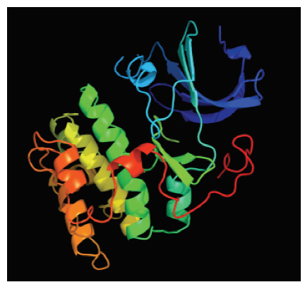
2.2.3 AKT2蛋白三级结构预测
PHYER2预测结果显示,AKT2蛋白具有α-螺旋及无规则卷曲等结构,三级结构整体呈晶体结构,与蛋白酶K相似度为99%,属于蛋白酶催化亚基(图7)。
图7
 新窗口打开|下载原图ZIP|生成PPT
图7AKT2蛋白3D结构预测
Fig. 73D structure model prediction of AKT2 protein
3 讨论
本文利用GO、KEGG和R语言等方法对差异丰度蛋白质进行聚类、功能注释和通路富集等生物信息学分析,分析结果对揭示绵羊胚胎骨骼肌生长发育关键窗口期、筛选调控蛋白具有重要意义。胚胎时期骨骼肌大部分由生肌节中的肌肉前体细胞发育而来,这些肌肉前体细胞会在初级生肌节中分化成单核肌肉细胞,初级生肌节最终生成脊椎动物早期的肌肉组织[24,25]。前期研究表明,绵羊胚胎骨骼肌纤维在胚胎期第85天至第105天增殖分化,在第105天至第135天增大增粗,而这些差异丰度蛋白质主要富集于能够调控肌纤维发生生长的代谢及氧化磷酸化等信号通路[8, 26-30]。本研究中,绵羊胚胎骨骼肌蛋白质组学数据R语言分析发现,cluster 5蛋白在胚胎发育第105天具有较高表达丰度(图1)。相关文献报道绵羊胚胎骨骼肌纤维在胚胎期第50天至第100天基本发育完成,并在第100天左右开始分化[31]。因此,初步判断D105N是调控绵羊胚胎骨骼肌发育转折点。KEGG分析发现,cluster 5蛋白质在PI3K-AKT信号通路中显著富集。由此推断,PI3K-AKT信号通路可能对胚胎时期骨骼肌发育转折及调控具有重要的作用。PI3K-AKT信号通路参与骨骼肌生长发育,能够调控细胞周期、细胞凋亡和蛋白质合成等生物过程[32]。研究表明,PI3K-AKT信号通路能够调控肌浆蛋白形成,促进肌肉分化和肥大[33]。本研究发现PI3K-AKT信号通路在绵羊胚胎骨骼肌发育转折过程中具有重要作用,AKT作为第二信使在该通路中扮演重要的角色。PI3K-AKT信号通路下游的转导因子AKT/PKB在调节个体发育、生长和细胞存活过程中发挥着重要作用[34]。ATK是一种保守的丝/苏氨酸蛋白激酶,可以调控动物胚胎发育及幼体生长,而ATK2是ATK的不同亚基也具有相同作用[35]。在正常生理条件下,PI3K-AKT信号通路由受体酪氨酸激酶(RTK)激活,并通过活化PI3K诱导PIP3激活AKT,上调下游靶基因从而调节细胞周期及分化。本研究中,RAC-β丝/苏氨酸蛋白激酶X1(ATK2)显著富集于PI3K-AKT信号通路,成肌调控因子Myostatin作为AKT的活化因子之一,也可以通过激活PI3K-AKT信号通路来调控肌肉生长[36,37,38]。因此,PI3K-AKT信号通路可以通过调控MSTN和MCK(肌酸激酶)骨骼肌发育分化标志分子表达来调控骨骼肌纤维发育及分化[39,40]。
AKT2蛋白在胞质比例较高,属于膜受体蛋白。由此可以推断,该蛋白可能在核膜上大量分布,并在蛋白质翻译时具有重要作用。而AKT2蛋白与蛋白酶催化亚基的三级结构具有较高的同源性,表明该蛋白可能是蛋白质翻译时重要的催化激活因子。同时,该蛋白的三级结构整体较为复杂,存在α-螺旋及无规则卷曲等结构,可能对配体或受体蛋白的识别和结合具有重要作用。该蛋白大量磷酸化修饰位点的发现表明,可逆磷酸化调控可能在实现AKT2蛋白质功能中起到重要作用。综上,AKT2蛋白不仅在PI3K-AKT信号通路中具有重要的信号传导及调控功能,还在绵羊胚胎骨骼肌发育分化时具有关键的调控作用,但AKT2蛋白调控肌纤维发育分化的分子机制还有待进一步验证和研究。
4 结论
通过GO二级注释、KEGG富集及R语言表达模式聚类等分析发现,蛋白质功能和富集通路均与个体发育和骨骼肌生长发育相关,第105天是绵羊胚胎骨骼肌纤维由增殖分化到增大增粗的转折点,PI3K-AKT信号通路对骨骼肌纤维生长发育转换具有调控作用。候选蛋白质生物信息学分析表明,ATK2具有重要催化调控功能,是调控PI3K-AKT信号通路信号传导的重要候选蛋白。参考文献 原文顺序
文献年度倒序
文中引用次数倒序
被引期刊影响因子
[本文引用: 1]
DOI:10.1016/s0070-2153(08)60758-9URLPMID:10635461 [本文引用: 1]
DOI:10.1016/j.crvi.2007.03.015URLPMID:17631448 [本文引用: 1]

Satellite cells, which lie under the basal lamina of muscle fibres, are marked by the expression of Pax7, and in many muscles of Pax3 also. A pure population of satellite cells, isolated from a Pax3(GFP/+) mouse line by flow cytometry, contribute very efficiently to skeletal muscle regeneration and also self-renew, thus demonstrating their role as muscle stem cells. Pax3/7 regulates the entry of these cells into the myogenic programme via the activation of the myogenic determination gene, MyoD. Pax7 is also essential for the survival of satellite cells. This dual role underlines the importance of ensuring that a tissue stem cell that has lost its myogenic instruction should not be left to run amok, with the potential risk of tissue deregulation and cancer. A somite-derived population of Pax3/Pax7 positive cells is responsible for muscle growth during development and gives rise to the satellite cells of postnatal muscles. In the absence of both Pax3 and Pax7, these cells die or assume other cell fates. Pax3/7 lies genetically upstream of both MyoD and Myf5, which determine the skeletal muscle fate of these cells. To cite this article: M. Buckingham, C. R. Biologies 330 (2007).
DOI:10.1038/nbt.2478URL [本文引用: 1]
We report the similar to 2.66-Gb genome sequence of a female Yunnan black goat. The sequence was obtained by combining short-read sequencing data and optical mapping data from a high-throughput whole-genome mapping instrument. The whole-genome mapping data facilitated the assembly of super-scaffolds >5x longer by the N50 metric than scaffolds augmented by fosmid end sequencing (scaffold N50 = 3.06 Mb, super-scaffold N50 = 16.3 Mb). Super-scaffolds are anchored on chromosomes based on conserved synteny with cattle, and the assembly is well supported by two radiation hybrid maps of chromosome 1. We annotate 22,175 protein-coding genes, most of which were recovered in the RNA-seq data of ten tissues. Comparative transcriptomic analysis of the primary and secondary follicles of a cashmere goat reveal 51 genes that are differentially expressed between the two types of hair follicles. This study, whose results will facilitate goat genomics, shows that whole-genome mapping technology can be used for the de novo assembly of large genomes.
DOI:10.1016/B978-0-12-385940-2.00001-2URLPMID:21621065 [本文引用: 1]
Muscle development, growth, and regeneration take place throughout vertebrate life. In amniotes, myogenesis takes place in four successive, temporally distinct, although overlapping phases. Understanding how embryonic, fetal, neonatal, and adult muscle are formed from muscle progenitors and committed myoblasts is an area of active research. In this review we examine recent expression, genetic loss-of-function, and genetic lineage studies that have been conducted in the mouse, with a particular focus on limb myogenesis. We synthesize these studies to present a current model of how embryonic, fetal, neonatal, and adult muscle are formed in the limb.
DOI:10.1002/1521-1878(200007)22:7<616::AID-BIES4>3.0.CO;2-RURLPMID:10878574 [本文引用: 1]
The members of the Six gene family were identified as homologues of Drosophila sine oculis which is essential for compound-eye formation. The Six proteins are characterized by the Six domain and the Six-type homeodomain, both of which are essential for specific DNA binding and for cooperative interactions with Eya proteins. Mammals possess six Six genes which can be subdivided into three subclasses, and mutations of Six genes have been identified in human genetic disorders. Characterization of Six genes from various animal phyla revealed the antiquity of this gene family and roles of its members in several different developmental contexts. Some members retain conserved roles as components of the Pax-Six-Eya-Dach regulatory network, which may have been established in the common ancestor of all bilaterians as a toolbox controlling cell proliferation and cell movement during embryogenesis. Gene duplications and cis-regulatory changes may have provided a basis for diverse functions of Six genes in different animal lineages.
DOI:10.3864/j.issn.0578-1752.2014.01.016URL [本文引用: 1]
【Objective】Skeletal satellite cells are activated by some specific stresses such as development and trauma, and differentiate and form myotubes to participate in the development or repair of skeletal muscle. FoxO1 negatively controls the genesis of skeletal muscle, but the molecular mechanisms by which FoxO1 funcions in the differentiation of satellite cells have not been reported so far. This experiment was conducted to explore the effects of FoxO1 on porcine skeletal muscle satellite cells differentiation, aiming to provide new theoretical reference for further research. 【Method】Extensor digitorum longus of 1 to 3-day-old piglets were used to isolate the skeletal muscle satellite cells and the cells were observed and pictures were taken by inverted microscope on day 2, day 4 and day 6, respectively. The cells were stained by immunofluorescence staining and DAPI nuclear staining on day 8 of differentiation, and observed under a fluorescence microscope. Meanwhile, the medium was replaced with differentiation medium containing 50 nmol•L-1 wortmannin (wortmannin, WM) differentiation medium when the cells density reached 70% -80% confluence, the cells were collected on day 0, day 4, and day 8, respectively. Total RNA and total protein were extracted, and Real-time qPCR and Western blotting were performed to measure the alterations in the expression of FOXO1 and myogenic differentiation marker genes caused by WM supplement. 【Result】Porcine skeletal muscle satellite cells became adherent to the dish bottem, spindle-shaped on day 2. Cell number increased on day 4 and some cells started to fuse. On day 6, cell started to grow with directivity. On day 8, cell further fused to form myotubes, further fused to form myotubes. There was no significant difference in mRNA expression level of FoxO1 between WM treatment group and the control (P>0.05), unphosphorylated FoxO1 increased significantly (P<0.05) with WM treatment, whereas phosphorylation level of FoxO1 dropped drastically (P<0.05). Although on day 8 the cells displayed an alveolate morphology after treated with WM, they failed to show directional growth and formation of myotubes. Moreover, Western blotting results demonstrated that WM decreased the protein level of MyoD (early myogenic marker), MyoG (middle-stage marker), and MyHC (late marker) significantly.【Conclusion】Results of the study suggest that inhibition of PI3K signaling pathway by WM blocks results in FoxO1 phosphorylation, suppression of porcine skeletal muscle satellite cell differentiation, delay of the formation of myotubes, and down-regulation of myogenic differentiation marker genes, such as MyoD, MyoG, and MyHC. Take together, blockade of PI3K signaling pathway suppresses porcine skeletal muscle satellite cell differentiation through the activation of FoxO1.
URLPMID:27508388 [本文引用: 2]
DOI:10.1016/0014-4886(72)90071-4URLPMID:4118074 [本文引用: 1]
[本文引用: 1]
[本文引用: 1]
URLPMID:28533755 [本文引用: 1]
URLPMID:29510239 [本文引用: 1]
DOI:10.1186/s12864-016-2464-1URL [本文引用: 1]
URLPMID:17093219 [本文引用: 1]
URLPMID:12713048 [本文引用: 1]
DOI:10.3864/j.issn.0578-1752.2020.03.015URL [本文引用: 1]
【Objective】The meat production of livestock, which is closely related to the development of skeletal muscle, is an important economic trait to measure the quality of livestock. For mammals, the skeletal muscle development depends on the growth and differentiation of embryonic myocyte, which has a significant impact on the subsequent growing potential. In this study, the developmental mode of skeletal muscle, the important transformation nodes, the formation of muscle fibers and the molecular regulation mechanism of transformation were mainly explored. 【Method】 Based on the previous research, the important nodes D85, D105 and D135 related to the myotube development were used in the experiment, and the longissimus dorsi muscles were sequenced by whole transcriptome sequencing. The differentially expressed (DE) circRNAs were screened by bioinformatics analysis and verified by quantitative real-time PCR (qRT-PCR). 【Result】 1 126 DE circRNAs were obtained by conditional screening (|log2| ≥1 and P≤0.05). The 3 groups were compared and many specific expressions of circRNA were found at each stage, but in the D85 vs D135 group, the amount was the most. 374 DE circRNAs were obtained, which contained 201 up-regulated and 173 down-regulated, and 44.7% of the DE genes were differentially expressed with a difference of more than 4 times. These DE circRNAs were subjected to run GO and KEGG functional analysis and targeted prediction, and they were enriched into some pathways, such as energy metabolism and signal transduction, which involved in muscle differentiation and muscle fiber development, including MAPK, PI3K-Akt, Ras, regulation of actin cytoskeleton and other signal transduction pathways. According to the results, it was confirmed that the DE circRNAs enriched during D85 to D105 were mostly associated with cell proliferation and survival, regulation of myocyte development and cell cycle, while D105 to D135 were mainly related to energy conversion, material transport, RNA transport, and DNA repair. By drawing co-expression visualization network with the targeted prediction results used by Cytoscape, the core regulatory transcripts, such as circRNA8239, circRNA19073, circRNA2765 and circRNA1616, were identified. In the D105 period, a key factor circRNA7527 that regulated the conversion of fast and slow muscle types was found, which targets the bta-miR-135a, bta-miR-615, and chi-miR-133a-5p to regulate the MEF2C gene. According to the differential expression and functional prediction in three comparison groups, 4 circRNAs related to muscle development and 4 target miRNA were selected for qRT-PCR, and the results showed that the gene expression trend was consistent with the sequencing data. 【Conclusion】 It was verified that the stabilization of the number of muscle fibers occurred between sheep embryos at D85 and D105, and muscle fiber hypertrophy happened during the D105 to D135 period, which lead to the conclusion that D105 was probably a key time point. In this study, we firstly constructed a circRNA map in sheep embryonic skeletal muscle development based on the whole transcriptome sequencing. The transcriptome differences at key stages were revealed, and multiple circRNAs and miRNAs targeting MEF2C that involved in the MAPK signaling pathway were found, which provided reference for livestock myofiber development research and other research on non-coding RNA.
[本文引用: 1]
[本文引用: 1]
URLPMID:18084642 [本文引用: 1]
[本文引用: 1]
[本文引用: 1]
URLPMID:10600390 [本文引用: 1]
DOI:10.1002/pmic.200300771URLPMID:15174133 [本文引用: 1]
Post-translational modifications (PTMs) occur on almost all proteins analyzed to date. The function of a modified protein is often strongly affected by these modifications and therefore increased knowledge about the potential PTMs of a target protein may increase our understanding of the molecular processes in which it takes part. High-throughput methods for the identification of PTMs are being developed, in particular within the fields of proteomics and mass spectrometry. However, these methods are still in their early stages, and it is indeed advantageous to cut down on the number of experimental steps by integrating computational approaches into the validation procedures. Many advanced methods for the prediction of PTMs exist and many are made publicly available. We describe our experiences with the development of prediction methods for phosphorylation and glycosylation sites and the development of PTM-specific databases. In addition, we discuss novel ideas for PTM visualization (exemplified by kinase landscapes) and improvements for prediction specificity (by using ESS--evolutionary stable sites). As an example, we present a new method for kinase-specific prediction of phosphorylation sites, NetPhosK, which extends our earlier and more general tool, NetPhos. The new server, NetPhosK, is made publicly available at the URL http://www.cbs.dtu.dk/services/NetPhosK/. The issues of underestimation, over-prediction and strategies for improving prediction specificity are also discussed.
DOI:10.1038/emboj.2013.79URLPMID:23584533 [本文引用: 1]
Glycosylation is the most abundant and diverse posttranslational modification of proteins. While several types of glycosylation can be predicted by the protein sequence context, and substantial knowledge of these glycoproteomes is available, our knowledge of the GalNAc-type O-glycosylation is highly limited. This type of glycosylation is unique in being regulated by 20 polypeptide GalNAc-transferases attaching the initiating GalNAc monosaccharides to Ser and Thr (and likely some Tyr) residues. We have developed a genetic engineering approach using human cell lines to simplify O-glycosylation (SimpleCells) that enables proteome-wide discovery of O-glycan sites using 'bottom-up' ETD-based mass spectrometric analysis. We implemented this on 12 human cell lines from different organs, and present a first map of the human O-glycoproteome with almost 3000 glycosites in over 600 O-glycoproteins as well as an improved NetOGlyc4.0 model for prediction of O-glycosylation. The finding of unique subsets of O-glycoproteins in each cell line provides evidence that the O-glycoproteome is differentially regulated and dynamic. The greatly expanded view of the O-glycoproteome should facilitate the exploration of how site-specific O-glycosylation regulates protein function.
URLPMID:9108376 [本文引用: 1]
URLPMID:12135925 [本文引用: 1]
The dorsomedial lip (DML) of the somite dermomyotome is the source of cells for the early growth and morphogenesis of the epaxial primary myotome and the overlying dermomyotome epithelium. We have used quail-chick transplantation to investigate the mechanistic basis for DML activity. The ablated DML of chick wing-level somites was replaced with tissue fragments from various mesoderm regions of quail embryos and their capacity to form myotomal tissue assessed by confocal microscopy. Transplanted fragments from the epithelial sheet region of the dermomyotome exhibited full DML growth and morphogenetic capacity. Ventral somite fragments (sclerotome), head paraxial mesoderm or non-paraxial (lateral plate) mesoderm tested in this assay were each able to expand mitotically in concert with the surrounding paraxial mesoderm, although no myogenic potential was evident. When ablated DMLs were replaced with fragments of the dermomyotome ventrolateral lip of wing-level somites or pre-somitic mesoderm (segmental plate), myotome development was evident but was delayed or otherwise limited in some cases. Timed DML ablation-replacement experiments demonstrate that DML activity is progressive throughout the embryonic period (to at least E7) and its continued presence is necessary for the complete patterning of each myotome segment. The results of serial transplantation and BrdU pulse-chase experiments are most consistent with the conclusion that the DML consists of a self-renewing population of progenitor cells that are the primary source of cells driving the growth and morphogenesis of the myotome and dermomyotome in the epaxial domain of the body.
DOI:10.1016/j.devcel.2004.07.019URLPMID:15469841 [本文引用: 1]
We have carried out a small pool expression screen for modulators of the Wnt/beta-catenin pathway and identified Xenopus R-spondin2 (Rspo2) as a secreted activator of this cascade. Rspo2 is coexpressed with and positively regulated by Wnt signals and synergizes with Wnts to activate beta-catenin. Analyses of functional interaction with components of the Wnt/beta-catenin pathway suggest that Rspo2 functions extracellularly at the level of receptor ligand interaction. In addition to activating the Wnt/beta-catenin pathway, Rspo2 overexpression blocks Activin, Nodal, and BMP4 signaling in Xenopus, raising the possibility that it may negatively regulate the TGF-beta pathway. Antisense Morpholino experiments in Xenopus embryos and RNAi experiments in HeLa cells reveal that Rspo2 is required for Wnt/beta-catenin signaling. In Xenopus embryos depleted of Rspo2, the muscle markers myoD and myf5 fail to be activated and later muscle development is impaired. Thus, Rspo2 functions in a positive feedback loop to stimulate the Wnt/beta-catenin cascade.
URLPMID:9753670
Activation of myogenesis in newly formed somites is dependent upon signals derived from neighboring tissues, namely axial structures (neural tube and notochord) and dorsal ectoderm. In explants of paraxial mesoderm from mouse embryos, axial structures preferentially activate myogenesis through a Myf5-dependent pathway and dorsal ectoderm preferentially through a MyoD-dependent pathway. Here we report that cells expressing Wnt1 will preferentially activate Myf5 while cells expressing Wnt7a will preferentially activate MyoD. Wnt1 is expressed in the dorsal neural tube and Wnt7a in dorsal ectoderm in the early embryo, therefore both can potentially act in vivo to activate Myf5 and MyoD, respectively. Wnt4, Wnt5a and Wnt6 exert an intermediate effect activating both Myf5 and MyoD equivalently in paraxial mesoderm. Sonic Hedgehog synergises with both Wnt1 and Wnt7a in explants from E8.5 paraxial mesoderm but not in explants from E9.5 embryos. Signaling through different myogenic pathways may explain the rescue of muscle formation in Myf5 null embryos, which do not form an early myotome but later develop both epaxial and hypaxial musculature. Explants of unsegmented paraxial mesoderm contain myogenic precursors capable of expressing MyoD in response to signaling from a neural tube isolated from E10.5 embryos, the developmental stage when MyoD is present throughout the embryo. Myogenic cells cannot activate MyoD in response to signaling from a less mature neural tube. Together these data suggest that different Wnt molecules can activate myogenesis through different pathways such that commitment of myogenic precursors is precisely regulated in space and time to achieve the correct pattern of skeletal muscle development.
URLPMID:15328533
URLPMID:32019949 [本文引用: 1]
[本文引用: 1]
[本文引用: 1]
DOI:10.1371/journal.pone.0119396URLPMID:25950587 [本文引用: 1]
MicroRNAs (miRNAs), which are short (22-24 base pairs), non-coding RNAs, play critical roles in myogenesis. Using Solexa deep sequencing, we detected the expression levels of 229 and 209 miRNAs in swine skeletal muscle at 90 days post-coitus (E90) and 100 days postnatal (D100), respectively. A total of 138 miRNAs were up-regulated on E90, and 31 were up-regulated on D100. Of these, 9 miRNAs were selected for the validation of the small RNA libraries by quantitative RT-PCR (RT-qPCR). We found that miRNA-21 was down-regulated by 17-fold on D100 (P<0.001). Bioinformatics analysis suggested that the transforming growth factor beta-induced (TGFbetaI) gene was a potential target of miRNA-21. Both dual luciferase reporter assays and western blotting demonstrated that the TGFbetaI gene was regulated by miRNA-21. Co-expression analysis revealed that the mRNA expression levels of miRNA-21 and TGFbetaI were negatively correlated (r = -0.421, P = 0.026) in skeletal muscle during the 28 developmental stages. Our results revealed that more miRNAs are expressed in prenatal than in postnatal skeletal muscle. The miRNA-21 is a novel myogenic miRNA that is involved in skeletal muscle development and regulates PI3K/Akt/mTOR signaling by targeting the TGFbetaI gene.
DOI:10.1038/ncb1101-1009URLPMID:11715022 [本文引用: 1]
Skeletal muscle is composed of multinucleated fibres, formed after the differentiation and fusion of myoblast precursors. Skeletal muscle atrophy and hypertrophy refer to changes in the diameter of these pre-existing muscle fibres. The prevention of atrophy would provide an obvious clinical benefit; insulin-like growth factor 1 (IGF-1) is a promising anti-atrophy agent because of its ability to promote hypertrophy. However, the signalling pathways by which IGF-1 promotes hypertrophy remain unclear, with roles suggested for both the calcineurin/NFAT (nuclear factor of activated T cells) pathway and the PtdIns-3-OH kinase (PI(3)K)/Akt pathway. Here we employ a battery of approaches to examine these pathways during the hypertrophic response of cultured myotubes to IGF-1. We report that Akt promotes hypertrophy by activating downstream signalling pathways previously implicated in activating protein synthesis: the pathways downstream of mammalian target of rapamycin (mTOR) and the pathway activated by phosphorylating and thereby inhibiting glycogen synthase kinase 3 (GSK3). In contrast, in addition to demonstrating that calcineurin does not mediate IGF-1-induced hypertrophy, we show that IGF-1 unexpectedly acts via Akt to antagonize calcineurin signalling during myotube hypertrophy.
URLPMID:11882383 [本文引用: 1]
URLPMID:18801898 [本文引用: 1]
DOI:10.1016/j.cellsig.2008.03.013URLPMID:18472397 [本文引用: 1]
Myostatin is a negative regulator of skeletal muscle growth and affects numerous genes expression involved in cell proliferation, differentiation and metabolism. However, the molecular mechanisms underlying myostatin-regulated genes expression remain to be elucidated. In this study, we showed that myostatin blocked the recruitment of p300 to the cyclin D1 promoter, resulting in the silence of cyclin D1 expression. Our data further demonstrated that myostatin decreased the protein level of p300 by inducing p300 degradation via the ubiquitin-proteasome system. In addition, we provided experimental evidence to show that myostatin-induced p300 degradation was mediated by the phosphatidylinositol 3-kinase/PTEN/Akt signaling pathway and this could be antagonized by IGF-1 or insulin. Results presented in this study uncovered an epigenetic control of genes expression in response to myostatin.
URLPMID:19357233 [本文引用: 1]
[本文引用: 1]
[本文引用: 1]
[本文引用: 1]
[本文引用: 1]
DOI:10.1128/mcb.23.14.4991-5004.2003URLPMID:12832484 [本文引用: 1]
In this report, we investigate the role of the RNA-binding protein HuR during skeletal myogenesis. At the onset of myogenesis in differentiating C2C12 myocytes and in vivo in regenerating mouse muscle, HuR cytoplasmic abundance increased dramatically, returning to a predominantly nuclear presence upon completion of myogenesis. mRNAs encoding key regulators of myogenesis-specific transcription (myogenin and MyoD) and cell cycle withdrawal (p21), bearing AU-rich regions, were found to be targets of HuR in a differentiation-dependent manner. Accordingly, mRNA half-lives were highest during differentiation, declining when differentiation was completed. Importantly, HuR-overexpressing C2C12 cells displayed increased target mRNA expression and half-life and underwent precocious differentiation. Our findings underscore a critical function for HuR during skeletal myogenesis linked to HuR's coordinate regulation of muscle differentiation genes.
