 ,, 李珣,广西大学动物科学技术学院,南宁 530004
Effects of GnIH on Autophagy and Apoptosis of Porcine Ovarian Granulosa Cells via p38MAPK Signaling Pathway
ZHANG Xin, HUO KongLin, SONG XingXing, ZHANG DuoNi, HU Wen, HU ChuanHuo,, LI Xun,College of Animal Science and Technology, Guangxi University, Nanning 530004通讯作者:
责任编辑: 林鉴非
收稿日期:2018-11-30接受日期:2020-03-11网络出版日期:2020-05-16
| 基金资助: |
Received:2018-11-30Accepted:2020-03-11Online:2020-05-16
作者简介 About authors
张鑫,Tel:15676119603;E-mail:2507854337@qq.com。

摘要
关键词:
Abstract
Keywords:
PDF (2774KB)元数据多维度评价相关文章导出EndNote|Ris|Bibtex收藏本文
本文引用格式
张鑫, 霍孔林, 宋星星, 张多妮, 胡文, 胡传活, 李珣. GnIH通过p38MAPK信号通路对猪卵巢颗粒细胞自噬与凋亡的影响[J]. 中国农业科学, 2020, 53(9): 1904-1912 doi:10.3864/j.issn.0578-1752.2020.09.016
ZHANG Xin, HUO KongLin, SONG XingXing, ZHANG DuoNi, HU Wen, HU ChuanHuo, LI Xun.
0 引言
【研究意义】卵巢闭锁是哺乳动物卵巢中普遍存在的现象[1]。卵巢闭锁对母猪的产子率有影响。促性腺激素抑制激素(gonadotropin-inhibitory hormone,GnIH)作为一种由下丘脑释放的性腺激素,我们猜测,对卵巢细胞活性有着一定的调控作用。为此,本试验可以为解决母猪的产子率以及同期发情[2]等问题提供参考。【前人研究进展】GnIH是TSUTSUI等于2000年从鹌鹑的下丘脑中分离的一种C末端具有RF酰胺结构的12肽[3]。后续研究发现哺乳动物中存在GnIH类似物–RF酰胺相关肽(RFRP),RFRP 基因主要编码两条有生物活性的短肽,分别为RFRP-1、RFRP-3,然而在大多数哺乳动物生殖系统中发挥主要作用的是RFRP-3[4],也是目前研究较为广泛的[5,6,7]。人类和其他物种的研究数据表明,RFRP-3可能参与病理变化,是影响排卵、卵泡发育和卵巢分泌类固醇的因素[8,9]。GnIH可以通过对下丘脑GnRH和垂体前叶的作用抑制促性腺激素释放[10]。GnIH在生殖调控中具有重要作用,在其他脊椎动物中也具有类似的作用[11]。细胞的活性决定各项生理机能,由此凋亡作为第一个细胞程序性死亡被认知[12],并成为哺乳动物组织细胞死亡的主要机制。但近来研究发现凋亡并不能单独对细胞死亡起决定性作用,由此自噬被发现[13],并探究出自噬与凋亡存在促进与抑制的双重作用。已有研究证明,肿瘤细胞中自噬可以使凋亡率下降[14]。自噬与凋亡也可以共同导致细胞死亡,当自噬或凋亡受到抑制,另外一种则会转变成细胞的死亡途径[15,16]。当自噬体积累到一定水平,则会对细胞的凋亡产生影响。卵母细胞质量是由卵泡闭锁这一正常的生理过程决定的,而细胞的自噬与凋亡可能参与调节卵泡闭锁[17]。研究表明,卵泡的发育和颗粒细胞的凋亡过程中都存在着细胞自噬[18],因此自噬可能参与颗粒细胞的凋亡;大鼠颗粒细胞的自噬可能与卵泡发育有关[19],CHOI在研究自噬与卵泡闭锁的关系中发现,在卵泡的发育过程中,自噬主要集中在卵巢颗粒细胞,并且与细胞的凋亡存在着相关性[20]。在卵泡闭锁的初期,颗粒细胞中有Beclin-1的表达,而Caspase-3则是分布在卵泡中;在中期,卵巢颗粒细胞中Beclin-1和Caspase-3均有表达;而在晚期,颗粒细胞中Caspase-3的表达水平较高[19]。LC3在大鼠卵泡颗粒细胞中表达量最高, 也表明自噬主要发生在颗粒细胞中, 而自噬导致的颗粒细胞死亡可能参与卵泡闭锁[21]。p38MAPK信号通路是细胞调节的主要信号通路之一[22],MAPK信号通路可能参与调节GCs的凋亡[23]。GnIH参与调节小鼠卵巢的发育及功能[24,25]。目前已证实,GnIH对母猪生殖有一定的调控作用[26]。MADDINENI于2008年发现GnIH能减少鸡卵巢颗粒细胞的活化,但是这种抑制作用能被外源FSH中和[27,28]。在睾丸细胞中,GnIH与GnIH-R均有表达[29],表明GnIH可能参与生殖细胞的分化。给成熟鹌鹑连续14 d注射GnIH,发现睾丸中精原细胞活性降低,并诱导细胞凋亡[30],提示GnIH可能通过诱导细胞凋亡从而调节性腺的发育。研究表明,GnIH对小鼠附睾组织细胞的凋亡和自噬有影响[31]。【本研究切入点】虽然目前已有关于GnIH在卵巢水平的相关研究。但是GnIH处理的pGCs通过p38MAPK信号通路对细胞凋亡与自噬的调控还未见报道。因此,我们提出猜测,GnIH可能通过p38MAPK信号通路对细胞凋亡与自噬产生影响。【拟解决的关键问题】首先探究了GnIH是否对p38MAPK信号通路的活化产生影响;进一步探究了GnIH是否对卵巢颗粒细胞的自噬和凋亡有影响;最后探究了GnIH是否是通过p38MAPK信号通路对自噬和凋亡产生的影响。1 材料与方法
1.1 材料和试剂
本试验于2018年3—10月在广西大学解剖组培实验室完成。DMEM/F12培养基、澳洲来源胎牛血清购自南京维森特生物技术有限公司。台盼蓝染液购自Gibco。其他试剂均为国产分析纯。Western细胞裂解液、超敏ECL化学发光试剂盒、BCA蛋白定量试剂盒购于上海碧云天生物技术有限公司,一抗GAPDH、p38、p-p38、Bcl-2、Caspase3、Bax、Beclin-1、LC3、Atg5和Atg12浓度均为1﹕2 000,二抗羊抗鼠、羊抗兔浓度为1﹕20 000均购自CST公司。GnIH由实验室于NCBI查找相关氨基酸序列,送至公司合成。
1.2 猪卵巢的采集
从广西本地屠宰场当天取回卵巢,将卵巢放置在含有两性霉素(青霉素/链霉素(1%))的生理盐水中(37℃),2 h内运输回广西大学解剖组培实验室。用生理盐水冲洗备用。1.3 猪卵巢颗粒细胞的体外培养
pGCs分离培养方法参考相关研究[32,33]进行。于37℃、5%CO2及饱和湿度的培养箱中进行成熟培养,培养48 h后,加入p38MAPK激活剂U-46619和不同浓度的GnIH(购自康肽生物(北京)科技有限公司),培养1 h后,收取细胞,每组试验平行3次重复。1.4 猪卵巢颗粒细胞蛋白的提取
弃去细胞瓶中培养液,加入PBS,洗2—3次,弃洗液;加入混有1%PMSF的细胞裂解液,静置30 min,每隔10 min轻轻晃动细胞瓶;裂解后,将细胞瓶内裂解液移入1.5 mL预冷的EP管,4℃、12 000 r/min离心5 min;BCA测浓度,分装,-80℃保存。1.5 Western blot检测
12%SDS-PAGE凝胶电泳,半干法转印至PVDF膜上,放入5%脱脂牛奶,室温封闭1—2 h,一抗p38、p-p38、Bcl-2、Caspase3、Bax、Beclin-1、LC3、Atg5和Atg12以及GAPDH,4℃过夜,二抗孵育1 h,每步完成后,PBST洗涤3次,每次15 min。ECL化学发光液显色,暗室曝光,扫描灰度值。试验重复3次,用 ImageJ进行灰度值分析,其中GAPDH作为标准化的内部对照。1.6 数据分析
所有数据均显示为平均数±标准误(Mean±SE)。使用SPSS 21.0统计软件进行单因素方差分析,LSD检验各组之间的统计学显著性。检验结果以*P<0.05为差异显著,**P<0.01为差异极显著。2 结果
2.1 GnIH对猪卵巢颗粒细胞p38MAPK信号通路的最佳作用时间
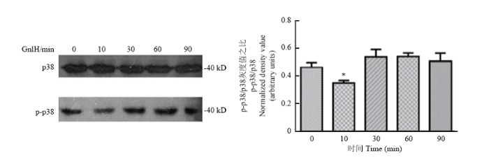
在GnIH梯度时间(0、10、30、60、90 min)孵育pGCs后,采用Western blot检测p38及p-p38的表达变化,检测灰度值。结果提示10 min与其他试验组比较,p38及p-p38显著降低(P<0.05)(图1)。图1
 新窗口打开|下载原图ZIP|生成PPT
图110-6 mol·L-1 GnIH(0、10、30、60、90 min)处理p38及p-p38的免疫印迹及统计学分析
用ImageJ进行灰度值分析。数值为平均值±S.E,与对照组比较,星号表示显著差异。*P<0.05;**P<0.01
Fig. 1Immunoblot and statistical analysis of p38 and p-p38 proteins with different time of RFRP-3 (0, 10, 30, 60, 90 min, n=3)
Densitometric quantification was performed using ImageJ with GAPDH as the internal control for normalization. Values are mean±S.E. Compared with control group, asterisk indicates significant difference. *P<0.05; **P<0.01
2.2 GnIH对p38MAPK信号通路的影响
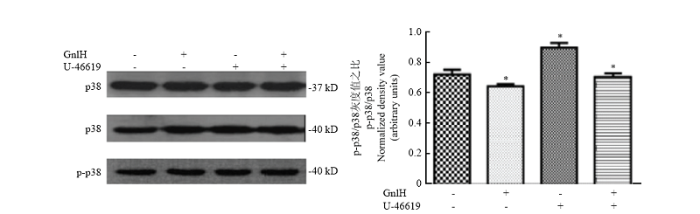
Western blot检测(空白、GnIH、p38激活剂、p38激活剂+GnIH)p38及p-p38的表达变化,检测灰度值。GnIH组与空白组比较,p38及p-p38的比值显著降低(P<0.05);p38激活剂组与空白组比较,p38及p-p38的比值显著升高(P<0.05);p38激活剂+GnIH组与p38激活剂比较,p38及p-p38的比值显著降低(P<0.05,图2)。图2
 新窗口打开|下载原图ZIP|生成PPT
图2按空白对照、GnIH、U-46619、U-46619+GnIH四组处理pGCs,Western blot检测p38及p-p38的免疫印迹及统计学分析
用ImageJ进行灰度值分析。数值为平均值±S.E,与对照组比较,星号表示显著差异。*P<0.05;**P<0.01
Fig. 2Immunoblot and statistical analysis of p38 and p-p38 proteins (control, GnIH, U-46619, GnIH+U-46619) (n=3)
Densitometric quantification was performed using ImageJ with GAPDH as the internal control for normalization. Values are mean±S.E. Compared with control group, asterisk indicates significant difference. *P<0.05; **P<0.01
2.3 不同浓度GnIH对猪卵巢颗粒细胞自噬和凋亡的影响
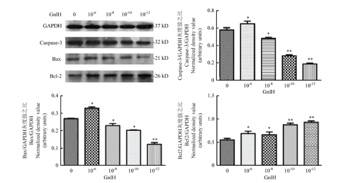
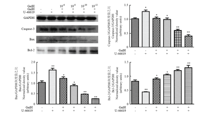
GnIH浓度梯度(0、10-6、10-8、10-10、10-12)处理pGCs,Western blot检测Caspase-3、Bax和Bcl-2,灰度值分析。caspase-3、Bcl-2、Bax和GAPDH的免疫印迹在大约32、26、21和37 kD。试验组与对照组相比均有显著性差异(P<0.05),结果提示,低剂量组Bcl-2的表达极显著升高(P<0.01),caspase-3与Bax的表达极显著降低(P<0.01,图3)。图3
 新窗口打开|下载原图ZIP|生成PPT
图3不同剂量GnIH(0、10-6、10-8、10-10、10-12)处理pGCs,Western blot检测caspase-3、Bax和Bcl-2的免疫印迹及统计学分析
用ImageJ进行灰度值分析。数值为平均值±S.E,与对照组比较,星号表示显著差异。*P<0.05;**P<0.01
Fig. 3RFRP-3 concentration gradient (0, 10-6, 10-8, 10-10, 10-12) treated with pGCs.Western blot caspase-3, Bax and Bcl-2 and statistical analysis (n=3)
Densitometric quantification was performed using ImageJ with GAPDH as the internal control for normalization. Values are mean±S.E. Compared with control group, asterisk indicates significant difference. *P<0.05; **P<0.01
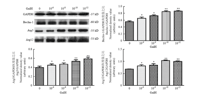
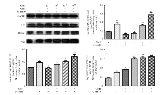
Western blot测定Beclin-1、Atg12和Atg5,灰度值分析。Beclin-1、Atg5和Atg12的免疫印迹分别位于60、55和53 kD。结果提示,低剂量组Beclin-1、Atg12和Atg5的表达极显著升高(P<0.01,图4)。
图4
 新窗口打开|下载原图ZIP|生成PPT
图4不同剂量GnIH(0、10-6、10-8、10-10、10-12)处理pGCs,Western blot检测Beclin-1、Atg12和Atg5的免疫印迹及统计学分析
用ImageJ进行灰度值分析。数值为平均值±S.E,与对照组比较,星号表示显著差异。*P<0.05;**P<0.01
Fig. 4RFRP-3 concentration gradient (0, 10-6, 10-8, 10-10, 10-12) treated with pGCs. Western blot Beclin-1, Atg12 and Atg5 and statistical analysis (n=3)
Densitometric quantification was performed using ImageJ with GAPDH as the internal control for normalization. Values are mean±S.E.M. Compared with control group, asterisk indicates significant difference. *P<0.05; **P<0.01
2.4 不同浓度GnIH通过p38MAPK信号通路对猪卵巢颗粒细胞自噬和凋亡的影响
p38激活剂孵育pGCs后,GnIH浓度梯度(0、10-6、10-8、10-10、10-12)处理pGCs,Western blot检测Caspase-3、Bax和Bcl-2,灰度值分析。结果提示,P38激活剂组与空白对照相比,存在显著性差异(P<0.05);经不同剂量GnIH处理后的试验组与p38激活剂组比,低剂量组Bcl-2的表达极显著升高(P<0.01);caspase-3与Bax的表达极显著降低(P<0.01,图5)。图5
 新窗口打开|下载原图ZIP|生成PPT
图5用p38激活剂孵育,用不同剂量GnIH(0、10-6、10-8、10-10、10-12)处理pGCs,Western blot检测caspase-3、Bax和Bcl-2的免疫印迹及统计学分析
用ImageJ进行灰度值分析。数值为平均值±S.E,与对照组比较,星号表示显著差异。*P<0.05;**P<0.01
Fig. 5After incubated with p38 activator, RFRP-3 concentration gradient (0, 10-6, 10-8, 10-10, 10-12) was treated with pGCs.Western blot caspase-3,Bax and Bcl-2 and statistical analysis (n=3)
Densitometric quantification was performed using ImageJ with GAPDH as the internal control for normalization. Values are mean±S.E. Compared with control group, asterisk indicates significant difference. *P<0.05; **P<0.01
Western blotting测定Beclin-1、LC3和Atg5,灰度值分析。结果显示,P38激活剂组与空白比较,存在显著性差异(P<0.05);经不同剂量GnIH处理后的试验组与p38激活剂组比较,均有显著性差异(P<0.05);低剂量组Beclin-1、LC3和Atg5的表达极显著升高(P<0.01,图6)。
图6
 新窗口打开|下载原图ZIP|生成PPT
图6用p38激活剂孵育,再用不同剂量GnIH(0、10-6、10-8、10-10、10-12)处理pGCs,Western blot检测Beclin-1、LC3和Atg5的免疫印迹及统计学分析(n=3)。用ImageJ进行灰度值分析
数值为平均值±S.E,与对照组比较,星号表示显著差异。*P<0.05;**P<0.01
Fig. 6After incubated with p38 activator, RFRP-3 concentration gradient (0, 10-6, 10-8, 10-10, 10-12) was treated with pGCs. Western blot Beclin-1,LC3 and Atg-5 and statistical analysis
Densitometric quantification was performed using ImageJ with GAPDH as the internal control for normalization. Values are mean±S.E. Compared with control group, asterisk indicates significant difference. *P<0.05; **P<0.01
3 讨论
相关研究表明,在GT1-7 细胞系上,mRFRP3预处理可使vip诱导的p38磷酸化受到抑制,此结果显示GnIH具有抑制p38磷酸化的作用[34]。本研究中,体外培养pGCs,加入GnIH孵育,检测其对p38MAPK信号通路活化的影响。当GnIH作用10 min时,对p38MAPK信号通路活化的影响最显著(P<0.05);加入p38激活剂后,GnIH可以有效抑制p38MAPK信号通路活化。与前人的结论一致,GnIH可以抑制p38磷酸化。已有研究表明,在卵巢颗粒细胞中存在着细胞自噬与凋亡。我们猜想GnIH可能与颗粒细胞的自噬和凋亡有关。结果提示,高浓度GnIH可以促进细胞的自噬和凋亡,随着GnIH浓度的降低,凋亡水平逐渐降低,而自噬水平逐渐升高。2017年DAVE等对动情前期小鼠卵巢进行了研究,研究发现,RFRP-1使Caspase-3、凋亡标记物明显减少[35]。与本试验结果一致。前期实验室已证明GnIH可以调控附睾的自噬与凋亡[33],GnIH对附睾凋亡出现了极显著的促进作用,而对自噬则出现了双向调控。与本试验结果不一致。猜测可能原因卵巢颗粒细胞是自噬的主要场所,有研究证明,细胞可以通过促进自噬来抑制凋亡的产生,从而提高细胞活性;附睾为体内试验,猜测动物体内对GnIH有神经激素调节作用,导致结果不同;通过试验验证,激素的浓度与作用时间均会对细胞的自噬与凋亡产生影响。
p38MAPK磷酸化水平升高,发挥凋亡促进的作用[36,37],研究证实,抑制p38MAPK信号通路可以通过阻止COCs扩散,从而影响GCs的生长,该结论在大鼠GCs和卵母细胞选择性敲除p38受体中也得到了证实[38];INAGAKI等发现大鼠GCs中存在p38受体,用p38抑制剂SB203580抑制大鼠GCs的p38受体,试验显示通过降低FSH,从而降低了雌二醇和孕激素的产生,充分证明p38及其受体在GCs中发挥重要作用,进而促进卵母细胞成熟。当p38MAPK信号通路被激活,能通过调节细胞形态和提高细胞存活率促进细胞分化[39]。本试验加入p38MAPK激活剂,结果提示,p38MAPK激活剂可以促进p38MAPK的磷酸化;p38MAPK激活剂可以促进卵巢颗粒细胞的凋亡和自噬,与前人研究一致。加入GnIH后,结果提示,不同浓度的GnIH对p38MAPK信号通路活化都具有抑制作用。我们进一步探究了p38MAPK信号通路活化后,GnIH对卵巢颗粒细胞自噬和凋亡的影响,从而探究GnIH是否通过p38MAPK信号通路对卵巢颗粒细胞的自噬和凋亡进行调控。通过检测自噬和凋亡的标志性蛋白,我们得出,随着GnIH浓度的降低,Caspase-3和Bax的表达呈下降趋势,Bcl-2的表达呈上调趋势,提示了GnIH通过抑制p38MAPK信号通路活化对pGCs的凋亡存在高浓度促进,低浓度抑制的作用;Beclin-1、Atg12和Atg5的表达呈上调趋势,提示了GnIH对通过抑制p38MAPK信号通路活化对自噬有促进作用,GnIH的浓度越低,促进作用越明显,且浓度低于10-10 mol·L-1,有极显著的促进作用(P<0.01)。以往研究表明,自噬的作用不仅通过增强caspase的活化导致细胞凋亡,还可以通过凋亡导致细胞死亡。通过应激反应,延缓caspase的激活,可以清除受损细胞[40],这些数据证明了细胞凋亡和自噬之间存在着明显的复杂性,有时甚至相互矛盾。
4 结论
本研究证明了促性腺激素抑制激素可以通过调控p38MAPK信号通路影响猪卵巢颗粒细胞的凋亡和自噬。自噬和凋亡的关系异常复杂,自噬和凋亡又存在很多交叉的信号通路。因此,本试验的结果为后续研究猪卵巢颗粒细胞自噬与凋亡的分子机制提供了参考,也为研究卵泡闭锁提供了新的思路。参考文献 原文顺序
文献年度倒序
文中引用次数倒序
被引期刊影响因子
[本文引用: 1]
[本文引用: 1]
[本文引用: 1]
[本文引用: 1]
DOI:10.1006/bbrc.2000.3350URLPMID:10964719 [本文引用: 1]

The neuropeptide control of gonadotropin secretion at the level of the anterior pituitary gland is primarily through the stimulatory action of the hypothalamic decapeptide, gonadotropin-releasing hormone (GnRH), which was originally isolated from mammals and subsequently from non-mammals. To date, however, an inhibitory peptide of gonadotropin release is unknown in vertebrates. Here we show, in a bird, that the hypothalamus also contains a novel peptide which inhibits gonadotropin release. Acetic acid extracts of quail brains were passed through C-18 reversed-phase cartridges, and then the retained material was subjected to the reversed-phase and cation-exchange high-performance liquid chromatography (HPLC). The peptide was isolated from avian brain and shown to have the sequence Ser-Ile-Lys-Pro-Ser-Ala-Tyr-Leu-Pro-Leu-Arg-Phe-NH(2). Cell bodies and terminals containing this peptide were localized immunohistochemically in the paraventricular nucleus and median eminence, respectively. This peptide inhibited, in a dose-related way, gonadotropin release from cultured quail anterior pituitaries. This is the first hypothalamic peptide inhibiting gonadotropin release reported in a vertebrate. We therefore term it gonadotropin-inhibitory hormone (GnIH).
DOI:10.1016/j.ygcen.2012.02.013URL [本文引用: 1]
A hypothalamic neuropeptide, gonadotropin-releasing hormone (GnRH), is the primary factor regulating gonadotropin secretion. An inhibitory hypothalamic neuropeptide for gonadotropin secretion was, until recently, unknown, although gonadal sex steroids and inhibin can modulate gonadotropin secretion. Findings from the last decade, however, indicate that GnRH is not the sole hypothalamic regulatory neuropeptide of vertebrate reproduction, with gonadotropin-inhibitory hormone (GnIH) playing a key role in the inhibition of reproduction. GnIH was originally identified in birds and subsequently in mammals and other vertebrates. GnIH acts on the pituitary and on GnRH neurons in the hypothalamus via a novel G protein-coupled receptor (GPR147). GnIH decreases gonadotropin synthesis and release, inhibiting gonadal development and maintenance. Such a down-regulation of the hypothalamo-pituitary-gonadal (HPG) axis may be conserved across vertebrates. Recent evidence further indicates that GnIH operates at the level of the gonads as an autocrine/paracrine regulator of steroidogenesis and gametogenesis. More recent evidence suggests that GnIH also acts both upstream of the GnRH system and at the level of the gonads to appropriately regulate reproductive activity across the seasons and during times of stress. The discovery of GnIH has fundamentally changed our understanding of hypothalamic control of reproduction. This review summarizes the discovery, progress and prospect of GnIH, a key regulator of vertebrate reproduction. (C) 2012 Elsevier Inc.
[本文引用: 1]
[本文引用: 1]
[本文引用: 1]
[本文引用: 1]
DOI:10.1210/en.2015-1532URLPMID:26259035 [本文引用: 1]
RFamide-related peptide-3 (RFRP-3) is a recently discovered neuropeptide that has been proposed to play a role in the stress response. We aimed to elucidate the role of RFRP-3 and its receptor, neuropeptide FF (NPFF1R), in modulation of stress and anxiety responses. To achieve this, we characterized a new NPFF1R antagonist because our results showed that the only commercially available putative antagonist, RF9, is in fact an agonist at both NPFF1R and the kisspeptin receptor (KISS1R). We report here the identification and pharmacological characterization of GJ14, a true NPFFR antagonist. In in vivo tests of hypothalamic-pituitary-adrenal (HPA) axis function, GJ14 completely blocked RFRP-3-induced corticosterone release and neuronal activation in CRH neurons. Furthermore, chronic infusion of GJ14 led to anxiolytic-like behavior, whereas RFRP-3 infusion had anxiogenic effects. Mice receiving chronic RFRP-3 infusion also had higher basal circulating corticosterone levels. These results indicate a stimulatory action of RFRP-3 on the HPA axis, consistent with the dense expression of NPFF1R in the vicinity of CRH neurons. Importantly, coinfusion of RFRP-3 and GJ14 completely reversed the anxiogenic and HPA axis-stimulatory effects of RFRP-3. Here we have established the role of RFRP-3 as a regulator of stress and anxiety. We also show that GJ14 can reverse the effects of RFRP-3 both in vitro and in vivo. Infusion of GJ14 causes anxiolysis, revealing a novel potential target for treating anxiety disorders.
DOI:10.1111/j.1365-2826.2009.01851.xURLPMID:19210295 [本文引用: 1]
Gonadotrophin-inhibitory hormone (GnIH) was discovered 8 years ago in birds. Its identification raised the possibility that gonadotrophin-releasing hormone (GnRH) is not the sole hypothalamic neuropeptide that directly influences pituitary gonadotrophin release. Initial studies on GnIH focused on the avian anterior pituitary as comprising the only physiological target of GnIH. There are now several lines of evidence indicating that GnIH directly inhibits pituitary gonadotrophin synthesis and release in birds and mammals. Histological studies on projections from hypothalamic GnIH neurones subsequently implied direct actions of GnIH within the brain and in the periphery. In addition to actions on the pars distalis via the median eminence, GnIH axons and terminals are present in multiple brain areas in birds, and the GnIH receptor is expressed on GnRH-I and -II neurones. Furthermore, we have demonstrated the presence of GnIH and its receptor in avian and mammalian gonads. Thus, GnIH can act directly at multiple levels: within the brain, on the pituitary and in the gonads. In sum, our data indicate that GnIH and its related peptides are important modulators of reproductive function at the level of the GnRH neurone, the gonadotroph and the gonads. Here, we provide an overview of the known levels of GnIH action in birds and mammals. In addition, environmental and physiological factors that are involved in GnIH regulation are reviewed.
DOI:10.1093/icb/icx085URLPMID:28992195 [本文引用: 1]
Based on research in protochordates and basal vertebrates, we know that communication across the first endocrine axes likely relied on diffusion. Because diffusion is relatively slow, rapid responses to some cues, including stress-related cues, may have required further local control of axis outputs (e.g., steroid hormone production by the gonads). Despite the evolution of much more efficient circulatory systems and complex nervous systems in vertebrates, production of many &quot;neuro&quot;transmitters has been identified outside of the hypothalamus across the vertebrate phylogeny and these neurotransmitters are known to locally regulate endocrine function. Our understanding of tissue-specific neuropeptide expression and their role coordinating physiological/behavioral responses of the whole organism remains limited, in part, due to nomenclature and historic dogma that ignores local regulation of axis output. Here, we review regulation of gonadotropin-inhibitory hormone (GnIH) across the reproductive axis in birds and mammals to bring further attention to context-dependent disparities and similarities in neuropeptide production by the brain and gonads. We find that GnIH responsiveness to cues of stress appears conserved across species, but that the response of specific tissues and the direction of GnIH regulation varies. The implications of differential regulation across tissues remain unclear in most studies, but further work that manipulates and contrasts function in different tissues has the potential to inform us about both organism-specific function and endocrine axis evolution.
DOI:10.1111/jne.12140URLPMID:24612072 [本文引用: 1]
Leptin, a permissive hormonal regulator of fertility, provides information about the body's energy reserves to the hypothalamic gonadotrophin-releasing hormone (GnRH) neuronal system that drives reproduction. Leptin does not directly act on GnRH neurones, and the neuronal pathways that it uses remain unclear. RFamide-related peptide-3 (RFRP-3) neurones project to GnRH neurones and primarily inhibit their activity. We tested whether leptin could act via RFRP-3 neurones to potentially modulate GnRH activity. First, the effects of leptin deficiency or high-fat diet-induced obesity on RFRP-3 cell numbers and gene expression were assessed in male and female mice. There was no significant difference in Rfrp mRNA levels or RFRP-3-immunoreactive cell counts in wild-type versus leptin-deficient ob/ob animals, or in low-fat versus high-fat diet fed wild-type mice. Second, the presence of leptin-induced signalling in RFRP-3 neurones was examined in male and female wild-type mice and rats. Dual label immunohistochemistry revealed leptin-induced phosphorylated signal transducer and activator of transcription-3 in close proximity to RFRP-3 neurones, although there was very little (2-13%) colocalisation and no significant differences between vehicle and leptin-treated animals. Furthermore, we were unable to detect leptin receptor mRNA in a semi-purified RFRP-3 cell preparation. Because GABA neurones form critical leptin-responsive GnRH inputs, we also determined whether RFRP-3 and GABA cells were colocalised. No such colocalisation was detected. These results support the concept that leptin has little or no effects on RFRP-3 neurones, and that these neurones are unlikely to be an important neuronal pathway for the metabolic regulation of fertility by leptin.
DOI:10.1016/j.peptides.2012.05.008URLPMID:22664321 [本文引用: 1]
Since its discovery, gonadotropin-inhibitory hormone (GnIH) has appeared to act as a key neuropeptide in the control of vertebrate reproduction. GnIH acts via the novel G protein-coupled receptor 147 (GPR147) to inhibit gonadotropin release and synthesis. To determine the physiological functions of GnIH in the pig, a study was conducted to clone and sequence the cDNA of the GnIH precursor and GPR147. Our results demonstrated that the cloned pig GnIH precursor cDNA encoded three LPXRF and that its receptor possessed typical transmembrane features. Subsequently, tissue expression studies revealed that GnIH was mainly expressed in the brain, corresponding largely with the tissue expression patterns of GPR147 in the pig. The expression patterns in the reproductive axis of the female pig across the estrous cycle were also systemically investigated. The hypothalamic levels of both GnIH and its receptor mRNA were lowest in estrus and peaked in the proestrus and diestrus phases. The highest pituitary GnIH mRNA level was detected in the metestrus, and its receptor displayed a somewhat similar pattern of expression to that of the ligand. However, the expression patterns of GnIH and GPR147 were negatively correlated in the ovary. Immunolocalization in the ovary during the estrous cycle revealed that the immunoreactivities of GnIH and GPR147 were mainly localized in the granulosa and theca cells of the antral follicles during proestrus and estrus and in the luteal cells during metestrus and diestrus. Taken together, this research provided molecular and morphological data for further study of GnIH in the pig.
DOI:10.1038/bjc.1972.33URLPMID:4561027 [本文引用: 1]
The term apoptosis is proposed for a hitherto little recognized mechanism of controlled cell deletion, which appears to play a complementary but opposite role to mitosis in the regulation of animal cell populations. Its morphological features suggest that it is an active, inherently programmed phenomenon, and it has been shown that it can be initiated or inhibited by a variety of environmental stimuli, both physiological and pathological.The structural changes take place in two discrete stages. The first comprises nuclear and cytoplasmic condensation and breaking up of the cell into a number of membrane-bound, ultrastructurally well-preserved fragments. In the second stage these apoptotic bodies are shed from epithelial-lined surfaces or are taken up by other cells, where they undergo a series of changes resembling in vitro autolysis within phagosomes, and are rapidly degraded by lysosomal enzymes derived from the ingesting cells.Apoptosis seems to be involved in cell turnover in many healthy adult tissues and is responsible for focal elimination of cells during normal embryonic development. It occurs spontaneously in untreated malignant neoplasms, and participates in at least some types of therapeutically induced tumour regression. It is implicated in both physiological involution and atrophy of various tissues and organs. It can also be triggered by noxious agents, both in the embryo and adult animal.
DOI:10.1126/science.8456305URLPMID:8456305 [本文引用: 1]
Sphingomyelin hydrolysis and ceramide generation have been implicated in a signal transduction pathway that mediates the effects of tumor necrosis factor-alpha (TNF-alpha) and other agents on cell growth and differentiation. In many leukemic cells, TNF-alpha causes DNA fragmentation, which leads to programmed cell death (apoptosis). C2-ceramide (0.6 to 5 microM), a synthetic cell-permeable ceramide analog, induced internucleosomal DNA fragmentation, which was inhibited by zinc ion. Other amphiphilic lipids failed to induce apoptosis. The closely related C2-dihydroceramide was also ineffective, which suggests a critical role for the sphingolipid double bond. The effects of C2-ceramide on DNA fragmentation were prevented by the protein kinase C activator phorbol 12-myristate 13-acetate, which suggests the existence of two opposing intracellular pathways in the regulation of apoptosis.
DOI:10.1093/toxsci/kfp101URLPMID:19451193 [本文引用: 1]
Accumulation of reactive oxygen species (ROS) such as hydrogen peroxide (H(2)O(2)) is an oxidative stress response, which induced various defense mechanisms or programmed cell death (PCD). As one of the major types of PCD, autophagy has been observed in response to several anticancer drugs and demonstrated to be responsible for cell death. To date, however, the exact mechanism by which ROS regulates autophagy is still poorly understood. Thus, the purposes of this study were to elucidate how H(2)O(2) exerts its cytotoxic effects on malignant glioma U251 cells and to uncover the molecular mechanism that might be involved. Here, we show that H(2)O(2)-induced autophagy and apoptosis in U251 cells are mediated through the Beclin 1 and Akt/mTOR pathways. Accumulation of ROS leads to changes in mitochondrial permeability with loss of mitochondrial membrane potential and disruption of mitochondrial dynamics at a transcriptional level of fission and fusion. Overexpression of cellular Bcl-2 partially inhibited autophagy through both the Beclin 1 and the Akt/mTOR pathways and led to recovery of mitochondrial dynamics. When autophagy was prevented at an early stage by 3-methyladenine, apoptosis significantly increased. Our data provide the first evidence that H(2)O(2) induces autophagy through interference with the Beclin 1 and Akt/mTOR signaling pathways and is regulated by the anti-apoptotic gene Bcl-2 in glioma U251 cells.
[本文引用: 1]
[本文引用: 1]
DOI:10.1038/cdd.2009.33URLPMID:19325568 [本文引用: 1]
It is not surprising that the demise of a cell is a complex well-controlled process. Apoptosis, the first genetically programmed death process identified, has been extensively studied and its contribution to the pathogenesis of disease well documented. Yet, apoptosis does not function alone to determine a cell's fate. More recently, autophagy, a process in which de novo-formed membrane-enclosed vesicles engulf and consume cellular components, has been shown to engage in a complex interplay with apoptosis. In some cellular settings, it can serve as a cell survival pathway, suppressing apoptosis, and in others, it can lead to death itself, either in collaboration with apoptosis or as a back-up mechanism when the former is defective. The molecular regulators of both pathways are inter-connected; numerous death stimuli are capable of activating either pathway, and both pathways share several genes that are critical for their respective execution. The cross-talk between apoptosis and autophagy is therefore quite complex, and sometimes contradictory, but surely critical to the overall fate of the cell. Furthermore, the cross-talk is a key factor in the outcome of death-related pathologies such as cancer, its development and treatment.
DOI:10.1016/j.fertnstert.2010.06.006URLPMID:20630503 [本文引用: 1]
This study evaluated the effect of autophagosome accumulation on apoptotic cell death in granulosa cells from developing follicles. Our results indicate that the accumulation of autophagosomes induces apoptotic cell death of granulosa cells through decreased bcl-2 expression and subsequent caspase activation.
[本文引用: 1]
DOI:10.1016/j.fertnstert.2009.11.021URL [本文引用: 2]
Objective
To investigate the involvement of autophagy in folliculogenesis and its correlation with apoptosis.Design
Animal model–based study.Setting
University medical center.Animal(s)
Twenty-one day-old female Sprague-Dawley rats.Intervention(s)
Ovaries obtained from established immature rat models primed with pregnant mare serum gonadotropin (PMSG) were used for the induction of follicular development and atresia. Granulosa cells isolated from developing follicles were cultured in serum-free condition with or without follicle-stimulating hormone.Main Outcome Measure(s)
Microtubule-associated light-chain protein 3 (LC3) and autophagic vacuoles were used as autophagic markers, and cleaved caspase-3 was used as an apoptotic marker in ovaries and/or granulosa cells.Result(s)
The LC3 protein was expressed mainly in granulosa cells during all developmental stages. In granulosa cells isolated from PMSG-primed immature rat ovaries, LC3-II expression showed a similar expression pattern to cleaved caspase-3. In addition, granulosa cells of atretic follicles that showed intense cleaved caspase-3 staining also showed intense LC3 immunoreactivity. An in vitro culture experiment revealed that the levels of LC3-II and cleaved caspase-3 proteins were gonadotropin-dependent. The induction and the gonadotropin dependency of granulosa cell autophagy were confirmed by the observation of autophagic vacuoles under transmission electron microscopy.Conclusion(s)
These preliminary results suggest that autophagy is induced mainly in granulosa cells during folliculogenesis and shows good correlation with apoptosis.DOI:10.1007/s10495-011-0626-9URLPMID:21739276 [本文引用: 1]
This study was designed to determine follicular atresia in the newborn and the prepubertal spiny mouse. We analyzed the processes of follicle loss using classical markers of apoptosis (TUNEL reaction, active caspase-3) and autophagy (Lamp1). Numerous small clear vacuoles and autophagosomes as well as strong Lamp1 staining were observed in dying oocytes of all follicle types, especially of the primordial and primary ones. Active caspase 3 and the TUNEL reaction were detected only in the granulosa cells of large secondary and antral follicles. The expression of apoptosis and autophagy markers was also changing during the prepubertal period. Western blot analysis indicated that at the moment of birth, females undergo an increased rate of follicular atresia mediated by autophagy, while apoptosis is the dominant form of ovarian atresia in consecutive postnatal days. On the basis of these observations, we concluded that apoptosis and autophagy are involved in follicular atresia and these processes are cell and developmental stage-specific.
DOI:10.1007/s00441-012-1327-6URL [本文引用: 1]
Follicular atresia in fish ovary provides an interesting model for studying autophagy and apoptosis. In order to improve knowledge of the mechanisms regulating ovarian regression, we investigated the immunolocalisation of various proteins involved in the complex network of autophagy and apoptosis. Females of three species of freshwater fish maintained in captivity were sampled after the reproductive period and the main events of follicular atresia were assessed by histology: splits in the zona radiata, yolk degradation and reabsorption, hypertrophy of the follicular cells, accumulation of autophagic vacuoles, closing of the follicular lumen and thickening of the theca. The interplay of apoptosis and autophagy was analysed by TUNEL in situ and by immunocytochemistry for caspase-3, bax, bcl-2, beclin-1 and cathepsin-D. During early and advanced stages of follicular regression, the actin cytoskeleton was well developed and labelling for bcl-2 and cathepsin-D were pronounced in the follicular cells at a stage when they were intensively involved in yolk phagocytosis. Immunofluorescence for beclin-1 was prevalent in the follicular cells, punctate labelling often surrounding autophagic vacuoles during the advanced stage of follicular regression, a critical step towards cell death. TUNEL-positive reaction and immunostaining for bax and caspase-3 demonstrated the participation of apoptosis in late follicular regression. Overall, this study provides evidence that autophagic and apoptotic proteins are activated in a coordinated fashion depending on the stage of follicular regression, with interplay between autophagy and apoptosis being essential in determining the fate of the cell during follicular atresia in fish ovary.
DOI:10.1262/jrd.50.47URLPMID:15007201 [本文引用: 1]
A number of endocrine and paracrine factors regulate the follicular growth and atresia, which are closely associated with granulosa cell survival and apoptosis. However, the molecular mechanisms underlying the intracellular events induced by these factors are poorly understood. Here, we describe the correlation of mitogen-activated protein kinase (MAPK) activities with granulosa cell survival and apoptosis, and the cellular functions of protein tyrosine phosphatases (PTPs) in these cells based on our recent data. MAPKs play key roles in various cellular responses because numerous extracellular stimuli are integrated into MAPKs. The protein phospho-Tyr level regulated by protein tyrosine kinases (PTKs) and PTPs is a major control mechanism for processes as diverse as cell survival, proliferation, differentiation, and metabolism. Although PTKs are critically involved in granulosa cell survival and proliferation, there are no reports indicating the roles of PTPs in the ovary except for ours. Information about MAPKs and PTPs in these cells will provide a basis for the understanding of the molecular mechanisms controlling the fate of follicles.
DOI:10.1095/biolreprod.103.026310URLPMID:15115730 [本文引用: 1]
The p38 MAPK is a member of the mitogen-activated protein kinase (MAPK) family that participates in a signaling cascade in response to cytokines and stress in somatic cells. The present study was designed to investigate the expression and possible function of p38 MAPK in porcine oocytes during maturation. In immunoblots, p38 MAPK was detected in oocytes and cumulus cells. Its activity was determined during oocyte maturation in vitro by the phosphorylation of its substrate, activated transcription factor 2. As ERK1/2, oocyte p38 MAPK became active around germinal vesicle breakdown (GVBD) and maintained activity until metaphase II (MII). Immunofluorescent microscopy showed phosphorylated p38 MAPK accumulated in the nucleus before GVBD and localized in the cytoplasm and around chromosomes from metaphase I (MI) to MII. In cultured cumulus-oocyte complexes, a specific inhibitor of p38 MAPK, SB203580, inhibited phosphorylation of p38 MAPK in cumulus cells and blocked both FSH-induced cumulus expansion and meiotic resumption of oocytes. During spontaneous meiotic resumption of denuded oocytes, SB203580 did not affect GVBD, but it significantly decreased the number of oocytes reaching MII and conversely increased the number of oocytes arrested at MI. These results suggest that p38 MAPK in porcine oocytes becomes active around GVBD, remains active through MI to MII, and has a role in MI-MII transition, and that cumulus p38 MAPK might be involved in FSH-induced meiotic resumption of oocytes.
DOI:10.1016/j.fertnstert.2010.03.052URLPMID:20452585 [本文引用: 1]
To evaluate the effects of gonadotropin-inhibitory hormone (GnIH) treatment on ovarian activity of mice.
[本文引用: 1]
[本文引用: 1]
[本文引用: 1]
[本文引用: 1]
DOI:10.1530/REP-07-0369URLPMID:18239054 [本文引用: 1]
Gonadotropin-inhibitory hormone (GnIH), an RFamide peptide, has been found to inhibit pituitary LH secretion in avian and mammalian species. The gene encoding a putative receptor for GnIH (GnIHR) was recently identified in the chicken and Japanese quail brain and pituitary gland. GnIHR appears to be a seven-transmembrane protein belonging to a family of G-protein-coupled receptors. In the present study, we have characterized the expression of GnIHR mRNA in the chicken ovary and demonstrate that GnIHR may exert an inhibitory effect on ovarian follicular development. By RT-PCR, we detected GnIHR mRNA in the chicken testis and in the ovary, specifically both thecal and granulosa cell layers. Real-time quantitative PCR analysis revealed greater GnIHR mRNA quantity in theca cells of prehierarchial follicles compared with that of preovulatory follicles. GnIHR mRNA quantity was significantly decreased in sexually mature chicken ovaries versus ovaries of sexually immature chickens. Estradiol (E(2)) and/or progesterone (P(4)) treatment of sexually immature chickens significantly decreased ovarian GnIHR mRNA abundance. Treatment of prehierarchial follicular granulosa cells in vitro with chicken GnIH peptide significantly decreased basal but not FSH-stimulated cellular viability. Collectively, our results indicate that the ovarian GnIHR is likely to be involved in ovarian follicular development. A decrease in ovarian GnIHR mRNA abundance due to sexual maturation or by E(2) and/or P(4) treatment would implicate an inhibitory role for GnIHR in ovarian follicular development. Furthermore, GnIH may affect follicular maturation by decreasing the viability of prehierarchial follicular granulosa cells through binding to GnIHR.
[本文引用: 1]
[本文引用: 1]
DOI:10.1016/0303-7207(92)90145-vURLPMID:1511789 [本文引用: 1]
Pig granulosa cells have been shown to synthesize insulin-like growth factor (IGF) I peptide in vitro, and this expression is regulated by gonadotropins via the cAMP pathway. By hybridizing an IGF I cDNA probe with total RNA isolated from pig granulosa cells cultured in vitro, we show that these cells contain two IGF I transcripts of about 0.9 kb and 9 kb in size. Treatment of the cells with gonadotropins (follicle-stimulating hormone, luteinizing hormone) or cAMP agonists (dibutyryl-cAMP, forskolin) induces an accumulation of the transcripts which can be abolished by transcriptional inhibitors, but not by translational inhibitors. We thus provide new evidence that pig granulosa cells are a site of IGF I synthesis, and we conclude that (1) gonadotropins increase IGF I mRNA levels; (2) the accumulation of IGF I mRNA results from an increased transcription; (3) the stimulation of IGF I gene transcription does not require ongoing protein synthesis; (4) these effects of follicle-stimulating hormone can be mimicked by cAMP agonists.
DOI:10.1016/j.toxicon.2009.05.002URLPMID:19463844 [本文引用: 1]
Fusarium mycotoxins, such as trichothecenes and zearalenone, are produced by molds and contaminate a large variety of grains and feedstuffs worldwide. Mycotoxins of Fusarium fungi include the trichothecenes, deoxynivalenol and T-2 toxin (T2), and zearalenone, and have been implicated in poor reproductive performance in pigs. However, direct ovarian effects of T2 toxin have not been reported. Therefore, porcine granulosa cells (GC) from small follicles (1-5mm) were cultured for 2 days in 5% fetal bovine serum and 5% porcine serum-containing medium followed by 2 days in serum-free medium containing various doses of FSH, insulin-like growth factor-I and T2 (at various doses/combinations) to evaluate the influence of T2 on steroid production and cell proliferation. T2 at 1, 3, 30 and 300 ng/mL completely inhibited FSH plus IGF-I-induced estradiol production, whereas 0.3 ng/mL of T2 inhibited estradiol production by 40%. Progesterone production was less sensitive to the inhibitory effects of T2 with 0.3 ng/mL having no effect and 1 ng/mL inhibiting progesterone production by only 30%. At 30 and 300 ng/mL, T2 completely inhibited FSH plus IGF-I-induced progesterone production. The impact of T2 on the dose-response to IGF-I (0, 3, 10 and 30 ng/mL) was also evaluated; T2 blunted the stimulatory effect of 3-30 ng/mL of IGF-I on steroid production and cell proliferation. Serum-induced granulosa cell proliferation was decreased (P&lt;0.05) by 40% after 1 day and by 56% after 2 days of T2 treatment. The present studies indicate for the first time that T2 may be able to alter in growth of the granulosa layer within ovarian follicles in addition to their effect on steroidogenesis. In conclusion, T2 has potent direct dose-dependent effects on granulosa cell proliferation and steroidogenesis. These direct ovarian effects could be one mechanism whereby contaminating Fusarium mycotoxins in feedstuffs could impact reproductive performance in swine.
DOI:10.1016/j.ygcen.2007.10.003URLPMID:18031743 [本文引用: 1]
Many hormones that are classified as neuropeptides are synthesized in vertebrate gonads in addition to the brain. Receptors for these hormones are also expressed in gonadal tissue; thus there is potential for a highly localized autocrine or paracrine effect of these hormones on a variety of gonadal functions. In the present study we focused on gonadotropin-inhibitory hormone (GnIH), a neuropeptide that was first discovered in the hypothalamus of birds. We present different lines of evidence for the synthesis of GnIH and its receptor in the avian reproductive system including gonads and accessory reproductive organs by studies on two orders of birds: Passeriformes and Galliformes. Binding sites for GnIH were initially identified via in vivo and in vitro receptor fluorography, and were localized in ovarian granulosa cells along with the interstitial layer and seminiferous tubules of the testis. Furthermore, species-specific primers produced clear PCR products of GnIH and GnIH receptor (GnIH-R) in songbird and quail gonadal and other reproductive tissues, such as oviduct, epididymis and vas deferens. Sequencing of the PCR products confirmed their identities. Immunocytochemistry detected GnIH peptide in ovarian thecal and granulosa cells, testicular interstitial cells and germ cells and pseudostratified columnar epithelial cells in the epididymis. In situ hybridization of GnIH-R mRNA in testes produced a strong reaction product which was localized to the germ cells and interstitium. In the epididymis, the product was also localized in the pseudostratified columnar epithelial cells. In sum, these results indicate that the avian reproductive system has the capability to synthesize and bind GnIH in several tissues. The distribution of GnIH and its receptor suggest a potential for autocrine/paracrine regulation of gonadal steroid production and germ cell differentiation and maturation.
DOI:10.7326/0003-4819-147-3-200708070-00008URLPMID:17679707 [本文引用: 1]
An assessment of the independent effectiveness of primary care interventions to increase the proper use of child safety seats, booster seats, and lap-and-shoulder belts to prevent motor vehicle occupant injuries (MVOIs) and to prevent alcohol-related MVOIs in adolescents and adults.
DOI:10.1016/j.theriogenology.2018.05.029URLPMID:29913425 [本文引用: 2]
RFamide-related peptide-3 (RFRP-3) and its receptor (GPR147) play an important role in reproduction regulation in mammals. To understand the role of RFRP-3 in male reproductive function of epididymis, we first investigated the expression changes in RFRP-3 and its receptor at different stages of development, that is, postnatal day 20 (P20), 40 (P40), 60 (P60) and 80 (P80). Our results showed that fluctuations in the expression of GnIH and GPR147 during postnatal development occurred, and the highest epididymal GnIH and GPR147 expression were both detected in P60. Subsequently, we further investigated the effect of RFRP-3 on the histology, apoptosis and autophagy of the epididymis in?vivo. For in?vivo study, male rats were treated intratesticularly with different doses of RFRP-3 (control, 0.1?μg, 1?μg, and 10?μg per day) for 7 days. Our results show that RFRP-3 caused dose-dependent histological changes in the epididymal duct, such as a decline in the number of spermatozoa and an increase in degenerated and vacuolated epididymal epithelial cells. Rats treated intratesticularly with RFRP-3 also showed dose-dependent effects on caspase-3 activation and the expression of apoptotic markers (whole caspase-3, cleaved caspase-3 and Bcl-2). However, the expression of autophagy markers (Beclin-1 and Atg5) exhibited a bidirectional, dose-dependent effect. It is concluded that RFRP-3 plays a regulatory role in male rat reproduction, possibly because RFRP-3 mediates the apoptosis and autophagy of the epididymis.
DOI:10.1096/fj.201500055URLPMID:26929433 [本文引用: 1]
Gonadotropin-inhibitory hormone (GnIH) acts as a negative regulator of reproduction by acting on gonadotropes and gonadotropin-releasing hormone (GnRH) neurons. Despite its functional significance, the molecular mechanism of GnIH action in the target cells has not been fully elucidated. To expand our previous study on GnIH actions in gonadotropes, we investigated the potential signal transduction pathway that conveys the inhibitory action of GnIH in GnRH neurons by using the GnRH neuronal cell line, GT1-7. We examined whether GnIH inhibits the action of kisspeptin and vasoactive intestinal polypeptide (VIP), positive regulators of GnRH neurons. Although GnIH significantly suppressed the stimulatory effect of kisspeptin on GnRH release in hypothalamic culture, GnIH had no inhibitory effect on kisspeptin stimulation of serum response element and nuclear factor of activated T-cell response element activities and ERK phosphorylation, indicating that GnIH may not directly inhibit kisspeptin signaling in GnRH neurons. On the contrary, GnIH effectively eliminated the stimulatory effect of VIP on p38 and ERK phosphorylation, c-Fos mRNA expression, and GnRH release. The use of pharmacological modulators strongly demonstrated the specific inhibitory action of GnIH on the adenylate cyclase/cAMP/protein kinase A pathway, suggesting a common inhibitory mechanism of GnIH action in GnRH neurons and gonadotropes.-Son, Y. L., Ubuka, T., Soga, T., Yamamoto, K., Bentley, G. E., Tsutsui, K. Inhibitory action of gonadotropin-inhibitory hormone on the signaling pathways induced by kisspeptin and vasoactive intestinal polypeptide in GnRH neuronal cell line, GT1-7.
DOI:10.1016/j.ygcen.2017.06.024URLPMID:28658602 [本文引用: 1]
Arg(R)-Phe(F)-amide related peptide-1 (RFRP-1) and -3 (RFRP-3) are known as mammalian orthologs of gonadotropin-inhibitory hormone (GnIH). In mammals, these RFRPs are expressed not only in the hypothalamus and but also in gonads. Inhibitory roles of the hypothalamic and gonadal RFRP-3 in reproduction have been documented in mammals. However, functional roles of the hypothalamic and gonadal RFRP-1 in reproduction are still unclear in mammals. Therefore, in vitro studies were conducted to elucidate the direct effect of RFRP-1, a mammalian GnIH ortholog, on ovarian activities, such as steroidogenesis, apoptosis, cell proliferation and metabolism in the cyclic mouse. The ovaries collected from the proestrus mice were cultured in vitro with different doses (Control, 1ng/ml, 10ng/ml and 100ng/ml) of RFRP-1 for 24h at 37°C. A significant dose-dependent increase in estradiol release from the ovary was detected after the treatment of RFRP-1. Therefore, changes in the ovarian activities, such as steroidogenic markers (luteinizing hormone receptors; LH-R and 3β-hydroxysteroid dehydrogenase; 3β-HSD), apoptotic markers [Poly(ADP-ribose) polymerase-1; PARP-1 and cysteine-aspartic protease; caspase-3], a cell proliferation marker (proliferating cell nuclear antigen; PCNA) and metabolic markers (GLUT-4; glucose uptake) were assessed by the treatment of RFRP-1 in the proestrus ovary. The densitometry analysis showed the treatment of RFRP-1 significantly increased the expressions of LH-R and 3β-HSD, steroidogenic markers. In contrast, the expressions of PCNA, a cell proliferation maker; PARP-1 and caspase-3, apoptotic markers were significantly decreased. Interestingly, RFRP-1 treatment further increases significantly glucose uptake and GLUT-4 receptor expression. These findings indicate that RFRP-1 possesses a stimulatory effect on ovarian steroidogenesis in the proestrus mouse. This is the first evidence showing the direct action of RFRP-1 on steroidogenesis in any vertebrate. In addition, RFRP-1 may also act directly on ovarian folliculogenesis as an inhibitory factor.
DOI:10.1517/14728222.2015.1012156URLPMID:25828376 [本文引用: 1]
Radix Glycyrrhiza has been used in China for thousand years to treat cancer. However, focus on its tumor-suppressing mechanism has been concentrated on its effect on tumor cell growth and apoptosis.
DOI:10.1007/s00210-016-1235-5URL [本文引用: 1]
DOI:10.1530/REP-11-0082URLPMID:21846809 [本文引用: 1]
During oogenesis, mammalian oocytes accumulate maternal mRNAs that support the embryo until embryonic genome activation. RNA-binding proteins (RBP) may regulate the stability and turnover of maternal and embryonic mRNAs. We hypothesised that varying embryo culture conditions, such as culture medium, oxygen tension and MAPK inhibition, affects regulation of RBPs and their targets during preimplantation development. STAU1, ELAVL1, KHSRP and ZFP36 proteins and mRNAs were detected throughout mouse preimplantation development, whereas Elavl2 mRNA decreased after the two-cell stage. Potential target mRNAs of RBP regulation, Gclc, Slc2a1 and Slc7a1 were detected during mouse preimplantation development. Gclc mRNA was significantly elevated in embryos cultured in Whitten's medium compared with embryos cultured in KSOMaa, and Gclc mRNA was elevated under high-oxygen conditions. Inhibition of the p38 MAPK pathway reduced Slc7a1 mRNA expression while inhibition of ERK increased Slc2a1 mRNA expression. The half-lives of the potential RBP mRNA targets are not regulated in parallel; Slc2a1 mRNA displayed the longest half-life. Our results indicate that mRNAs and proteins encoding five RBPs are present during preimplantation development and more importantly, demonstrate that expression of RBP target mRNAs are regulated by culture medium, gas atmosphere and MAPK pathways.
DOI:10.1210/en.2008-0851URLPMID:19022884 [本文引用: 1]
Roles of the p38-MAPK pathway in steroidogenesis were investigated using coculture of rat granulosa cells with oocytes. Activin and FSH readily phosphorylated p38 in granulosa cells. Activin effect on p38 phosphorylation was abolished by a selective activin receptor-like kinase-4, -5, and -7 inhibitor, SB431542. SB431542 decreased FSH-induced estradiol but had no effect on progesterone production with a marginal cAMP reduction, suggesting that endogenous activin is primarily involved in estradiol synthesis. FSH-induced p38 activation was not affected either by SB431542 or follistatin, suggesting that FSH activates p38 not through the endogenous activin. Bone morphogenetic protein (BMP)-2 and BMP-4 also enhanced FSH-induced p38 phosphorylation, which was augmented by oocyte action. A specific p38 inhibitor, SB203580, decreased FSH-induced estradiol production. However, FSH-induced cAMP accumulation was not changed by SB203580, suggesting that p38 activation is linked to estradiol synthesis independently of cAMP. BMP-2 and BMP-4 inhibited FSH- and forskolin (FSK)-induced progesterone and cAMP synthesis regardless of oocyte action. BMP-2, BMP-4, and activin increased FSH-induced estradiol production, which was enhanced in the presence of oocytes. In contrast to activin that enhanced FSK-induced estradiol, BMP-2 and BMP-4 had no effects on FSK-induced estradiol production, suggesting that BMP-2 and BMP-4 directly activate FSH-receptor signaling. Given that activin increased, but BMP-2 and BMP-4 decreased, FSH-induced cAMP, the effects of BMP-2 and BMP-4 on estradiol enhancement appeared to be diverged from the cAMP-protein kinase A pathway. Thus, BMP-2 and BMP-4 differentially regulate steroidogenesis by stimulating FSH-induced p38 and suppressing cAMP. The former is involved in estradiol production and enhanced by oocyte action, whereas the latter leads to reduction of progesterone synthesis.
DOI:10.1016/j.bbamcr.2013.06.001URLPMID:23770045 [本文引用: 1]
Apoptosis and necrosis are the two major modes of cell death, the molecular mechanisms of which have been extensively studied. Although initially thought to constitute mutually exclusive cellular states, recent findings reveal cellular contexts that require a balanced interplay between these two modes of cellular demise. Several death initiator and effector molecules, signaling pathways and subcellular sites have been identified as key mediators in both processes, either by constituting common modules or alternatively by functioning as a switch allowing cells to decide which route to take, depending on the specific situation. Importantly, autophagy, which is a predominantly cytoprotective process, has been linked to both types of cell death, serving either a pro-survival or pro-death function. Here we review the recent literature that highlights the intricate interplay between apoptosis, necrosis and autophagy, focusing on the relevance and impact of this crosstalk in normal development and in pathology. This article is part of a Special Section entitled: Cell Death Pathways.
