Expression Differences and Functional Analysis of Exosomes microRNA in Porcine Mature and Atretic Follicles
CHEN HuiFang,, HUANG QiLiang, HU ZhiChao, PAN XiaoTing, WU ZhiSheng, BAI YinShan,*School of Life Science and Engineering, Foshan University, Foshan 528231, Guangdong
Abstract 【Objective】 To explore the regulatory role of follicular fluid Exosomes (EXs) miRNA in follicular development and atresia, the difference of miRNA expression between mature follicular fluid Exosomes (mffEXs) and atretic follicular fluid Exosomes (affEXs) were analyzed. 【Method】In this study, the follicular fluid of 4-6 mm porcine mature development and atresia follicles was extracted. Then EXs were identified by particle size analysis and Western Blot detection, respectively. the sequencing analysis of the characteristic EXs carried miRNA and functional enrichment analysis were carried out, and then the key signal pathways and differential genes were screened. Finally, mffEXs and affEXs were used as additives for granular cell culture, and Q-PCR detection technology was used to analyze the expression of key genes to verify and analyze the regulatory functions of EXs miRNA in the two types of follicular fluid in follicular development. 【Result】This study successfully separated mffEXs and affEXs. The sequencing results showed that compared with mffEXs, 90 miRNAs in affEXs were up-regulated and 220 miRNAs were down-regulated, indicating that the level of miRNA expression in follicular fluid could directly regulate follicular development. KEGG enrichment analysis showed that the differential signaling pathways of the two types of follicles were mainly concentrated in the signal pathways, such as Ras, cAMP, P53 and MAPK, which involved in the regulation of biological functions, such as oocyte development, meiosis, and granulosa cell cycle. In atretic follicles, the up-regulated expression of ssc-let-7a and ssc-miR-133a-3p potentially targeted and regulated cyclin-dependent kinase (CDK1) and insulin growth factor (IGF1), which inhibited G1 and G2/M Phase operation, and steroid hormone metabolism promoted the obstruction of granular cell cycle and the apoptosis of granular cells, causing follicular atresia; down-regulated ssc-miR-21-5p potentially targeted tumor suppressor gene (P53) and inhibited cell cycle operation to promote the apoptosis of granular cells. mffEXs and affEXs were added to granular cells cultured in vitro, and Q-PCR results showed that CDK1 was significantly up-regulated in mffEXs, while P53 was significantly down-regulated, indicating the reliability of the sequencing analysis results. These results all showed that changes in miRNA expression levels in affEXs promoted granular cell apoptosis and cell cycle arrest, causing follicular atresia. 【Conclusion】 Porcine affEXs carry miRNAs increased the regulation of CDK1, IGF1 and P53 gene expression, and inhibited the cell cycle of granulosa cells and steroid hormone metabolism and other signal pathways, causing granulosa cell apoptosis and follicular atresia. Keywords:porcine;mature follicular fluid;atretic follicular fluid;EXs;miRNA
PDF (1674KB)元数据多维度评价相关文章导出EndNote|Ris|Bibtex收藏本文 本文引用格式 陈慧芳, 黄绮亮, 胡智超, 潘晓婷, 吴志胜, 白银山. 外泌体microRNA在猪成熟和闭锁卵泡中的表达差异及功能分析. 中国农业科学, 2021, 54(21): 4664-4676 doi:10.3864/j.issn.0578-1752.2021.21.015 CHEN HuiFang, HUANG QiLiang, HU ZhiChao, PAN XiaoTing, WU ZhiSheng, BAI YinShan. Expression Differences and Functional Analysis of Exosomes microRNA in Porcine Mature and Atretic Follicles. Scientia Agricultura Sinica, 2021, 54(21): 4664-4676 doi:10.3864/j.issn.0578-1752.2021.21.015
采用Trizol(Life teachnologies)传统法提取mffEXs和affEXs总RNA,通过NanoDrop(Nanodrop 2000,Thermo,USA)以及Agilent 2100(Agilent Technologies, Palo Alto, CA, USA)对提取的总RNA的浓度和完整性进行检测,并构建小RNA文库,使用Illumina HiSeqTM 2000(Illumina, San Diego, CA, USA)进行测序,该部分由广州基迪奥生物科技有限公司完成。
1.6 miRNA的差异分析与靶基因预测
评估miRNA测序质量,计算RNA的长度分布,过滤掉原始数据中低质量序列(质量值<20的碱基数超过1个或含N的序列),获得高质量的Reads。通过计算TPM(Tags per million)的表达量[24],筛选出mffEXs和affEXs中差异表达的miRNA。使用RNAhybrid(v2.1.2)、Miranda(v3.3a)和TargetScan(Version:7.0)进行miRNA靶基因预测和功能分析,并将获得的靶基因用Cytoscape软件绘制成可视化互作网络图。
1.7 GO和KEGG通路富集分析
筛选出卵泡液EXs中差异表达miRNA对应的靶基因,进行GO( http://www.geneontology.org/)和KEGG(kyoto encyclopedia of genesand genomes; http://www.kegg.jp/kegg)显著性富集分析[25],分别描述GO的分子功能(molecular function)、细胞组分(cellular component)和生物进程(biological process),并选择与卵母细胞发育相关的主要调节基因和信号通路进行分析。
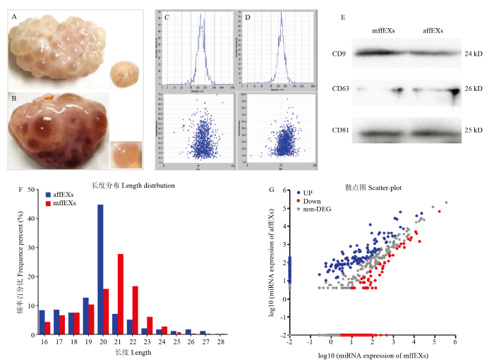
A:闭锁卵泡的形态观察结果;B:成熟卵泡的形态观察结果;C:mffEXs粒径分析结果;D:affEXs粒径分析结果;E:Westem blot检测结果;F:miRNA的序列长度分布;G:mffEXs和affEXs中miRNA的表达差异结果 Fig. 1Isolation and detection of exosomes and sequencing of miRNA
A: Morphological observation of atretic follicles; B: Morphological observation of mature follicles; C: Particle size analysis results of mffexs; D: Particle size analysis results of affexs; E: Results of Westem blot analysis; F: Sequence length distribution of miRNA; G: Differential expression of miRNA in mffEXs and affEXs
A:添加mffEXs培养的颗粒细胞结果;B:添加affEXs培养颗粒细胞的结果;C和D:Q-PCR检测CDK1和P53表达结果 Fig. 5Regulatory effects of mffEXs and affEXs on pig granulosa cells
A: Results of granulosa cells cultured with mffEXs; B: Results of affEXs culture of granulosa cells; C&D: Expression results of CDK1 and P53 detected by Q-PCR
HU JH, TANGT, TAN XS, ZENGZ, WUJ. The advance of exosomes regulating oocyte development in ovarian follicle Chinese Agricultural Science Bulletin, 2019(27):153-157. (in Chinese) [本文引用: 1]
TAOW, SUNL, SHIH, CHENGY, JIANGD, FUB, CONTE MA, GAMMERDINGER WJ, KOCHER TD, WANGD. Integrated analysis of miRNA and mRNA expression profiles in tilapia gonads at an early stage of sex differentiation BMC Genomics, 2016, 17(1):328. DOI:10.1186/s12864-016-2636-zURL [本文引用: 1]
SANGQ, YAOZ, WANGH, FENGR, WANGH, ZHAOX, XINGQ, JINL, HEL, WUL, WANGL. Identification of microRNAs in human follicular fluid: characterization of microRNAs that govern steroidogenesis in vitro and are associated with polycystic ovary syndrome in vivo The Journal of Clinical Endocrinology and Metabolism, 2013, 98(7):3068-3079. doi: https://www.chinaagrisci.com/article/2021/0578-1752/10.1210/jc.2013-1715. URL [本文引用: 1]
WILLIS GR, CONNOLLYK, LADELLK, DAVIES TS, GUSCHINA IA, RAMJID, MINERSK, PRICE DA, CLAYTONA, JAMES PE, REES DA. Young women with polycystic ovary syndrome have raised levels of circulating annexin v-positive platelet microparticles Human Reproduction, 2014, 29(12):2756-2763. DOI:10.1093/humrep/deu281URL [本文引用: 1]
SØRENSEN AE, WISSING ML, ENGLUND A LM, DALGAARD LT. MicroRNA species in follicular fluid associating with polycystic ovary syndrome and related intermediary phenotypes The Journal of Clinical Endocrinology & Metabolism, 2016, 101(4):1579-1589. DOI:10.1210/jc.2015-3588URL [本文引用: 1]
ZHAN XS, LUO HN, LUO DZ, CHEN SF, WANG BY, BAI YS, CHEN ZS, LIU CY, JI HQ. Effects of exosomes derived from canine umbilical cord mesenchymal stem cells on proliferation, migration and apoptosis of vascular endothelial cells Journal of Clinical Rehabilitative Tissue Engineering Research, 2019, 23(29):4637-4643. doi: https://www.chinaagrisci.com/article/2021/0578-1752/10.3969/j.issn.2095-4344.1808. (in Chinese) [本文引用: 1]
HUJ, TANGT, ZENGZ, WUJ, TAN XS, YANJ. The expression of small RNAs in exosomes of follicular fluid altered in human polycystic ovarian syndrome PeerJ, 2020, 8:e8640. DOI:10.7717/peerj.8640URL [本文引用: 1]
LÖTVALLJ, HILL AF, HOCHBERGF, BUZÁS EI, VIZIO DD, GARDINERC, GHO YS, KUROCHKIN IV, MATHIVANANS, QUESENBERRYP. Minimal experimental requirements for definition of extracellular vesicles and their functions: A position statement from the International Society for Extracellular Vesicles Journal of Extracellular Vesicles, 2014, 3(1):26913. DOI:10.3402/jev.v3.26913URL [本文引用: 1]
AL-DOSSARY AA, STREHLER EE, MARTIN-DELEON PA. Expression and secretion of plasma membrane Ca2+-ATPase 4a (PMCA4a) during murine Estrus: Association with oviductal exosomes and uptake in sperm PLoS ONE, 2013, 8(11):e80181. DOI:10.1371/journal.pone.0080181URL [本文引用: 1]
MASOUMI-DEHGHIS, BABASHAHS, SADEGHIZADEHM. microRNA-141-3p-containing small extracellular vesicles derived from epithelial ovarian cancer cells promote endothelial cell angiogenesis through activating the JAK/STAT3 and NF-κB signaling pathways Journal of Cell Communication and Signaling, 2020, 14(2):233-244. doi: https://www.chinaagrisci.com/article/2021/0578-1752/10.1007/s12079-020-00548-5. URL [本文引用: 1]
MACHTINGERR, RODOSTHENOUS RS, ADIRM, MANSOURA, RACOWSKYC, BACCARELLI AA, HAUSERR. Extracellular microRNAs in follicular fluid and their potential association with oocyte fertilization and embryo quality: An exploratory study Journal of Assisted Reproduction and Genetics, 2017, 34(4):525-533. doi: https://www.chinaagrisci.com/article/2021/0578-1752/10.1007/s10815-017-0876-8. URL [本文引用: 2]
LIANGM, YAOG, YINM, LÜM, TIANH, LIUL, LIANJ, HUANGX, SUNF. Transcriptional cooperation between p53 and NF-κB p65 regulates microRNA-224 transcription in mouse ovarian granulosa cells Molecular and Cellular Endocrinology, 2013, 370(1/2):119-129. doi: https://www.chinaagrisci.com/article/2021/0578-1752/10.1016/j.mce.2013.02.014. URL
DA SILVEIRA JC, VEERAMACHANENI DN, WINGER QA, CARNEVALE EM, BOUMA GJ. Cell-secreted vesicles in equine ovarian follicular fluid contain miRNAs and proteins: A possible new form of cell communication within the ovarian follicle Biology of Reproduction, 2012, 86(3):71. doi: https://www.chinaagrisci.com/article/2021/0578-1752/10.1095/biolreprod.111.093252. [本文引用: 2]
PIETRO CD. Exosome-mediated communication in the ovarian follicle Journal of Assisted Reproduction and Genetics, 2016, 33(3):303-311. DOI:10.1007/s10815-016-0657-9URL [本文引用: 1]
SALILEW-WONDIMD, AHMADI, GEBREMEDHNS, SAHADEVANS, HOSSAIN MD, RINGSF, HOELKERM, THOLENE, NEUHOFFC, LOOFTC, SCHELLANDERK, TESFAYED. The expression pattern of microRNAs in granulosa cells of subordinate and dominant follicles during the early luteal phase of the bovine estrous cycle PLoS ONE, 2014, 9(9):e106795. doi: https://www.chinaagrisci.com/article/2021/0578-1752/10.1371/journal.%20pone.0106795. URL [本文引用: 2]
ZHOUJ, LUO RB, TANG CF, QU SL. Effect of Bcl-2 protein family and p53 gene on regulating and controlling cell apoptosis Journal of Clinical Rehabilitative Tissue Engineering Research, 2007(10):1950-1952.(in Chinese) [本文引用: 1]
LVC, YU WX, WANGY, YI DJ, ZENGM, XIAOH. MiR-21 in extracellular vesicles contributes to the growth of fertilized eggs and embryo development in mice Bioscience Reports, 2018, 38(4): BSR20180036. [本文引用: 1]
ZHANGX, LIZ, XUANZ, XU PH, WANG WZ, CHENZ, WANGS, SUN GL, XU JH, XU ZK. Novel role of miR-133a-3p in repressing gastric cancer growth and metastasis via blocking autophagy-mediated glutaminolysis Journal of Experimental & Clinical Cancer Research, 2018, 37(1):320. [本文引用: 1]
HANY, WANG SM, WANG YZ, ZENG SM. IGF-1 inhibits apoptosis of porcine primary granulosa cell by targeting degradation of BimEL International Journal of Molecular Sciences, 2019, 20(21):5356. DOI:10.3390/ijms20215356URL [本文引用: 1]
SANTONOCITOM, VENTOM, GUGLIELMINO MR, BATTAGLIAR, WAHLGRENJ, RAGUSAM, BARBAGALLOD, BORZÌP, RIZZARIS, MAUGERIM, SCOLLOP, TATONEC, VALADIH, PURRELLOM, DI PIETROC. Molecular characterization of exosomes and their microRNA cargo in human follicular fluid: BIoinformatic analysis reveals that exosomal microRNAs control pathways involved in follicular maturation Fertility and Sterility, 2014, 102(6): 1751-61.e1. doi: https://www.chinaagrisci.com/article/2021/0578-1752/10.1016/j.fertnstert.2014.08.005. [本文引用: 1]
 ,, 黄绮亮, 胡智超, 潘晓婷, 吴志胜, 白银山

 新窗口打开|下载原图ZIP|生成PPT
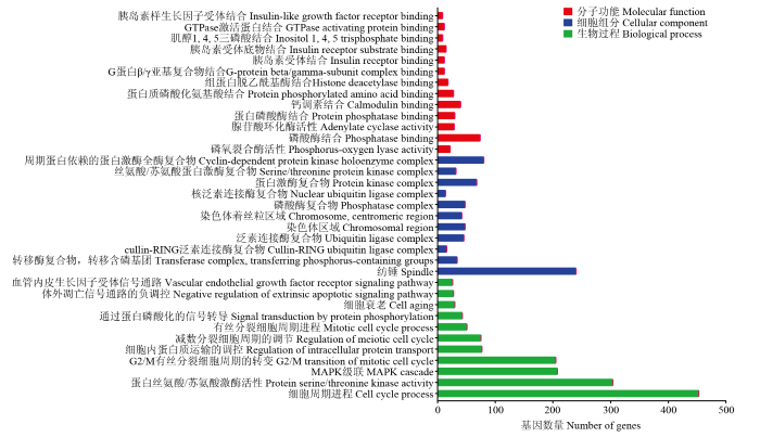 新窗口打开|下载原图ZIP|生成PPT
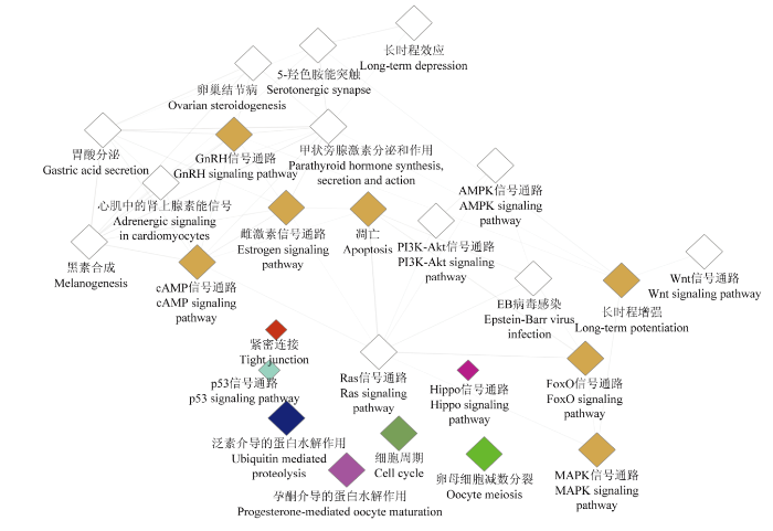 新窗口打开|下载原图ZIP|生成PPT
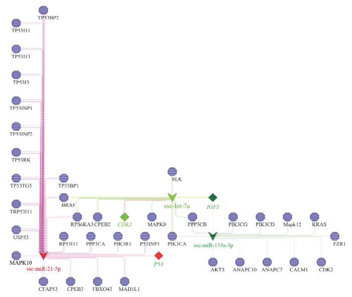 新窗口打开|下载原图ZIP|生成PPT
 新窗口打开|下载原图ZIP|生成PPT