1.College of Physics and Information Engineering, Shanxi Normal University, Linfen 041004, China 2.Key Laboratory of Spectral Measurement and Analysis of Shanxi Province, Shanxi Normal University, Linfen 041004, China 3.State Key Laboratory of Quantum Optics and Quantum Optics Devices, Institute of Laser Spectroscopy, Shanxi University, Taiyuan 030006, China 4.Department of Radiology, First Clinical Medical College, Shanxi Medical University, Taiyuan 030001, China
Fund Project:Project supported by the National Natural Science Foundation of China (Grant Nos. 61805134, 11504216, 61527824, 61675119) and the Applied Basic Research Program in Shanxi Province, China (Grant No. 201801D221016).
Received Date:13 March 2019
Accepted Date:11 April 2019
Available Online:01 June 2019
Published Online:20 June 2019
Abstract:The dipole orientation of single-molecule plays an important role in improving the fluorescence collection efficiency and promises to have applications in super-resolution imaging, protein folding, and F?rster resonance energy transfer between fluorophores. However, these applications are realized usually by precisely manipulating the orientation of the dipole moment of single molecules. Here, the dipole orientation of 1,1′-dioctadecyl-3,3,3′,3′,-tetramethylindodicarbocyanine (DiD) single molecules with the permanent dipole moment of 14.9 D is manipulated by using an external electric field of 3500 V/mm. Single DiD molecules are prepared by using mixed solvent of chloroform and dimethyl sulfoxide. The dipole orientation of single molecules is manipulated by an external electric field during the evaporation of solvent. The fluorescence of single molecules is measured by splitting the fluorescence collected by an objective into the S-polarized and P-polarized beams, and the fluorescence polarization of single molecules can be calculated by measuring the intensities of two orthogonal channels (IS and IP). The distribution of dipole orientation angle (α) for single DiD molecules in poly-(methyl methacrylate) (PMMA) film is analyzed statistically, and its changes are compared under different electric fields. It is found that the dipole orientation angle α of single DiD molecules in the PMMA film without applying electric field obeys a single-peak Gaussian distribution with the most probable value of 41°, which results from the fluorescence dichroism signal of the high numerical aperture objective. Applying a perpendicular electric field to the surface of single-molecule sample, the distribution of dipole orientation angle α of single DiD molecules can be still fitted by a single-peak Gaussian function with the most probable value of 44.2°. The dipole orientation of single DiD molecules under the perpendicular electric field changes little. However, by applying a parallel electric field to the surface of single-molecule sample, the dipole orientation angle α of single DiD molecules changes prominently. It obeys a two-peak Gaussian distribution with the most probable values of ~ 32° and 55.5°, indicating that the orientation polarization of the dipole moment occurs to the single DiD molecules in PMMA film. The dipole orientation of single polar molecules tends to the parallel electric field in this case. Keywords:single molecule/ dipole orientation/ electric field manipulation/ polarization property
全文HTML
--> --> --> 1.引 言单分子光学探测消除了系综平均效应, 已广泛应用于物理、化学、生物、材料等领域[1-3]. 单分子的偶极取向[4-6]作为单分子的重要物理参数之一, 决定着单分子荧光辐射的偏振特性及空间分布, 从而影响单分子的荧光收集效率[7]及基于单分子荧光成像的分辨率[8]. 生物体系中蛋白质的折叠、肌动蛋白的运动涉及到生物大分子的构象变化, 可以通过跟踪标记的荧光分子的取向变化获得其动力学过程[9]. 分子与分子间偶极-偶极相互作用引起的共振能量转移过程也极大地依赖于两个单分子相对的偶极取向[10]. 另外, 利用单分子作为载体产生单光子是制备单光子源的有效手段之一[11], 基于单光子源的偏振编码是量子密钥分发的重要方案[12]. 单分子的荧光偏振状态取决于其偶极取向, 因此单分子偶极取向的有效操控可为基于单分子的单光子源在量子密钥分发中的应用提供基础. 人们已经利用宽场散焦成像[13]和扫描共聚焦成像[14]实现了单分子偶极取向的有效测量, 但是对单分子偶极取向的确定性操控至今仍然是一个难题. Huang等[15]在低温下通过脉冲扫描隧道显微镜的金属尖针, 将金属针尖放置在氯铝酞菁单分子上方并施加正向偏压脉冲, 将单分子面内朝上的氯原子转变成面内朝下, 改变了单分子的偶极取向, 从而实现了单分子二进制位的读写操作. Sandoghdar课题组[16]通过选取合适的主客体组合, 发现处于对-三联苯晶体中的terrylene单分子的偶极取向被固定在特定方向, 其荧光偏振方向垂直于玻璃基底, 随后获得了高达96%的单分子荧光收集效率[17]. 尽管如此, 该方法所获得的分子取向依赖于特定分子与晶体的组合, 不便于推广应用于其他分子. 近年来, 利用电场实现单分子操控成为人们的研究热点. Shaik课题组[18,19]用外部定向电场对分子偶极进行选择从而实现了分子的手性识别; Sajadi等[20]利用强太赫兹电场脉冲耦合到分子偶极矩, 共振激发极性液体分子使其重新定向振动; Kato等[21]研究了电场下单分子的超滞后电极化效应. 本文利用外部电场作用于掺杂在聚甲基丙烯酸甲酯薄膜中的极性单分子, 在溶剂挥发过程中对极性单分子的偶极取向实现了有效操控. 通过测量单分子的荧光偏振方向, 研究了外电场对极性单分子偶极取向的极化特性的影响. 2.实 验实验中所用的分子1,1′-dioctadecyl-3,3,3′,3′,-tetramethylindodicarbocya-nine (DiD, molecular probes)是极性染料分子, 分子结构见图1(a), 图中红色箭头显示了DiD分子的固有偶极取向. DiD分子的固有偶极矩高达14.9 D (1 D = 3.33564 × 10⁻³⁰ C·m), 其极易于被电场操控. DiD分子的吸收峰位于640 nm, 荧光发射峰位于660 nm. 将DiD分子溶解到氯仿溶剂中, 稀释至~10–9 mol/L; 10 mg的聚甲基丙烯酸甲酯(poly-(methyl methacrylate), PMMA, 分子量15000, Sigma-Aldrich)溶解到1 mL的氯仿和二甲基亚砜溶液(Dimethylsulfoxide, DMSO)混合液(1 : 1混合)中; 单分子溶液与溶解有PMMA的氯仿/DMSO溶液混合, 制备单分子浓度约为10–10 mol/L的混合液; 将混合液以300 r/min的转速旋转涂覆于玻璃基片上. 由于DMSO为难挥发溶剂, 旋转涂覆后单分子样品溶剂并不会完全挥发. 将旋涂有DiD分子的玻璃基片放置于图1(b)所示的装置中, 分别施加与单分子样品表面垂直或平行的匀强电场(场强3500 V/mm), 测量电场作用下单分子荧光偏振响应特性. 电场作用过程在真空(< 20 kPa)干燥箱中进行, 作用时间设定为90 min, 长于溶剂挥发所需要的时间(约60 min). 实验中, 选用氯仿和DMSO混合液作为溶剂, 减缓溶剂挥发速度, 保证在样品制备过程中电场对单分子的有效操控. 溶剂完全挥发后, 单分子被固定于PMMA薄膜中. 图 1 (a) DiD分子的结构式, 红色箭头表示其固有偶极取向; (b)垂直于单分子样品表面电场和平行于样品表面电场操控单分子示意图 Figure1. (a) Structure of DiD dye molecule with its dipole orientation indicated by a red arrow; (b) schematic of single-molecule sample manipulated by applying a perpendicular or parallel electric field to the surface of single-molecule sample, respectively.
3.结果与讨论在共聚焦荧光成像系统上通过逐点扫描的方式对单分子样品进行荧光成像, 实验中每个像素点的采样时间是10 ms. 图2(a)显示了掺杂在PMMA聚合物薄膜中未通过电场作用时DiD分子在18 μm × 18 μm (100像素 × 100像素)区域里的分布, 每个亮点来自不同DiD分子的荧光. 由于单分子所处的纳米局部环境及单分子偶极取向的差异, 使得每个单分子的荧光强度略有不同. 为了获取单分子荧光偏振方向的信息, 在得到分子荧光成像后, 通过三维纳米位移平台将单个DiD分子移动到激光聚焦区域, 测量单个分子在S偏振和P偏振方向上荧光强度随时间的变化. 图 2 DiD单分子的偶极取向与偏振测量 (a)在18 μm × 18 μm区域内DiD单分子的荧光成像; (b)任意偶极取向的DiD单分子的S偏振及P偏振方向荧光探测示意图, 其中Obj是物镜, PBS是偏振分束棱镜; (c)成像图(a)中红色圆圈标记的DiD分子的S和P偏振方向的荧光强度轨迹图; (d)荧光偏振方向α随时间的变化; (e) DiD分子光漂白前荧光偏振方向的统计, 最可几值为48.8° Figure2. Fluorescence measurement of single DiD molecules: (a) Fluorescence image of single DiD molecules in 18 μm × 18 μm area; (b) schematic view of the S-polarized and P-polarized fluorescence of arbitrary dipole moment for single-molecule (Obj, objective; PBS, polarized beam splitter); (c) fluorescence trajectories of single DiD molecule indicated by the red circle in panel (a) in S and P polarization; (d) the relationship between fluorescence polarization and time; (e) the statistics of fluorescence polarization with the most probable value being 48.8°.
式中, θ和α与图2(a)一致; A, B, C是由高数值孔径物镜引入的与角度无关的常数, 可能引起所测量偏振方向的并不是均匀随机分布[25]. 对未经取向极化的DiD单分子而言, 其荧光偏振方向α分布呈现出以41°为中心的高斯分布. 外电场作用于单分子引起其能量的变化[26]可以简写为
$\Delta E = \mu \times {F_{{\rm{ext}}}} \times \sin \delta ,$
其中δ是单分子的偶极取向与外电场方向的夹角. 在外电场的作用下, 单分子的偶极取向会逐渐偏向于电场的方向(δ → 0), 这就是对单分子产生取向极化的过程. 电场强度越大, δ越小, 单分子偶极取向极化的偏转角度Δδ与电场强度有关[27]. 电场方向与单分子的偶极取向之间存在三种情况, 如图4所示, 其中红色箭头是分子的偶极取向. 第一种情况如图4(a): 外电场与分子偶极取向平行(δ = 0或180°), 电场并不会改变分子的偶极取向. 第二种情况如图4(b): 外电场方向与偶极取向垂直(δ = 90°), 两个偶极取向的分子等量地向电场方向偏转, 如图中蓝色箭头所示, 此时分子偶极取向的变化Δδ是一致的. 图4(a)和图4(b)中极少部分特殊偶极取向的分子并不会引起偏振方向α的双峰分布. 除了上述两种特殊情况外, 绝大部分随机分布的分子偶极取向与电场方向夹角如图4(c): 在与电场方向夹角δ较小的单分子被完全取向极化前, 两个分子还是等量地趋向电场方向. 由于夹角δ较小的单分子更容易偏转到电场方向, 而夹角δ 较大的单分子要想与电场方向一致还需要更强的电场, 在这种情况下, 两个分子的偶极取向极化过程不同, 存在两个夹角δ, 导致如图3(c)中平行电场作用下单分子偶极取向的双峰分布现象. 当垂直于x-y平面电场作用于单分子时, 电场主要改变的是分子偶极矩的θ角, 不会影响单分子偶极取向在x-y平面的投影角度, 也就是与光轴垂直平面的单分子的荧光偏振取向α分布, 所以垂直于单分子样品平面电场作用下单分子偶极取向分布基本不变. 当平行电场作用于单分子时, 引起分子的荧光偏振方向的双峰分布. 这也说明了电场并没有使得所有的DiD单分子的偶极取向与电场方向完全一致. 图 4 单分子偶极取向在外电场作用下的极化示意图 (a) 电场方向与分子偶极取向同向; (b) 电场方向垂直于单分子偶极取向; (c)电场作用于任意取向单分子 Figure4. Simplified scheme of the polarization of the dipole orientation of single-molecule under the influence of external electric field. The directions of the electric field are parallel (a), perpendicular (b), and arbitrary (c) to the dipole orientation of single-molecule, respectively.
 图 1 (a) DiD分子的结构式, 红色箭头表示其固有偶极取向; (b)垂直于单分子样品表面电场和平行于样品表面电场操控单分子示意图
 图 2 DiD单分子的偶极取向与偏振测量 (a)在18 μm × 18 μm区域内DiD单分子的荧光成像; (b)任意偶极取向的DiD单分子的S偏振及P偏振方向荧光探测示意图, 其中Obj是物镜, PBS是偏振分束棱镜; (c)成像图(a)中红色圆圈标记的DiD分子的S和P偏振方向的荧光强度轨迹图; (d)荧光偏振方向α随时间的变化; (e) DiD分子光漂白前荧光偏振方向的统计, 最可几值为48.8°
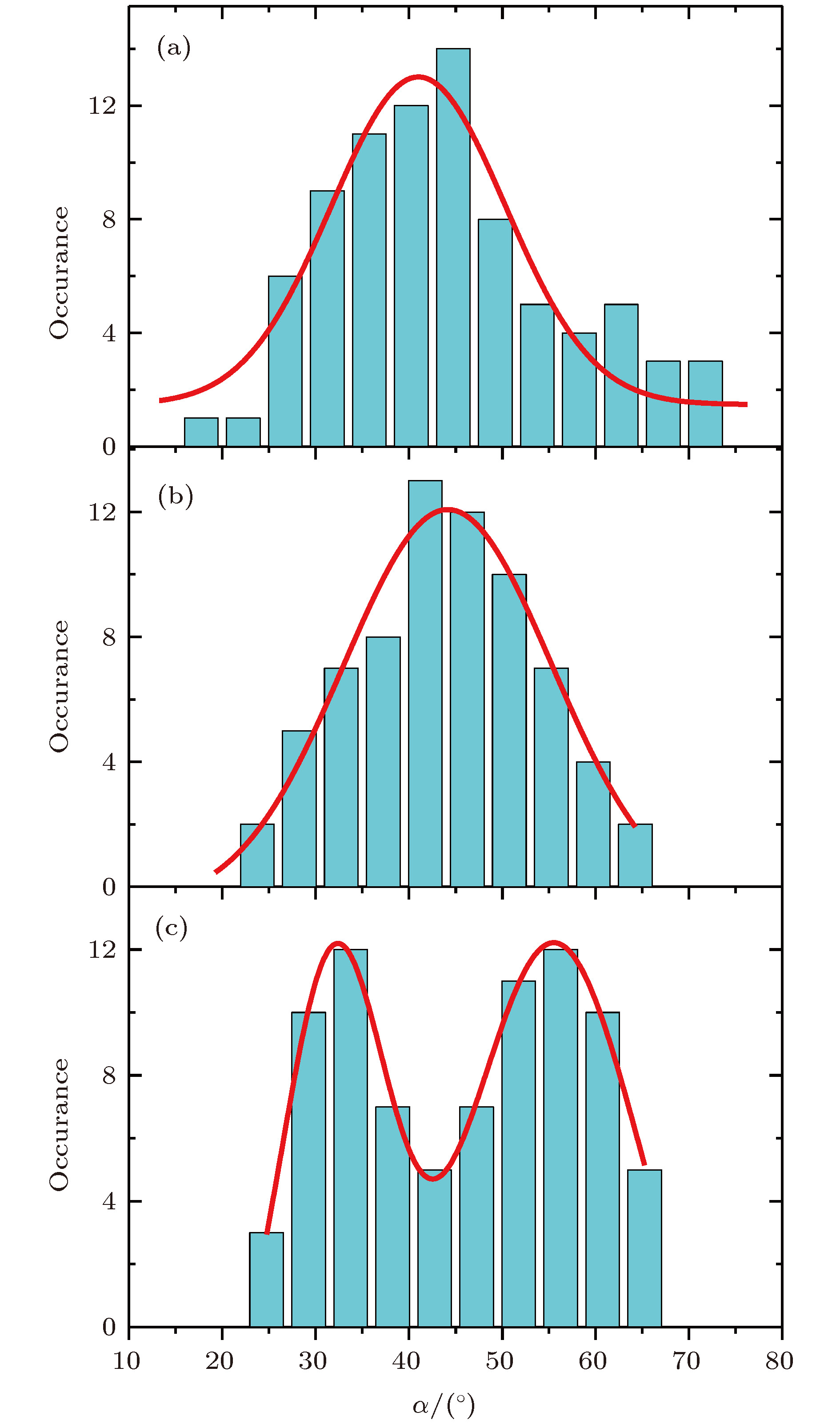 图 3 DiD单分子在不同情况下取向极化的效果 (a)未加电场; 3500 V/mm的(b)垂直电场取向极化和(c)平行电场取向极化; 荧光的偏振方向α的统计峰值分别是 (a) 41.0° ± 21.9°, (b) 44.2° ± 26.3°, (c) 32.0° ± 13.5°和55.5° ± 21.6°
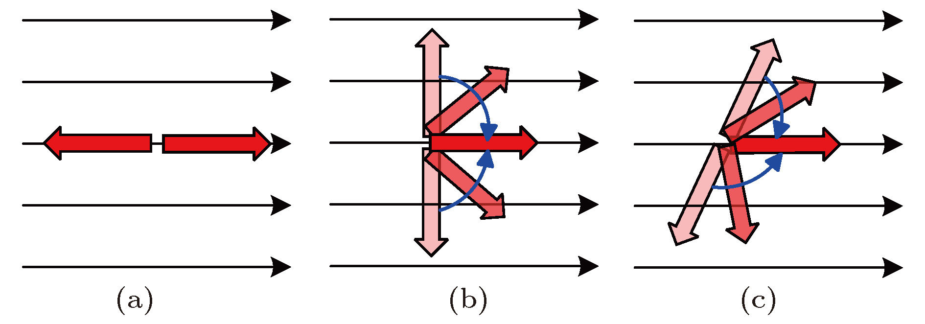 图 4 单分子偶极取向在外电场作用下的极化示意图 (a) 电场方向与分子偶极取向同向; (b) 电场方向垂直于单分子偶极取向; (c)电场作用于任意取向单分子