房凌旭1, 李龙1, 卢中一2,3, 李猛2


1. 深圳市南山区蛇口人民医院口腔科, 广东 深圳 518067;
2. 深圳大学高等研究院, 深圳市海洋微生物组工程重点实验室, 广东 深圳 518060;
3. 深圳大学光电工程学院, 光电子器件与系统教育部重点实验, 广东 深圳 518060
收稿日期:2020-09-23;修回日期:2021-01-29;网络出版日期:2021-02-02
基金项目:深圳市南山区卫生科技计划(2020057);深圳市基础研究计划重点项目(JCYJ20200109105010363);广东省基础与应用基础研究基金(2019A1515110089);广东省普通高校创新团队项目(自然科学)(2020KCXTD023)
作者简介:房凌旭, 口腔医学硕士, 深圳市蛇口人民医院口腔科门诊主治医师。主要从事口腔微生物学相关研究, 探讨口腔微生物群落与口腔及全身系统性疾病的关联性, 关注口腔耐药组及其风险性评估。发表学术期刊论文2篇。主持1项深圳市南山区卫生科技计划项目, 另参与多项国家级和省部级项目.
*通信作者:李猛。Tel: +86-755-26979250;E-mail: limeng848@szu.edu.cn.
摘要:口腔微生物是人体微生物组的重要组成部分,其群落组成丰富且独特。现有研究显示,口腔微生物与龋病、牙周炎等口腔健康问题有直接的联系,因而具有重要的研究价值。随着高通量测序技术的发展,人们对口腔中未培养微生物多样性的认识不断加深,这进一步催生对微生物分离培养技术需求的增加。为此,本文将围绕口腔未培养微生物及其分离培养策略的研究进展,首先介绍口腔中未培养微生物的研究现状;其次分析口腔微生物分离培养中可能的限制因素;最后综述微生物分离培养技术发展及其在口腔未培养微生物研究中的应用。全文旨在为口腔未培养微生物的分离培养提供思路和技术参考。
关键词:口腔未培养微生物微生物分离培养技术人体健康
Research strategies on isolation and cultivation of uncultivated oral microorganisms
Fang Lingxu1, Li Long1, Lu Zhongyi2,3, Li Meng2


1. Department of Stomatology, Shenzhen Shekou People's Hospital, Shenzhen 518067, Guangdong Province, China;
2. Shenzhen Key Laboratory of Marine Microbiome Engineering, Institute for Advanced Study, Shenzhen University, Shenzhen 518060, Guangdong Province, China;
3. Key Laboratory of Optoelectronic Devices and Systems of Ministry of Education and Guangdong Province, College of Optoelectronic Engineering, Shenzhen University, Shenzhen 518060, Guangdong Province, China
Received: 23 September 2020; Revised: 29 January 2021; Published online: 2 February 2021
*Corresponding author: Meng Li, Tel: +86-755-26979250; E-mail: limeng848@szu.edu.cn.
Foundation item: Supported by the Health Science and Technology Project of Shenzhen Nanshan District (2020057), by the Shenzhen Science and Technology Program (JCYJ20200109105010363), by the Basic and Applied Basic Research of Guangdong Province (2019A1515110089) and by the Innovation Team Project of Universities in Guangdong Province (2020KCXTD023)
Abstract: Representing a fundamental part of the human microbiota, the oral microbial community is characterized by its diverse and unique composition. Oral diseases like dental caries and periodontitis are directly associated with oral microorganisms. Therefore, it is crucial to deepen our understanding of oral microbiota. Advances in high-throughput sequencing technology provide extensive information of the diversity of oral uncultivated microorganisms, prompting an increasing need for microbial isolation and cultivation techniques. This review presents recent research progress on uncultivated oral microorganisms and lists factors that possibly hinder isolation attempts. In addition, advances in methodologies and techniques used for culturing previously uncultured microorganisms and their applications in oral microbiology studies are summarized, giving valuable insights into various aspects of uncultivated oral microorganisms.
Keywords: oral microbial communitymicrobial cultivation techniqueshuman healthy
口腔是人体中除肠道外微生物定殖的主要场所,因其具有的开放性和动态性等特点使口腔微生物成为人体多样化的微生物群落之一[1]。据统计,健康人口腔中定殖的细菌超过700种[2],而定殖的真菌也超过100种[3];此外,口腔中也存在古菌,但主要以产甲烷菌古菌属Methanogens为主[4-6]。通常认为,口腔中微生物与宿主和谐共存,维持口腔稳态;但当口腔环境改变,其中微生物群落平衡的破坏可导致严重的口腔健康问题,例如在膳食中摄入过多碳水化合物可促进Streptococcus mutans和Lactobacillus acidophilus等产酸和耐酸的细菌生长,进而改变口腔微生物群落结构,并最终损坏牙釉质诱发龋病[7-8]。因此,对于口腔微生物群落的研究是防治口腔疾病的重要前提。
在早期的口腔微生物研究中,****们主要采用传统的微生物分离培养技术,对口腔微生物多样性的认识受限于“所见所得”;例如,Clarke等从多份早期龋齿样品中分离到Streptococcus mutans,提示了该菌在龋齿形成中的潜在作用[9]。此后,基于16S rRNA基因和ITS (Internal Transcribed Spacer)等分子标志物的高通量测序技术的蓬勃发展极大加深了人们对口腔微生物特别是对口腔中未培养微生物多样性的认识[3, 10]。例如,eHOMD数据库(expanded Human Oral Microbiome Database)显示,口腔中只有57%的菌种被正式命名,13%已获得纯培养的菌种未得到正式命名,另有约30%的菌种未获得纯培养物,这从一个侧面提示未培养微生物是制约口腔微生物研究工作的“瓶颈”[11]。时至今日,分离培养口腔中微生物的工作仍不能被替代,这是因为:(1) 微生物的形态结构、生理生化、功能基因和细胞内调控通路等重要研究内容无法离开微生物纯培养[12-14];(2)现有高通量测序技术在获得样品微生物基因组完整度和准确度上仍有局限性[15-16];(3)病原菌诊断、致病力模型的建立和病原菌药物敏感性试验均需要纯培养物[13, 17-18]。鉴于以上原因,发展微生物分离培养技术,提高口腔样品中微生物的培养率,将未培养微生物转变成可培养微生物是口腔研究工作的重要方向。
1 口腔中未培养微生物研究现状 1.1 细菌 细菌是口腔中最丰富的微生物,其群落结构的改变及其与人体免疫系统的相互作用被认为是造成牙周炎和龋齿等口腔问题的主要原因[19]。基于16S rRNA基因测序结果显示,口腔中分布着超过20种细菌门,其中丰度较高的细菌主要来自Firmicutes、Bacteroidetes、Actinobacteria、Proteobacteria、Fusobacteria、Spirochaetes和Synergistetes等[20-21]。此外,在eHOMD中显示(截止2020年8月28日),未培养细菌的种类主要集中在细菌属Bergeyella、Treponema、Saccharibacteria (TM7)、Fretibacterium、Veillonellaceae (G-1)、Absconditabacteria (SR-1)和Gracilibacteria (GN02)等(表 1)[22]。
表 1. 口腔中的主要未培养细菌类群 Table 1. Major uncultivated bacteria groups in oral cavity
| Phylum | Genus | Unculturable species | Percentage rate/%* |
| Bacteroidetes | Bergeyella | HMT-322/-900/-907/-931 | 50.0 |
| Spirochaetes | Treponema | HMT-246/-247/-508/-518/-252/-254/-256/-253/-517/-951/-235/-242/-226/-238/-230/-239/-234/-227/-236/-228/-231/-237 | 34.4 |
| Saccharibacteria (TM7) | Saccharibacteria (TM7) | HMT-356/-870/-355/-351/-350/-347/-954/-349/-869/-346/-348/-352/-488 | 86.7 |
| Synergistetes | Fretibacterium | HMT-359/-362/-360/-358/-361 | 62.5 |
| Firmicutes | Veillonellaceae (G-1) | HMT-132/-150/-135/-918/-148/-145 | 66.7 |
| Absconditabacteria (SR1) | Absconditabacteria (SR-1) | HMT-875/-345/-874 | 100.0 |
| Gracilibacteria (GN02) | Gracilibacteria (GN02) | HMT-873/-996/-871/-872 | 100.0 |
| *Percentage rate=uncultivated oral bacterial species/known oral bacterial species (data are collected from expanded Human Oral Microbiome Database: http://www.homd.org/). | |||
表选项
1.2 真菌 真菌群落是口腔微生物的重要组成部分,特别是部分真菌具有条件致病性,可造成免疫力低下人群口腔黏膜感染[23]。此外,口腔中真菌也可以引起其他口腔健康问题,其作用方式可能与口腔细菌群落失调有关;例如,Bertolini等发现白色念珠菌可以促进Streptococcus oralis形成生物膜,并且增强了该菌对口腔组织的侵染能力[24]。Ghannoum等研究发现,健康人口腔中可检测到的真菌属高达85个,其中丰度较高的包括Candida、Cladosporium、Aureobasidium、Saccharomyces、Aspergillus和Cryptococcus等[3]。此外,未培养的真菌属有11个,例如Glomus等。
1.3 古菌 口腔中古菌的种类较少,并且这些古菌与牙周炎、龋齿等口腔健康问题是否存在直接的联系仍需进一步研究。目前,口腔中可检测到的古菌种类包括Methanobrevibacter oralis、Methanobrevibacter smithii、Methanobrevibacter massiliense、Methanosarcinia mazei、Methanobacterium congolense、Methanoculleus bourgensis和Nitrososphaera evergladensis等,其中M. massiliense和N. evergladensis仍未获得纯培养[25-28]。
2 口腔微生物分离培养中的限制因素 2.1 氧气条件和物理媒介相关的限制因素 厌氧环境是决定口腔微生物分离培养率的重要条件;例如,口腔中存在的细菌属Veillonella和Peptostreptococcus中部分种是专性厌氧菌,古菌属Nitrososphaera和Methanobrevibacter也被认为是专性厌氧菌[29-31]。口腔中存在厌氧菌生存的条件;例如牙周炎患者牙周袋深部被认为是一种微氧或无氧环境,因而分离这些位置的微生物时应注意厌氧培养。此外,许多微生物的生长需要附着在物理媒介表面,其原因包括:(1)在物理媒介表面生长的微生物有利于形成生物膜,从而增强了对物理性伤害和化学性伤害的抵抗力[32];(2)物体媒介表面增强微生物群体的群体感应能力(quorum-sensing),对微生物群落结构和生存均具有重要调节作用[33];(3)在物理媒介上的着生可提高微生物对营养物质的吸附和获取能力等[34-35]。
2.2 微生物生长特点和培养基成分相关的限制因素 丰度较低或生长缓慢可能是导致口腔中部分微生物难培养的重要原因。例如16S rRNA基因测序显示,Saccharibacteria (TM7)和Absconditabacteria (SR-1)等在健康人口腔微生物群落中占比通常不超过1%;由此可见常规的培养方法可能使这些细菌在分离培养初期难以有效富集[36]。此外,培养基成分可能是限制口腔微生物分离培养的因素,这一方面体现在不恰当的培养基成分可能抑制微生物生长,例如琼脂对部分细菌具有毒性,可能导致部分细菌生长受到抑制[37],而磷酸盐和琼脂混合物在高温灭菌后产生的活性氧可抑制对氧化环境敏感微生物的生长[38]。另一方面体现在培养基中缺少的特定成分抑制部分微生物生长;例如Tannerella forsythia需要N-乙酰胞壁酸才能生长,因此在富集口腔样品中的Tannerella属细菌可考虑添加该成分[39]。此外,Tian等通过对比PYG培养基(peptone-yeast extract-glucose medium)和BMM培养基(basal medium mucin medium)等培养效果后发现口腔样品中细菌菌落变化与培养基成分有关,反映了培养基的选择对初期富集口腔微生物时具有重要作用[40]。
2.3 体外条件对口腔环境模拟的限制因素 人体口腔是一个复杂的系统,因而在体外难以准确模拟,这无疑限制了口腔微生物的分离培养。首先,口腔中存在复杂的微生物间相互作用。例如,牙周袋中的Methanobrevibacter和Treponema存在代谢共生的关系[26];而在其他例子中,牙菌斑中常见细菌属Veillonella可利用由Streptococcus mutans产生的乳酸作为主要碳源[41]。此外,He等发现Saccharibacteria (TM7)等难以培养的主要原因是其缺失合成必需氨基酸的通路,因而只能与其他宿主(如Actinobacteria)等共生[42-43]。其次,人体内环境(例如组织及免疫系统)可能是部分微生物生长的重要条件。例如,在部分牙周炎患者中Saccharibacteria (TM7)丰度显著高于健康人群,在细菌群落中占比高达21%[44],暗示口腔炎症反应可能促进了Saccharibacteria (TM7)的生长。另一项动物模型实验表明,抗炎症反应治疗可减轻牙周细菌负荷,这提示人体免疫反应本身可影响口腔微生物群落结构[45]。目前对于宿主因素影响口腔微生物生长的一种解释是,炎症反应组织释放的氨基酸、铁离子或其他成分对特定微生物生长具有促进作用[46]。
3 微生物分离培养技术研究进展 本课题组前期系统梳理了环境微生物分离培养技术[47],并积极摸索未培养/难培养微生物在富集培养过程中的影响因素;此外,我们也将目光转移到不同来源的样品,尝试从这些样品中分离到更多的未培养微生物。人体口腔是连接环境和人体的重要“桥梁”,其中的微生物受自然环境和体内环境双重因素作用,是本课题组关注的方向之一。本节将围绕口腔微生物分离培养中限制因素,针对性介绍微生物分离培养技术,并且对其中已应用于口腔微生物分离培养的研究进行分析,以期为口腔中未培养微生物的分离培养提供新策略。
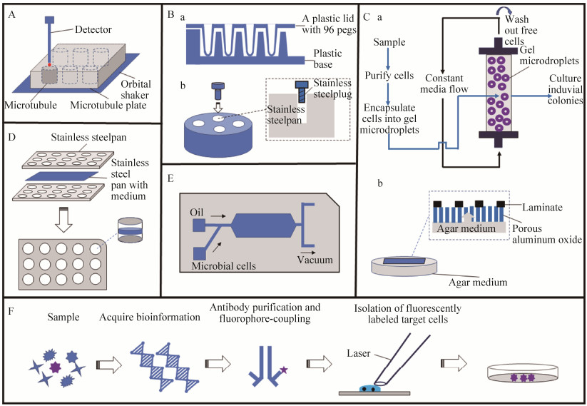
3.1 微型生物反应器 生物反应器是一种可以控制温度、压力和氧浓度等条件参数,从而模拟微生物生长环境的培养装置[48-53]。早期的微生物反应器体积较大且缺乏检测器,因而在微生物分离培养中应用较少。随着制造工艺和感应技术的不断进步,现已出现微型化的生物反应器,为高通量富集培养厌氧微生物提供了可能。该装置是由多个平行的微型生物反应器嵌合至摇床上,同时配备有多种传感器以实时监测反应器中微生物生长状态(图 1-A)。目前具有代表性的微型生物反应器包括RAMOS (respiration activity monitoring system)和BioLector等。RAMOS可以在线监测微生物培养过程中传氧速率(oxygen transfer rate)和呼吸商(respiratory quotient)等指标,有利于对厌氧微生物在培养过程中生长的连续监测[54]。BioLector配备的传感器可检测溶氧压(dissolved oxygen tension)、光密度和荧光强度等,因而可监测培养过程中氧气浓度和微生物生长状况[55]。因此,上述两种反应器为口腔中厌氧微生物分离培养提供了应用参考。
 |
| 图 1 微生物分离培养技术总结 Figure 1 Summary of isolation and cultivation techniques for microorganisms. A: Microbioreactor; B: Calgary biofilm device (a) and constant depth film fermenter (b); C: Gel embedding technique (a) and a million-well growth chip technique (b); D: Minitrap chip; E: Microfluidic culture technique; F: Reverse genomics method. |
| 图选项 |
3.2 物理媒介培养技术 物理媒介培养技术是针对部分微生物需要在支撑物表面着生生长这一情况而提出的。其中,玻璃、陶瓷、金属和多孔聚合物均可作为物理媒介以促进微生物的初期富集培养[34]。随着****们逐渐认识到生物膜可能是促进口腔微生物生长的重要因素,近年来体外生物膜形成技术作为新型的物理媒介被逐渐应用于口腔微生物的分离培养研究中。卡尔加里生物膜装置(Calgary biofilm device)是较早用于培养口腔样品中微生物的一种人工生物膜模型装置。该装置由上下两部分组成,上部分是由96个塑料钉组成的盖子,下部分是与上部分嵌合的底座;生物膜在上部塑料钉与底座接合的部位形成(图 1-Ba)[56]。Thompson等通过该装置从牙菌斑中成功分离到了Proteobacteria、Actinobacteria、Firmicutes和Bacteroidetes中部分未培养的菌种[57]。此外,定深膜发酵器(constant depth film fermenter)是另一种体外生物膜形成装置,该装置主体由一个柱形的不锈钢圆盘组成,其中分布的孔可被配套的不锈钢插头插入,而产生的空腔间隙用以构建生物膜(图 1-Bb)。例如,McBain等利用该装置从人类唾液中富集到Proteobacteria、Firmicutes和Bacteroidetes中部分菌种,证明了该装置在口腔微生物分离培养中的应用价值[58]。
3.3 单细胞分选和高通量分选技术 在常规微生物分离培养中通常难以获得生长缓慢或丰度较低的微生物,因而早期的****们尝试将借助显微镜和毛细吸管(或微型针)直接从样品中挑取目标微生物;随后的发展中,该技术逐渐被光镊技术取代,这是因为光镊技术可结合荧光染色的方法剔除死亡细胞,从而提高微生物细胞的分离效率[59-60]。Huber等从美国黄石公园热水塘样品中利用光镊技术直接分离出嗜热古菌,这被认为是单细胞分选的成功应用[61]。此外,激光压力弹射技术也被认为是一种重要的单细胞分选技术,该技术的操作方法是将待分拣的细胞样品均匀分散于聚乙烯膜上,随后利用氮原子激光切割聚乙烯膜以实现对单细胞的分拣[62]。
近年来,微生物高通量分选技术的发展为口腔微生物分离培养研究提供了全新的发展契机,例如凝胶微滴包埋技术。该技术首先用寡营养培养基富集样品中微生物;然后将富集的微生物样品包埋入凝胶微滴,并制备凝胶柱;接着利用循环流动的液体培养基分离凝胶柱中被包埋的微滴;最后将上述微滴批量接种至培养基中进行微生物分选(图 1-Ca)。Zengler等借助该技术从环境样品中分离出大量先前未获得纯培养的微生物,证明了该技术在微生物分离培养中的应用价值[63]。此外,Ingham等开发了一种以平板培养法为基础的生长芯片,该芯片是一块表面被蚀刻的多孔氧化铝,其表面分布着7–400万个可用于培养微生物的微型分隔;当该芯片插入培养基后,培养基成分可经铝膜孔隙从下层进入分隔为微生物提供营养(图 1-Cb)。该芯片的主要优势是通过提供大量平行的微型分隔以低成本的方式为样品中微生物的高通量分选培养提供了可能[64]。
3.4 原位培养技术 原位培养技术设计的初衷是为了克服实验室条件下无法模拟特殊生境条件的困境。例如,Kaeberlein等使用一种扩散盒技术(diffusion chambers)对海洋微生物进行富集;该技术中使用的扩散盒装置类似三明治,中间琼脂凝胶层被两层半透性的聚碳酸酯膜夹住。该研究组证实,通过这种扩散盒技术可使微生物总获得率比传统方法提高约40%[65]。此后,结合高通量理论方法,该扩散盒技术演变为一种分离芯片技术(isolation chip),该芯片可看作由多个微小扩散盒集成的盘;这些微小扩散盒两端覆盖有滤膜,可将待培养的微生物样品限制在盒内,进而可在原始生境下进行富集培养。已有研究显示,将该芯片可成倍数的提高微生物富集培养效率(约提高5–200倍)[66]。值得一提的是,Sizova等根据分离芯片技术开发了一种专用于口腔微生物分离培养的Minitrap芯片。这种芯片由具有中间孔隙的3个不锈钢片贴合而成,该芯片可固定于口腔内部以实现对口腔微生物的原位富集培养(图 1-D)[67]。Sizova等提出Minitrap芯片在口腔微生物分离培养研究中具有如下优点:(1)该方法适于富集培养在口腔内部才具有生长活性的特殊微生物;(2)该方法实现了微生物单细胞的长期培养,有利于对口腔中低丰度或生长缓慢微生物的富集。
3.5 共培养技术 通过共培养技术富集和分离口腔中未培养微生物已被证明是一种行之有效的方法。例如,Saccharibacteria (TM7)首次从口腔样品中被分离即是通过与一株Actinomyces odontolyticus共培养完成[42]。此外,Vartoukian等利用与牙菌斑细菌共培养的方式,从牙周炎患者的牙周袋中分离到1株未获得过纯培养的Synergistete[68]。目前,共培养技术在微生物分离培养研究中得到不断发展,其中具有代表性的是单菌落共培养技术。该技术中所用到的培养基有3层,其中上面2层琼脂含量为0.04%,并且这两层中间被0.02 μm的滤膜隔开,而最下面琼脂含量为1.5%。在实际应用中,两种可能具有共生关系的微生物被分别接种至上下的0.04%琼脂层,从而使两种微生物在滤膜处实现共生生长[69]。此外,微流培养技术(microfluidic culture technique)是一种具有高通量特点的共培养方法,该技术工作的原理是利用微油滴对经前期富集培养的微生物样品进行随机分离,并将分离后的微小群落批量接种至培养基中实现分离培养(图 1-E)[70]。
3.6 定向富集分离技术 定向富集技术设计初衷是提高样品中低丰度微生物的获得率。这其中具有代表性的是序列引导分离(sequence-guiding isolation)技术,其流程包括:首先将微生物样品接种至固体培养基中批量培养,随后冲洗培养基表面菌落并提取菌落总DNA,利用特异性引物检测培养物中目标微生物,对于含有目标微生物批次的培养物进行转接,并进一步分离培养。目前已知Acidobacteria、Verrucomicrobia和Thauera中部分菌种是通过该方式获得[71]。此外,最近被提出的反向基因组方法(reverse genomics)也可以看作一种定向富集分离培养技术,其原理是借助高通量测序(或已有基因组数据库)寻找目标微生物特异性靶点,然后制备标记有荧光的靶点抗体,接着以该抗体标记样品中目标微生物,并通过荧光分选方式获得目标微生物,最后完成目标微生物的纯培养(图 1-F)。最近,Cross等利用该技术从人体口腔唾液中获得Absconditabacteria (SR-1)和Saccharibacteria (TM7)的培养物,显示出该技术在口腔未培养微生物研究中的重要应用价值[43]。
4 展望 随着高通量测序技术的发展,人们从未如此认识到口腔微生物的多样性及其与人体健康的密切联系[72]。虽然宏基因组技术和单细胞测序技术可以直接解析口腔微生物群落结构,并分析口腔微生物群落变化与人体健康的联系;但是利用分离培养技术获得口腔微生物的纯培养物,一方面是认识口腔微生物多样性的重要手段,另一方面也是建立口腔病原微生物对人体致病模型,从而增强我们防治口腔乃至人体系统性健康问题的重要前提。
然而,目前诸多挑战限制了对口腔微生物的分离培养研究,这包括:(1)由于实验人员操作水平的不同和培养条件控制精度较低,现有技术在实验室条件下对口腔微生物分离培养重复性差;(2)许多微生物可能存在共生现象;(3)口腔中厌氧微生物的高通量培养仍需加强;(4)现有的分子或表型鉴定技术灵敏度较低,直接影响了对微生物新种的鉴定。
基于以上原因,微生物分离培养技术发展可能有以下趋势:(1)发展智能化微生物分离培养系统,实现高效自动化操作及实时培养监测[73];(2)发展高通量微生物共培养或群落培养技术;(3)发展适用于厌氧微生物的高通量培养的技术及装备;(4)发展高灵敏的微生物检测试剂盒或芯片技术,加强对新型微生物的鉴定能力。
References
| [1] | The Human Microbiome Project Consortium. Structure, function and diversity of the healthy human microbiome. Nature, 2012, 486(7402): 207-214. DOI:10.1038/nature11234 |
| [2] | Aas JA, Paster BJ, Stokes LN, Olsen I, Dewhirst FE. Defining the normal bacterial flora of the oral cavity. Journal of Clinical Microbiology, 2005, 43(11): 5721-5732. DOI:10.1128/JCM.43.11.5721-5732.2005 |
| [3] | Ghannoum MA, Jurevic RJ, Mukherjee PK, Cui F, Sikaroodi M, Naqvi A, Gillevet PM. Characterization of the oral fungal microbiome (mycobiome) in healthy individuals. PLoS Pathogens, 2010, 6(1): e1000713. DOI:10.1371/journal.ppat.1000713 |
| [4] | Dewhirst FE, Chen T, Izard J, Paster BJ, Tanner ACR, Yu WH, Lakshmanan A, Wade WG. The human oral microbiome. Journal of Bacteriology, 2010, 192(19): 5002-5017. DOI:10.1128/JB.00542-10 |
| [5] | Horz HP, Conrads G. Methanogenic Archaea and oral infections-ways to unravel the black box. Journal of Oral Microbiology, 2011, 3(1): 5940. DOI:10.3402/jom.v3i0.5940 |
| [6] | Matarazzo F, Ribeiro AC, Feres M, Faveri M, Mayer MPA. Diversity and quantitative analysis of Archaea in aggressive periodontitis and periodontally healthy subjects. Journal of Clinical Periodontology, 2011, 38(7): 621-627. DOI:10.1111/j.1600-051X.2011.01734.x |
| [7] | Bowen WH, Burne RA, Wu H, Koo H. Oral biofilms: pathogens, matrix, and polymicrobial interactions in microenvironments. Trends in Microbiology, 2018, 26(3): 229-242. DOI:10.1016/j.tim.2017.09.008 |
| [8] | Baker JL, Bor B, Agnello M, Shi WY, He XS. Ecology of the oral microbiome: beyond bacteria. Trends in Microbiology, 2017, 25(5): 362-374. DOI:10.1016/j.tim.2016.12.012 |
| [9] | Clarke JK. On the bacterial factor in the aetiology of dental caries. British Journal of Experimental Pathology, 1924, 5(3): 141. |
| [10] | Bik EM, Long CD, Armitage GC, Loomer P, Emerson J, Mongodin EF, Nelson KE, Gill SR, Fraser-Liggett CM, Relman DA. Bacterial diversity in the oral cavity of 10 healthy individuals. The ISME Journal, 2010, 4(8): 962-974. DOI:10.1038/ismej.2010.30 |
| [11] | Escapa IF, Chen T, Huang YM, Gajare P, Dewhirst FE, Lemon KP. New insights into human nostril microbiome from the expanded Human Oral Microbiome Database (eHOMD): a resource for the microbiome of the human aerodigestive tract. Msystems, 2018, 3(6): e00187-18. |
| [12] | Deming JW, Baross JA. Survival, Dormancy, and Nonculturable Cells in Extreme Deep-Sea Environments. Nonculturable Microorganisms in the Environment, 2000: 147-197. DOI:10.1007/978-1-4757-0271-2_10 |
| [13] | Austin B. The value of cultures to modern microbiology. Antonie Van Leeuwenhoek, 2017, 110(10): 1247-1256. DOI:10.1007/s10482-017-0840-8 |
| [14] | Mayumi D, Mochimaru H, Tamaki H, Yamamoto K, Yoshioka H, Suzuki Y, Kamagata Y, Sakata S. Methane production from coal by a single methanogen. Science: New York, N Y, 2016, 354(6309): 222-225. DOI:10.1126/science.aaf8821 |
| [15] | Da Cunha V, Gaia M, Gadelle D, Nasir A, Forterre P. Lokiarchaea are close relatives of Euryarchaeota, not bridging the gap between prokaryotes and eukaryotes. PLoS Genetics, 2017, 13(6): e1006810. DOI:10.1371/journal.pgen.1006810 |
| [16] | Steen AD, Crits-Christoph A, Carini P, DeAngelis KM, Fierer N, Lloyd KG, Cameron Thrash J. High proportions of bacteria and Archaea across most biomes remain uncultured. The ISME Journal, 2019, 13(12): 3126-3130. DOI:10.1038/s41396-019-0484-y |
| [17] | Tumbarski Y, Georgiev V, Nikolova R, Pavlov A. Isolation, identification and antibiotic susceptibility of Curtobacterium flaccumfaciens strain pm_yt from sea daffodil (Pancratium maritimum l.) shoot cultures. Journal of Microbiology, Biotechnology and Food Sciences, 2018, 7(6): 623-627. DOI:10.15414/jmbfs.2018.7.6.623-627 |
| [18] | Landete JM, Peirotén á, Medina M, Arqués JL, Rodríguez-Mínguez E. Virulence and antibiotic resistance of enterococci isolated from healthy breastfed infants. Microbial Drug Resistance, 2018, 24(1): 63-69. DOI:10.1089/mdr.2016.0320 |
| [19] | Lamont RJ, Koo H, Hajishengallis G. The oral microbiota: dynamic communities and host interactions. Nature Reviews Microbiology, 2018, 16(12): 745-759. DOI:10.1038/s41579-018-0089-x |
| [20] | Siqueira JF, R??as IN. The oral microbiota in health and disease: an overview of molecular findings. Methods in Molecular Biology: Clifton, N J, 2017, 1537: 127-138. |
| [21] | Keijser BJF, Zaura E, Huse SM, van der Vossen JMBM, Schuren FHJ, Montijn RC, ten Cate JM, Crielaard W. Pyrosequencing analysis of the oral microflora of healthy adults. Journal of Dental Research, 2008, 87(11): 1016-1020. DOI:10.1177/154405910808701104 |
| [22] | Balachandran M, Cross KL, Podar M. Single-cell genomics and the oral microbiome. Journal of Dental Research, 2020, 99(6): 613-620. DOI:10.1177/0022034520907380 |
| [23] | Farah C, Lynch N, McCullough M. Oral fungal infections: an update for the general practitioner. Australian Dental Journal, 2010, 55: 48-54. DOI:10.1111/j.1834-7819.2010.01198.x |
| [24] | Bertolini M, Dongari-Bagtzoglou A. The relationship of Candida albicans with the oral bacterial microbiome in health and disease. Oral Mucosal Immunity and Microbiome, 2019: 69-78. DOI:10.1007/978-3-030-28524-1_6 |
| [25] | Kulik EM, Sandmeier H, Hinni K, Meyer J. Identification of archaeal rDNA from subgingival dental plaque by PCR amplification and sequence analysis. FEMS Microbiology Letters, 2001, 196(2): 129-133. DOI:10.1111/j.1574-6968.2001.tb10553.x |
| [26] | Lepp PW, Brinig MM, Ouverney CC, Palm K, Armitage GC, Relman DA. Methanogenic Archaea and human periodontal disease. PNAS, 2004, 101(16): 6176-6181. DOI:10.1073/pnas.0308766101 |
| [27] | Huynh HTT, Pignoly M, Nkamga VD, Drancourt M, Aboudharam G. The repertoire of Archaea cultivated from severe periodontitis. PLoS One, 2015, 10(4): e0121565. DOI:10.1371/journal.pone.0121565 |
| [28] | Li CL, Jiang YT, Liu DL, Qian JL, Liang JP, Shu R. Prevalence and quantification of the uncommon Archaea phylotype Thermoplasmata in chronic periodontitis. Archives of Oral Biology, 2014, 59(8): 822-828. DOI:10.1016/j.archoralbio.2014.05.011 |
| [29] | Lebre PH, de Maayer P, Cowan DA. Xerotolerant bacteria: surviving through a dry spell. Nature Reviews Microbiology, 2017, 15(5): 285-296. DOI:10.1038/nrmicro.2017.16 |
| [30] | Gabani P, Prakash D, Singh OV. Bio-signature of ultraviolet-radiation-resistant extremophiles from elevated land. American Journal of Microbiological Research, 2014, 2(3): 94-104. DOI:10.12691/ajmr-2-3-3 |
| [31] | Miller TL. Methanobrevibacter//Whitman WB, Bergey's Manual of Systematics of Archaea and Bacteria, 2015, 1-14. |
| [32] | Muras A, Otero A. Breaking bad: understanding how bacterial communication regulates biofilm-related oral diseases. Trends in Quorum Sensing and Quorum Quenching, 2020: 175-185. |
| [33] | Konaklieva MI, Plotkin BJ. Chemical communication——do we have a quorum?. Mini Reviews in Medicinal Chemistry, 2006, 6(7): 817-825. DOI:10.2174/138955706777698589 |
| [34] | Alain K, Querellou J. Cultivating the uncultured: limits, advances and future challenges. Extremophiles, 2009, 13(4): 583-594. DOI:10.1007/s00792-009-0261-3 |
| [35] | Algburi A, Comito N, Kashtanov D, Dicks LMT, Chikindas ML. Control of biofilm formation: antibiotics and beyond. Applied and Environmental Microbiology, 2017, 83(3): e02508-e02516. |
| [36] | Podar M, Abulencia CB, Walcher M, Hutchison D, Zengler K, Garcia JA, Holland T, Cotton D, Hauser L, Keller M. Targeted access to the genomes of low-abundance organisms in complex microbial communities. Applied and Environmental Microbiology, 2007, 73(10): 3205-3214. DOI:10.1128/AEM.02985-06 |
| [37] | Pham VHT, Kim J. Cultivation of unculturable soil bacteria. Trends in Biotechnology, 2012, 30(9): 475-484. DOI:10.1016/j.tibtech.2012.05.007 |
| [38] | Kato S, Yamagishi A, Daimon S, Kawasaki K, Tamaki H, Kitagawa W, Abe A, Tanaka M, Sone T, Asano K, Kamagata Y. Isolation of previously uncultured slow-growing bacteria by using a simple modification in the preparation of agar media. Applied and Environmental Microbiology, 2018, 84(19): 00807-18. DOI:10.1128/aem.00807-18 |
| [39] | Wyss C. Dependence of proliferation of Bacteroides forsythus on exogenous N-acetylmuramic acid. Infection and Immunity, 1989, 57(6): 1757-1759. DOI:10.1128/IAI.57.6.1757-1759.1989 |
| [40] | Tian Y, He X, Torralba M, Yooseph S, Nelson KE, Lux R, McLean JS, Yu G, Shi W. Using DGGE profiling to develop a novel culture medium suitable for oral microbial communities. Molecular Oral Microbiology, 2010, 25(5): 357-367. DOI:10.1111/j.2041-1014.2010.00585.x |
| [41] | Marsh PD. Dental plaque: biological significance of a biofilm and community life-style. Journal of Clinical Periodontology, 2005, 32: 7-15. DOI:10.1111/j.1600-051X.2005.00790.x |
| [42] | He XS, McLean JS, Edlund A, Yooseph S, Hall AP, Liu SY, Dorrestein PC, Esquenazi E, Hunter RC, Cheng GH, Nelson KE, Lux R, Shi WY. Cultivation of a human-associated TM7 phylotype reveals a reduced genome and epibiotic parasitic lifestyle. Proceedings of the National Academy of Sciences of the United States of America, 2015, 112(1): 244-249. DOI:10.1073/pnas.1419038112 |
| [43] | Cross KL, Campbell JH, Balachandran M, Campbell AG, Cooper SJ, Griffen A, Heaton M, Joshi S, Klingeman D, Leys E, Yang Z, Parks JM, Podar M. Targeted isolation and cultivation of uncultivated bacteria by reverse genomics. Nature Biotechnology, 2019, 37(11): 1314-1321. DOI:10.1038/s41587-019-0260-6 |
| [44] | Liu B, Faller LL, Klitgord N, Mazumdar V, Ghodsi M, Sommer DD, Gibbons TR, Treangen TJ, Chang YC, Li S, Stine OC, Hasturk H, Kasif S, Segrè D, Pop M, Amar S. Deep sequencing of the oral microbiome reveals signatures of periodontal disease. PLoS One, 2012, 7(6): e37919. DOI:10.1371/journal.pone.0037919 |
| [45] | Lee CT, Teles R, Kantarci A, Chen T, McCafferty J, Starr JR, Brito LCN, Paster BJ, van Dyke TE. Resolvin E1 reverses experimental periodontitis and dysbiosis. Journal of Immunology, 2016, 197(7): 2796-2806. DOI:10.4049/jimmunol.1600859 |
| [46] | Hajishengallis G. The inflammophilic character of the periodontitis-associated microbiota. Molecular Oral Microbiology, 2014, 29(6): 248-257. DOI:10.1111/omi.12065 |
| [47] | Sun YH, Liu Y, Pan J, Wang FP, Li M. Perspectives on cultivation strategies of Archaea. Microbial Ecology, 2020, 79(3): 770-784. DOI:10.1007/s00248-019-01422-7 |
| [48] | Zhang Y, Henriet JP, Bursens J, Boon N. Stimulation of in vitro anaerobic oxidation of methane rate in a continuous high-pressure bioreactor. Bioresource Technology, 2010, 101(9): 3132-3138. DOI:10.1016/j.biortech.2009.11.103 |
| [49] | Houghton JL, Seyfried WE, Banta AB, Reysenbach AL. Continuous enrichment culturing of thermophiles under sulfate and nitrate-reducing conditions and at deep-sea hydrostatic pressures. Extremophiles, 2007, 11(2): 371-382. DOI:10.1007/s00792-006-0049-7 |
| [50] | Postec A, Lesongeur F, Pignet P, Ollivier B, Querellou J, Godfroy A. Continuous enrichment cultures: insights into prokaryotic diversity and metabolic interactions in deep-sea vent chimneys. Extremophiles, 2007, 11(6): 747-757. DOI:10.1007/s00792-007-0092-z |
| [51] | Li Y, Zhang YB, Liu YW, Zhao ZQ, Zhao ZS, Liu ST, Zhao HM, Quan X. Enhancement of anaerobic methanogenesis at a short hydraulic retention time via bioelectrochemical enrichment of hydrogenotrophic methanogens. Bioresource Technology, 2016, 218: 505-511. DOI:10.1016/j.biortech.2016.06.112 |
| [52] | Pillot G, Frouin E, Pasero E, Godfroy A, Combet-Blanc Y, Davidson S, Liebgott PP. Specific enrichment of hyperthermophilic electroactive Archaea from deep-sea hydrothermal vent on electrically conductive support. Bioresource Technology, 2018, 259: 304-311. DOI:10.1016/j.biortech.2018.03.053 |
| [53] | Imachi H, Nobu MK, Nakahara N, Morono Y, Ogawara M, Takaki Y, Takano Y, Uematsu K, Ikuta T, Ito M, Matsui Y, Miyazaki M, Murata K, Saito Y, Sakai S, Song CH, Tasumi E, Yamanaka Y, Yamaguchi T, Kamagata Y, Tamaki H, Takai K. Isolation of an archaeon at the prokaryote-eukaryote interface. Nature, 2020, 577(7791): 519-525. DOI:10.1038/s41586-019-1916-6 |
| [54] | Kensy F, Zimmermann HF, Knabben I, Anderlei T, Trauthwein H, Dingerdissen U, Büchs J. Oxygen transfer phenomena in 48-well microtiter plates: Determination by optical monitoring of sulfite oxidation and verification by real-time measurement during microbial growth. Biotechnology and Bioengineering, 2005, 89(6): 698-708. DOI:10.1002/bit.20373 |
| [55] | Wewetzer SJ, Kunze M, Ladner T, Luchterhand B, Roth S, Rahmen N, Klo? R, Costa E Silva A, Regestein L, Büchs J. Parallel use of shake flask and microtiter plate online measuring devices (RAMOS and BioLector) reduces the number of experiments in laboratory-scale stirred tank bioreactors. Journal of Biological Engineering, 2015, 9: 9. DOI:10.1186/s13036-015-0005-0 |
| [56] | Ceri H, Olson ME, Stremick C, Read RR, Morck D, Buret A. The Calgary Biofilm Device: new technology for rapid determination of antibiotic susceptibilities of bacterial biofilms. Journal of Clinical Microbiology, 1999, 37(6): 1771-1776. DOI:10.1128/JCM.37.6.1771-1776.1999 |
| [57] | Thompson H, Rybalka A, Moazzez R, Dewhirst FE, Wade WG. In vitro culture of previously uncultured oral bacterial phylotypes. Applied and Environmental Microbiology, 2015, 81(24): 8307-8314. DOI:10.1128/AEM.02156-15 |
| [58] | McBain AJ, Sissons C, Ledder RG, Sreenivasan PK, De Vizio W, Gilbert P. Development and characterization of a simple perfused oral microcosm. Journal of Applied Microbiology, 2005, 98(3): 624-634. DOI:10.1111/j.1365-2672.2004.02483.x |
| [59] | Ashkin A, Dziedzic JM, Yamane T. Optical trapping and manipulation of single cells using infrared laser beams. Nature, 1987, 330(6150): 769-771. DOI:10.1038/330769a0 |
| [60] | Fr?hlich J, K?nig H. New techniques for isolation of single prokaryotic cells. FEMS Microbiology Reviews, 2000, 24(5): 567-572. DOI:10.1016/S0168-6445(00)00045-0 |
| [61] | Huber R, Burggraf S, Mayer T, Barns SM, Rossnagel P, Stetter KO. Isolation of a hyperthermophilic archaeum predicted by in situ RNA analysis. Nature, 1995, 376(6535): 57-58. DOI:10.1038/376057a0 |
| [62] | Schütze K, P?sl H, Lahr G. Laser micromanipulation systems as universal tools in cellular and molecular biology and in medicine. Cellular and Molecular Biology: Noisy-Le-Grand, France, 1998, 44(5): 735-746. |
| [63] | Zengler K, Walcher M, Clark G, Haller I, Toledo G, Holland T, Mathur EJ, Woodnutt G, Short JM, Keller M. High-throughput cultivation of microorganisms using microcapsules. Methods in Enzymology, 2005, 397: 124-130. |
| [64] | Ingham CJ, Sprenkels A, Bomer J, Molenaar D, van den Berg A, van Hylckama Vlieg JET, de Vos WM. The micro-Petri dish, a million-well growth chip for the culture and high-throughput screening of microorganisms. PNAS, 2007, 104(46): 18217-18222. DOI:10.1073/pnas.0701693104 |
| [65] | Kaeberlein T, Lewis K, Epstein SS. Isolating "uncultivable" microorganisms in pure culture in a simulated natural environment. Science, 2002, 296(5570): 1127-1129. DOI:10.1126/science.1070633 |
| [66] | Berdy B, Spoering AL, Ling LL, Epstein SS. In situ cultivation of previously uncultivable microorganisms using the ichip. Nature Protocols, 2017, 12(10): 2232-2242. DOI:10.1038/nprot.2017.074 |
| [67] | Sizova MV, Hohmann T, Hazen A, Paster BJ, Halem SR, Murphy CM, Panikov NS, Epstein SS. New approaches for isolation of previously uncultivated oral bacteria. Applied and Environmental Microbiology, 2012, 78(1): 194-203. DOI:10.1128/AEM.06813-11 |
| [68] | Vartoukian SR, Palmer RM, Wade WG. Cultivation of a Synergistetes strain representing a previously uncultivated lineage. Environmental Microbiology, 2010, 12(4): 916-928. DOI:10.1111/j.1462-2920.2009.02135.x |
| [69] | Tanaka Y, Benno Y. Application of a single-colony coculture technique to the isolation of hitherto unculturable gut bacteria. Microbiology and Immunology, 2015, 59(2): 63-70. DOI:10.1111/1348-0421.12220 |
| [70] | Park J, Kerner A, Burns MA, Lin XN. Microdroplet-enabled highly parallel co-cultivation of microbial communities. PLoS One, 2011, 6(2): e17019. DOI:10.1371/journal.pone.0017019 |
| [71] | Stevenson BS, Eichorst SA, Wertz JT, Schmidt TM, Breznak JA. New strategies for cultivation and detection of previously uncultured microbes. Applied and Environmental Microbiology, 2004, 70(8): 4748-4755. DOI:10.1128/AEM.70.8.4748-4755.2004 |
| [72] | Wade W, Thompson H, Rybalka A, Vartoukian S. Uncultured Members of the Oral Microbiome. The Journal of the California Dental Association, 2016: 447-456. |
| [73] | Chen ZL, Ma CX, Xing XH, Zhang C. Micro-cultivation system in microbiology: frontiers and prospects. Chinese Journal of Biotechnology, 2019, 35(7): 1151-1161. (in Chinese) 陈政霖, 马春玄, 邢新会, 张翀. 微生物微培养系统研究现状与展望. 生物工程学报, 2019, 35(7): 1151-1161. |
