全文HTML
--> --> -->前视型探头可分为两种, 即非谐振扫描内窥探头和光纤谐振扫描内窥探头. 通过旋转一对互成一定角度的GRIN透镜[5]或通过微机电系统控制反射镜或透镜[6,7]实现非谐振扫描. 但是非谐振扫描内窥探头通常体积较大, 不利于探头的小型化. 光纤谐振扫描探头通常结构更紧凑, 更容易实现小型化. 光纤谐振扫描的扫描方式一般可分为光栅扫描[8-10]、螺旋扫描[11-16]和Lissajous扫描[17-21]三种. 实现快速光栅扫描需要很高的驱动电压, 安全性较差. 螺旋扫描的照明密度分布不均匀, 中心区域照明密度比边缘区域更高, 容易对组织造成损伤. 与螺旋扫描相比, Lissajous扫描能够提供更好的照明均匀性, 可避免对组织的潜在损伤[22]. 压电陶瓷(piezoelectric, PZT)管或PZT双晶片通常作为光纤谐振扫描探头的驱动器. 早期, Liu等[11]开发了一种由PZT管驱动的、外径为2.4 mm的小型化前视型内窥探头, 能够在时域光学相干层析(TD-OCT)系统中实现快速横向扫描成像. 2011年Zhang等[10]开发了一种紧凑型光纤谐振扫描前视探头, 通过将光纤反向安装在PZT管的近端并在空心PZT管内进行扫描, 有效地减少了探头的总体刚性长度. 2012年, Park等[7]提出了一种基于硅微结构的非对称光纤悬臂和PZT管驱动的Lissajous光纤扫描探头. 但是, PZT管需要较高的驱动电压才能产生足够大的扫描范围, 不利于活体成像应用. Wu等[18]提出了基于非对称光纤悬臂和PZT双晶片作为驱动器的前视型光纤谐振扫描探头, 可在低电压驱动下实现足够大的扫描范围, 但是由于其尺寸较大, 无法应用于实际的内窥成像.
本文提出了一种基于非对称光纤悬臂的小型化预标定Lissajous扫描光纤探头. 研究了Lissajous扫描轨迹的填充率与波瓣数的定量关系, 提出了根据填充率选择Lissajous扫描正交谐振频率的方法. 对非对称光纤悬臂进行数值模拟, 确定了与正交谐振频率对应的结构参数. 对全封装的Lissajous扫描光纤探头进行轨迹预标定, 研究了扫描轨迹的稳定性和可重复性. 结合实验室搭建的扫频OCT (SS-OCT)系统, 研究了探头成像的旋转稳定性, 对生物组织进行了验证性成像实验.
2.1.Lissajous扫描正交谐振频率选择方法
Lissajous扫描运动是两个正交简谐振动的合运动, 其扫描轨迹形状由两个简谐振动的振幅、频率和初相位共同决定. Lissajous扫描的运动方程可以描述为 图 1 (a)模拟的Lissajous扫描轨迹图; (b)考虑光斑大小的扫描填充情况; (c)填充率与波瓣数关系曲线
Figure1. (a) Simulated Lissajous scanning trajectory; (b) scanning pattern considering the spot size; (c) relation curve of filling rate vs. side-lobe number.
2
2.2.非对称光纤悬臂的仿真与数值模拟
图2(a)为实现Lissajous扫描所基于的非对称光纤悬臂的结构示意图. 用于扫描成像的主光纤近端固定在PZT双晶片上表面的中间位置, 远端附加一段毛细金属管. 附加的毛细金属管可降低非对称光纤悬臂的谐振频率以匹配SS-OCT系统的成像速度. 在PZT双晶片下表面边缘处固定一段附加光纤, 通过连接光纤黏接到主光纤, 组成刚性框架面BCDE. 图2(b)是非对称光纤悬臂的受力分析图, PZT双晶片提供的驱动力F垂直于其表面, 经非对称光纤悬臂分解为正交方向上的两个分力F1和F2, F1垂直于刚性框架面BCDE, F2在刚性框架面内. 将对应于非对称光纤悬臂正交谐振频率的正弦信号合成PZT双晶片的驱动信号, 用于驱动主光纤的自由端进行Lissajous扫描.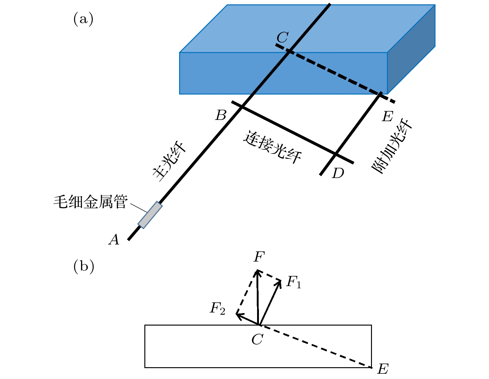 图 2 (a)非对称光纤悬臂结构示意图; (b)非对称光纤悬臂受力分析图
Figure2. (a) Schematic of the asymmetric fiber cantilever; (b) force analysis of the asymmetric fiber cantilever.
根据悬臂谐振理论, 单根光纤悬臂的谐振频率可由公式

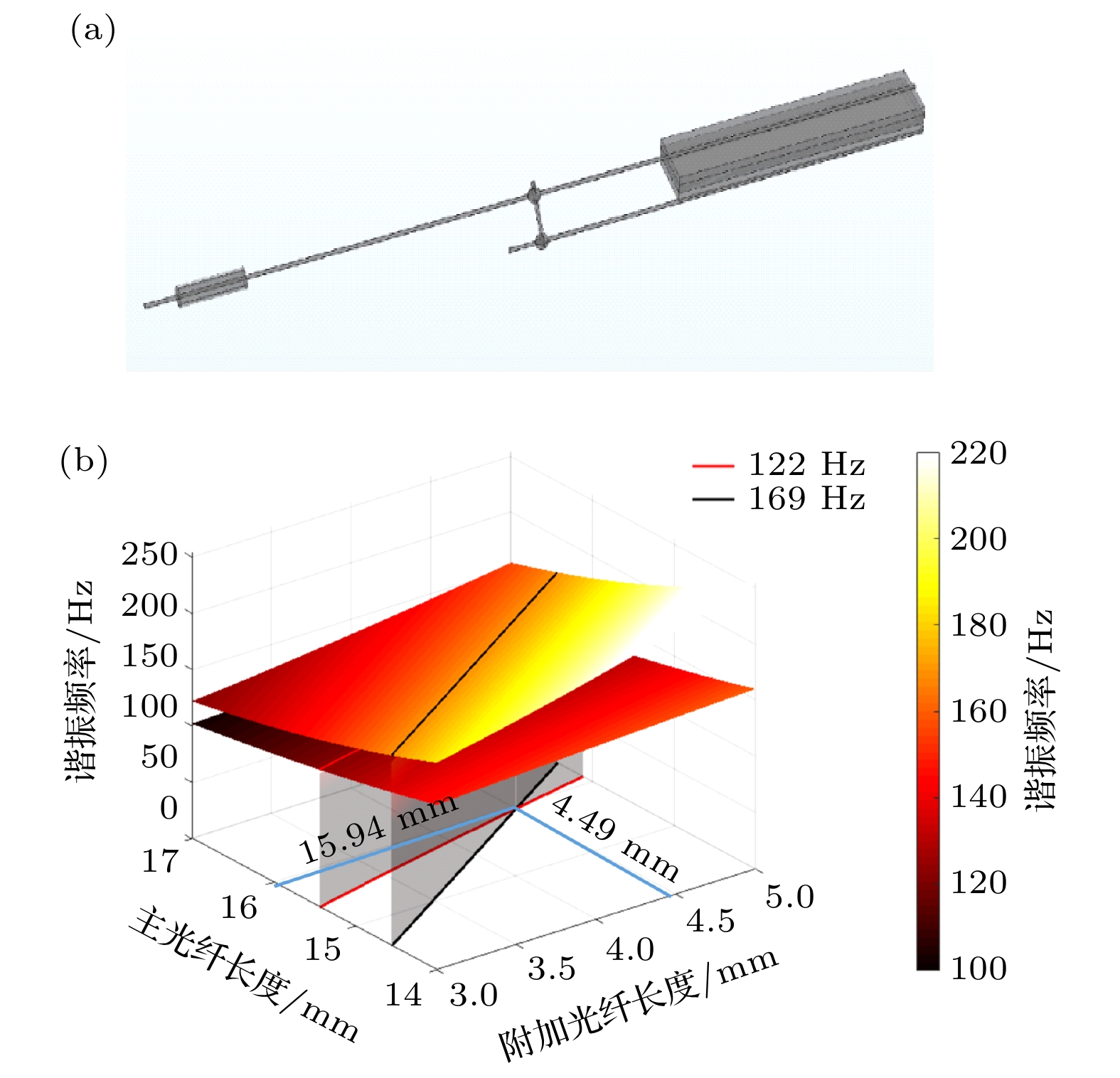 图 3 (a) COMSOL中仿真的非对称光纤悬臂结构示意图; (b)主光纤和附加光纤长度与正交谐振频率的关系图
Figure3. (a) Simulated probe structure in COMSOL; (b) the relationship between the length of the main fiber and the auxiliary fiber and the orthogonal resonance frequency.
 图 4 (a)全封装的Lissajous扫描光纤探头结构示意图; (b)全封装探头的实物照片
Figure4. (a) Schematic of the fully packaged Lissajous scanning fiber probe; (b) photograph of the fully packaged probe.
为了对Lissajous扫描轨迹进行预标定, 搭建了基于位置敏感探测器(position sensitive detector, PSD)的Lissajous扫描轨迹预标定系统, 使用He-Ne激光器将出射的激光耦合进光纤探头, 通过探头前端的GRIN透镜聚焦到PSD上. 由PSD输出的Lissajous扫描轨迹位置信息被数据采集卡采集. 通过计算机对采集到的位置信息数据进行预标定. 基于预标定后的位置信息可以将采集到的OCT轴向信息对应到正确的横向扫描位置处, 实现Lissajous扫描的图像重建.
内窥SS-OCT系统示意图如图5所示. 扫频光源(Axsun technology)的中心波长为1310 nm, 扫描速率为50 kHz, 扫描谱宽为135 nm. 系统实测的轴向分辨率为10.3 μm, 成像帧速率为1帧/s, 最大信噪比为110 dB. 由光源发出的扫频激光经分光比90/10的光纤耦合器1分为两路, 分别进入样品臂和参考臂. 进入样品臂的光经内窥探头照射样品, 进入参考臂的光经准直镜照射到平面镜上. 从样品臂和参考臂返回的干涉光在被平衡探测器探测后, 由数据采集卡采集并传输至计算机进行后续处理.
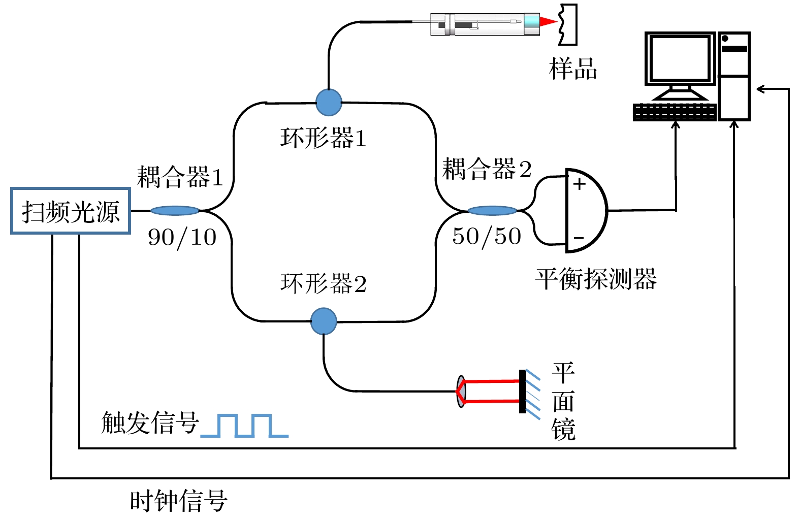 图 5 内窥SS-OCT系统示意图
Figure5. Schematic of the endoscopic SS-OCT system.
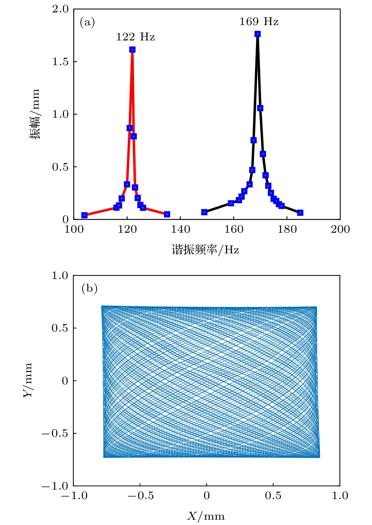 图 6 (a) Lissajous扫描光纤探头的振幅-频率响应曲线; (b)预标定的Lissajous扫描轨迹重建结果
Figure6. (a) Amplitude-frequency response curves of the Lissajous scanning fiber probe; (b) the reconstructed Lissajous scanning trajectory by pre-calibration.
为了研究Lissajous扫描光纤探头扫描的稳定性与可重复性, 通过预标定系统多次独立采集扫描轨迹的位置信息数据. 图7(a)—(f)展示了独立采集的6次扫描轨迹的前1500个位置信息数据的重建结果, 其中红色*号代表了扫描轨迹的起始位置. 由图7可见, 多次独立采集的扫描轨迹起始位置相同, 扫描路径一致. 计算了多次独立采集的位置信息数据与预标定位置信息数据对应坐标间的差值, 其差值最大值为13 μm, 方差约为0.016. 验证了探头扫描具有良好的稳定性和可重复性.
 图 7 (a)—(f) 6次独立实验的前1500个点的扫描轨迹重建结果
Figure7. (a)–(f) Reconstructed scanning trajectory of the first 1500 points from the 6 independent experiments
为了进一步验证Lissajous扫描光纤探头成像的旋转稳定性, 将探头接入实验室搭建的SS-OCT系统. 以探头中轴线为旋转轴分别旋转0°, 90°, 180°和270°, 对1元硬币上的字母A进行OCT成像. 图像重建采用预标定的扫描轨迹位置信息. 图8(a)是用相机拍摄的1元硬币及字母A的照片, 图8(b), (c), (d), (e)分别为探头在旋转0°, 90°, 180°, 270°状态下采集重建的OCT表面成像结果. 在探头绕自身中轴线旋转不同角度下采集到的字母A的OCT数据均能正确重建, 验证了探头成像具有良好的旋转稳定性.
 图 8 (a) 1元硬币及字母A的照片; (b)—(e)探头旋转0°, 90°, 180°和270°对字母A的OCT表面成像结果
Figure8. (a) Photograph of the 1 Yuan coin and the letter A; (b)–(e) the en-face OCT images of the letter A with the probe rotating to the angle of 0°, 90°, 180° and 270°.
为了验证所研制探头的成像性能, 应用基于Lissajous扫描光纤探头的内窥SS-OCT系统对生物组织进行了数据采集和图像重建. 首先实验中选取橘子果粒作为样品, 使用预标定的Lissajous扫描轨迹位置信息进行图像重建. 图9展示了重建的橘子果粒组织的OCT成像结果, 其中图9(a)为橘子果粒的实物照片, 红色方框为扫描光纤探头的成像范围. 图9(b)为橘子果粒组织的二维OCT横截面图像, 可以清晰分辨出橘子果粒组织内部的网格状细胞结构, 验证了研制的Lissajous扫描光纤探头具有良好的成像性能.
 图 9 (a)橘子果粒的实物照片; (b)橘子果粒组织的二维OCT横向截面图像
Figure9. (a) Photograph of the orange grain; (b) two-dimensional OCT cross-sectional image of orange grain tissue.
应用基于Lissajous扫描光纤探头的内窥SS-OCT系统对带有牙结石的成人磨牙进行了内窥成像. 磨牙牙体由牙釉质和牙本质组成, 牙釉质是人体最坚硬、钙化程度最高的组织, 牙本质的钙化程度比牙釉质稍低, 其散射系数大于牙釉质. 牙结石由人日常饮食堆积在牙齿附近的食物残渣矿化形成, 其主要组成为磷酸钙. 图10(a)和图10(b)分别为重建的磨牙健康牙体区域的二维OCT横截面图像和三维OCT图像, 可以清晰分辨出牙釉质、牙本质等健康牙体内部的分层结构. 图10(c)和图10(d)分别为重建的牙结石区域的二维OCT横截面图像和三维OCT图像, 可以看出牙结石内部不存在类似健康牙齿的分层结构. 研制的Lissajous扫描光纤探头可用于区分健康牙体和牙结石结构.
 图 10 (a)磨牙的二维OCT横截面图像; (b)磨牙的三维OCT图像; (c)牙结石的二维OCT横截面图像; (d)牙结石的三维OCT图像
Figure10. (a) Two-dimensional OCT cross sectional image of the health molar tooth tissue; (b) three-dimensional OCT image of the health molar tooth tissue; (c) two-dimensional OCT cross sectional image of the dental calculus; (d) three-dimensional OCT image of the dental calculus.
