Establishment and Preliminary Application of Lawsonia intracellularis IPMA Antigen Detection Method Based on SodC Monoclonal Antibody
LI MinXue,1, LI JianNan1, ZHOU Hong1, XIAO Ning1, LIN HuiXing1, MA Zhe1, FAN HongJie,1,21College of Veterinary Medicine, Nanjing Agricultural University, Nangjing 210095 2Jiangsu Co-innovation Center for the Prevention and Control of Important Animal, Yangzhou University Yangzhou, Yangzhou 225009 Jiangsu
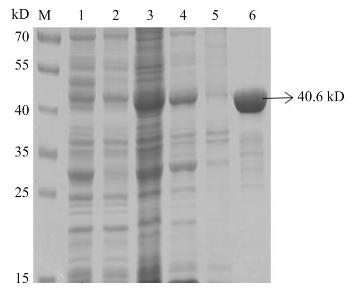
Abstract 【Objective】 Lawsonia intracellularis (L. intracellularis) is an enteric pathogenic bacteria that causes porcine proliferative enteropathy (PPE), which mainly shows the decline of animal welfare and causes serious economic losses to the world swine industries. The objective of this study was to prepare monoclonal antibodies against SodC of L. intracellularis, and to establish an immunoperoxidase monolayer assay (IPMA) method for detecting L. intracellularis base the monoclonal antibody, while test its application in clinical practice, so as to provide a scientific and effective means for the diagnosis of L. intracellularis. 【Method】In this study, the commercial live attenuated L. intracellularis vaccine was selected as target strain. The sodc gene was amplified by PCR and cloned into the prokaryotic expression vector pGex-6p-1. The recombinant plasmid pGex-6p-1-sodc was confirmed to be constructed successfully and induced expression of recombinant SodC protein. The reactivity of the recombinant protein was analyzed by Western Blot. The primary antibody was a mouse anti-GST labeled antibody. BALB/c mice aged 4-6 weeks were immunized with purified SodC protein, and hybridoma cells were screened by conventional cell fusion, limited dilution and indirect ELISA, then ascites were prepared. It was confirmed that two monoclonal antibodies had good specificity through indirect immunofluorescence (IFA). Using the monoclonal antibody as the primary antibody, a method of IPMA for detecting L. intracellularis was developed, and the specificity, sensitivity and repeatability of the method were evaluated. The optimized IPMA method was used to detect ileal tissue samples from pig farms in Jiangsu Province and to evaluate the clinical value of the method. 【Result】After purification, the concentration of SodC protein was higher, and it specifically bound to the antibody against GST tag, indicating that the protein had good regenicity. After three times of subcloning, two strains positive hybrid tumor cells were screened, named 1D6 and 1F7, respectively. The titers of two monoclonal antibodies were both reached 1﹕204 000 by ELISA. The subclass identification results of antibodies showed the subclass of 1D6 was IgA,and subclass of 1F7 was IgG3. The result IFA showed that 1D6 and 1F7 had specific reaction with L. intracellularis, but did not cross-react with S. Cholerasuis, PEDV and TGEV. The ascites of the two monoclonal antibodies were both 1﹕1 024 000 by ELISA; IFA confirmed that the two monoclonal antibodies had good specificity. The optimized IPMA reaction conditions showed that when the dilution ratio of the primary antibody was 1:800 for 45 min, and the dilution ratio of the secondary antibody was 1﹕2 500 for 1h, the established IPMA exhibited the best performance. The specificity and sensitivity tests showed that S. Cholerasuis, PEDV, TGEV, PCV2 and PRV were all negative, and the minimum detection limit was 103L.intracellularis. The optimized IPMA method was used to detect the ileum tissue samples from pig farms in the surrounding areas of Jiangsu Province. A total of 92 positive samples were detected from 146 samples of ileal tissues. The positive rates of 3 different pig farms were 65.6%, 68.1% and 53.7%, respectively, and the overall positive rate was 63.0%. 82 positive samples were detected by PCR method. and the positive coincidence rate of the two methods was 94.6%. These results indicated that this method had clinical value. 【Conclusion】The monoclonal antibodies against the recombinant SodC protein were successfully prepared, and the IPMA method for L. intracellularis was established with good specificity and sensitivity, and the clinical samples were tested. In summary, these results further proved that the IPMA had certain clinical value, and provided an effective technical means for the isolation and identification of L. intracellularis in the laboratory, localization in infected cells, epidemiological investigation and quarantine. Keywords:Lawsonia intracellularis;SodC protein;monoclonal antibody;an immunoperoxidase monolayer assay (IPMA)
PDF (1989KB)元数据多维度评价相关文章导出EndNote|Ris|Bibtex收藏本文 本文引用格式 李敏雪, 李剑男, 周红, 肖宁, 蔺辉星, 马喆, 范红结. 基于SodC单克隆抗体的胞内劳森菌IPMA抗原检测方法的建立及应用. 中国农业科学, 2021, 54(20): 4478-4486 doi:10.3864/j.issn.0578-1752.2021.20.020 LI MinXue, LI JianNan, ZHOU Hong, XIAO Ning, LIN HuiXing, MA Zhe, FAN HongJie. Establishment and Preliminary Application of Lawsonia intracellularis IPMA Antigen Detection Method Based on SodC Monoclonal Antibody. Scientia Acricultura Sinica, 2021, 54(20): 4478-4486 doi:10.3864/j.issn.0578-1752.2021.20.020
A:1D6与LI感染细胞反应;B:1F7与LI感染细反应;C-E:单抗分别与S.Cholerasuis、PEDV、TGEV反应;F:阴性对照 Fig. 3Specificity identification of monoclonal antibodies
A: 1D6 monoclonal antibody reacts with bacterial infected cells; B: 1F7 monoclonal antibody reacts with healthy cells; C-E:monoclonal antibody reacts with S. Cholerasuis, PEDV and TGEV, respectively; F: Negative control
A:单抗与LI感染细胞反应;B-F:单抗分别S. Cholerasuis、PEDV、TGEV、PCV2 、PRV感染细胞反应 Fig. 6The specificity test of IPMA
A: Monoclonal antibody against SodC reacts with LI-infected cells; B: Mouse monoclonal antibodies against SodC react with S. Cholerasuis, PEDV, TGEV, PCV2,PRV infected cells
YINH F, YANGX Y, ZHANGX P, ZHENGX T, LINZ, HUANGW B. Isolation and detection of Lawsonia intracellularis of porcine intestinal mucosa Journal of Longyan Univercity, 2012, 30(5):55-60. (in Chinese) [本文引用: 1]
STEGEH, JENSENT K, MØLLERK, VESTERGAARDK, BAEKBOP, JORSALS E. Infection dynamics of Lawsonia intracellularis in pig herds Veterinary Microbiology, 2004, 104(3/4):197-206. DOI:10.1016/j.vetmic.2004.09.015URL [本文引用: 1]
LOUBLIERC, CERRIS, GRYSPEERDTA, AMORYH, BAUWENSC, CESARINIC. High seroprevalence against Lawsonia intracellularis among adult horses in Belgium Journal of Equine Veterinary Science, 2020, 95:103304. DOI:10.1016/j.jevs.2020.103304URL [本文引用: 3]
TONGX Y. Epidemiology, clinical symptoms and control measures of ileitis in pigs Modern Animal Husbandry Science & Technology, 2019(5):90-91. (in Chinese) [本文引用: 1]
MCORISTS, MACINTYREN, STOKESC R, LAWSONG H. Immunocytological responses in porcine proliferative enteropathies Infection and Immunity, 1992, 60(10):4184-4191. DOI:10.1128/iai.60.10.4184-4191.1992URL [本文引用: 1]
LAWSONG H K, GEBHARTC J. Proliferative enteropathy Journal of Comparative Pathology, 2000, 122(2-3):77-100. DOI:10.1053/jcpa.1999.0347URL
KINOSHITAY, NIWAH, UCHIDA-FUJIIE, NUKADAT. Genotyping of equine Lawsonia intracellularis sampled in Japan by using multilocus variable-number tandem repeat analysis Journal of Equine Veterinary Science, 2021, 96:103311. DOI:10.1016/j.jevs.2020.103311URL
JACOBSONM, FELLSTRÖMC, JENSEN-WAERNM. Porcine proliferative enteropathy: an important disease with questions remaining to be solved Veterinary Journal, 2010, 184(3):264-268. DOI:10.1016/j.tvjl.2009.05.010URL
余彦国, 张瑾. 猪增生性肠炎的研究进展 畜牧兽医杂志, 2009, 28(3):33-36.
YUY G, ZHANGJ. Research advance in porcine proliferative enteritis Journal of Animal Sciaence and Veterinary Medicine, 2009, 28(3):33-36. (in Chinese)
LIUP H, WANGJ, ZHANGW Y. Advances in pathogenic characteristics of porcine proliferative enteritis Shanghai Journal of Animal Husbandry and Veterinary Medicine, 2005(1):12-13. (in Chinese) [本文引用: 1]
VISSCHERC, KRUSEA, SANDERS, KELLERC, MISCHOKJ, TABELINGR, HENNEH, DEITMERR, KAMPHUESJ. Experimental studies on effects of diet on Lawsonia intracellularis infections in fattening boars in a natural infection model Acta Veterinaria Scandinavica, 2018, 60(1):22. DOI:10.1186/s13028-018-0378-4URL [本文引用: 1]
LEITEF L, ABRAHANTEJ E, VASQUEZE, VANNUCCIF, GEBHARTC J, WINKELMANN, MUELLERA, TORRISONJ, RAMBOZ, ISAACSONR E. A cell proliferation and inflammatory signature is induced by Lawsonia intracellularis infection in swine bioRxiv, 2018, DOI: 10.1101/384230. [本文引用: 1]
WATTANAPHANSAKS, GEBHARTC, OLINM, DEENJ. Measurement of the viability of Lawsonia intracellularis Canadian Journal of Veterinary Research-revue Canadienne De Recherche Veterinaire, 2005, 69(4):265-271. [本文引用: 1]
OBRADOVICM R, WILSONH L. Immune response and protection against Lawsonia intracellularis infections in pigs Veterinary Immunology and Immunopathology, 2020, 219:109959. DOI:10.1016/j.vetimm.2019.109959URL [本文引用: 1]
WUY Y, YANGD D, GAOD S, LIY T, CHANGH T, WANGC Q, ZHAOJ. Prokaryotic expression of LsaA protein of Lawsonia intracellularis and development of a LsaA-based indirect ELISA for antibody detection Acta Veterinaria et Zootechnica Sinica, 2018, 49(4):786-793. (in Chinese) [本文引用: 1]
BIESTERH E, SCHWERTEL H. Intestinal adenoma in swine American Journal of Pathology, 1931, 7:175-185. [本文引用: 1]
GUOJ C, QINZ H, ZHANGY Y, PUW J, RENS N, ZHANGH J, MAC Q, LIG Q, BAIA Q. Development of PCR diagnostic method and clinical sample detection for pig proliferative enteropathy Progress in Veterinary Medicine, 2014, 35(4):89-93. (in Chinese) [本文引用: 1]
QUX Y, YAOH C, ZHANGX, HUANGT, LIX H. Epidemiological survey of porcine ileitis on intensive swine farms in East China Chinese Journal of Preventive Veterinary Medicine, 2012, 34(11):873-877. (in Chinese) [本文引用: 1]
HUANGZ, SONGC X, WANGH W, FANGS H. Serological investigation of porcine proliferative enteronitis in five provinces of South China Veterinary Science in China, 2006, 36(9):748-751. (in Chinese) [本文引用: 1]
LIUX J. Construction of recombinant S.Cholerasuis expressing Brucella sodc gene and establishment of PCR detection method for identification of Brucella [D]. Wuhan:Huazhong Agricultural University, 2010. (in Chinese) [本文引用: 1]
VANNUCCIF A, FOSTERD N, GEBHARTC J. Laser microdissection coupled with RNA-seq analysis of porcine enterocytes infected with an obligate intracellular pathogen (Lawsonia intracellularis) BMC Genomics, 2013, 14:421. DOI:10.1186/1471-2164-14-421URL [本文引用: 1]
BENGTSSONR J, MACINTYREN, GUTHRIEJ, WILSONA D, FINLAYSONH, MATIKAO, PONG-WONGR, SMITHS H, ARCHIBALDA L, AIT-ALIT. Lawsonia intracellularis infection of intestinal crypt cells is associated with specific depletion of secreted MUC2 in goblet cells Veterinary Immunology and Immunopathology, 2015, 168(1/2):61-67. DOI:10.1016/j.vetimm.2015.08.005URL [本文引用: 1]
SUNN, LIUX, CHENQ, LIUY, WENY J, CHENGS P. Establishment and primary application of PCR method for detection of Lawsonia intracellularis Chinese Journal of Veterinary Science, 2017, 37(1):36-39. (in Chinese) [本文引用: 1]
PENGZ, TUZ Q, LIANGW, HUR M, WANGH N, SHOUJ, DONGC, LIZ H, CHENH C. Development and preliminary application of a nested PCR method for detection of Lawsonia intracellularis in feces of pigs Chinese Journal of Veterinary Science, 2017, 37(1):40-46. (in Chinese) [本文引用: 1]
PEDERSENK S, STEGEH, JENSENT K, GUEDESR, STÅHLM, NIELSENJ P, HJULSAGERC, LARSENL E, ANGENØ. Diagnostic performance of fecal quantitative real-time polymerase chain reaction for detection of Lawsonia intracellularis-associated proliferative enteropathy in nursery pigs Journal of Veterinary Diagnostic Investigation, 2013, 25(3):336-340. DOI:10.1177/1040638713480499URL [本文引用: 1]
LIY N, WANGJ C, WANGJ F, LIUL B, ZHANGR X, SHIR H, HANQ G, SUNJ G, YUANW Z. A real-time loop-mediated isothermal amplification method for rapid detection of Lawsonia intracellularis in porcine fecal samples Journal of Microbiological Methods, 2018, 151:62-65. DOI:10.1016/j.mimet.2018.06.002URL [本文引用: 1]
RICHTERB, LADINIGA, NEDOROSTN, WEISSENBÖCKH. A TaqMan quantitative polymerase chain reaction assay for the detection of Lawsonia intracellularis in fecal and tissue samples from pigs Journal of Veterinary Diagnostic Investigation, 2010, 22(1):70-73. DOI:10.1177/104063871002200112URL [本文引用: 1]
ZHENGX T, HUANGC Q, HUANGQ C, DAIA L, TANX J. Establishment of SYBR Green Ⅰ real-time PCR assay for detection of Lawsonia intracellularis Journal of Northwest A&F University (Natural Science Edition), 2015, 43(12):29-34. (in Chinese) [本文引用: 1]
BAIA Q, GUOJ C, QINZ H, PUW J, MAC Q, LIG Q, ZHANGH J. Development of a TaqMan quantitative polymerase chain reaction assay for detecting Lawsonia intracellularis Acta Veterinaria et Zootechnica Sinica, 2014, 45(10):1733-1738. (in Chinese) [本文引用: 1]
WUY Y, TIANK Y, ZHANGY H, GUOH F, LIN, WANGZ, ZHAOJ. Rapid and visual detection of Lawsonia intracellularis with an improved recombinase polymerase amplification assay combined with a lateral flow dipstick BMC Veterinary Research, 2019, 15(1):97. DOI:10.1186/s12917-019-1841-9URL [本文引用: 1]
LIJ M, ZHUW, QINH G, JINGZ Q, LIAOY Y, YUY J, XIAOM, YUANG, XIEH L. IFA and IPMA for HCLV titer detection Chinese Journal of Veterinary Drug, 2013, 47(10):35-38. (in Chinese) [本文引用: 1]
ZHANGJ L, LIUW X, CHENW Y, LIC C, XIEM M, BUZ G. Development of an immunoperoxidase monolayer assay for the detection of antibodies against peste des petits ruminants virus based on BHK-21 cell line stably expressing the goat signaling lymphocyte activation molecule PLoS One, 2016, 11(10):e0165088. DOI:10.1371/journal.pone.0165088URL [本文引用: 1]
DIREKSINK, JOOH, GOYALS M. An immunoperoxidase monolayer assay for the detection of antibodies against swine influenza virus Journal of Veterinary Diagnostic Investigation, 2002, 14(2):169-171. DOI:10.1177/104063870201400215URL [本文引用: 1]
GUEDESR M C, GEBHARTC J, WINKELMANN L, MACKIE-NUSSR A. A comparative study of an indirect fluorescent antibody test and an immunoperoxidase monolayer assay for the diagnosis of porcine proliferative enteropathy Journal of Veterinary Diagnostic Investigation, 2002, 14(5):420-423. DOI:10.1177/104063870201400512URL [本文引用: 2]
ZHUY N, ZHANGX, ZHUX D, CHENJ F, SHIH Y, SHID, FENGL. Development of immunoperoxidase monolayer assay detecting transmissible gastroenteritis virus in situ Chinese Journal of Preventive Veterinary Medicine, 2017, 39(3):237-239. (in Chinese) [本文引用: 1]
HUERTAB, ARENASA, CARRASCL, MALDONADOA, TARRADASC,, CARBONERO, PEREAA. Comparison of diagnostic techniques for porcine proliferative enteropathy (Lawsonia intracellularis infection) Journal of Comparative Pathology, 2003, 129(2-3):179-185. DOI:10.1016/S0021-9975(03)00030-6URL [本文引用: 1]
WELLENBERGG J, ROOIJE M A V, MAISSANJ, OIRSCHOTJ T V. Evaluation of newly developed immunoperoxidase monolayer assays for detection of antibodies against bovine herpesvirus 4 Clinical & Diagnostic Laboratory Immunology, 1999, 6(4):447. [本文引用: 1]
KROLLJ J, ROOFM B, HOFFMANL J, DICKSONJ S, HARRISD L. Hank. Proliferative enteropathy:a global enteric disease of pigs caused by Lawsonia intracellularis Animal Health Research Reviews, 2005, 6(2):173-197. DOI:10.1079/AHR2005109URL [本文引用: 1]
LAWSONG H, MCORISTS, JASNIS, MACKIER A. Intracellular bacteria of porcine proliferative enteropathy: cultivation and maintenance in vitro Journal of Clinical Microbiology, 1993, 31(5):1136-1142. DOI:10.1128/jcm.31.5.1136-1142.1993URL [本文引用: 1]
MIRAJKARN S, KELLEYM R, GEBHARTC J. Draft genome sequence of Lawsonia intracellularis strain E40504, isolated from a horse diagnosed with equine proliferative enteropathy Genome Announcements, 2017, 5(19). [本文引用: 1]
MCORISTS, GEBHARTC J, BOSWORTHB T. Evaluation of porcine ileum models of enterocyte infection by Lawsonia intracellularis Canadian Journal of Veterinary Research, 2006, 70(2):155-159. [本文引用: 1]
OHY S, LEEJ B, MCORISTS. Microarray analysis of differential expression of cell cycle and cell differentiation genes in cells infected with Lawsonia intracellularis Veterinary Journal, 2010, 184(3):340-345. DOI:10.1016/j.tvjl.2009.03.018URL [本文引用: 1]
VANNUCCIF A, WATTANAPHANSAKS, GEBHARTC J. An Alternative Method for Cultivation of Lawsonia intracellularis Journal of Clinical Microbiology, 2012, 50(3):1070-1072. DOI:10.1128/JCM.05976-11URL [本文引用: 1]
 ,1, 李剑男1, 周红1, 肖宁1, 蔺辉星1, 马喆1, 范红结

 新窗口打开|下载原图ZIP|生成PPT
 新窗口打开|下载原图ZIP|生成PPT
 新窗口打开|下载原图ZIP|生成PPT
 新窗口打开|下载原图ZIP|生成PPT
 新窗口打开|下载原图ZIP|生成PPT
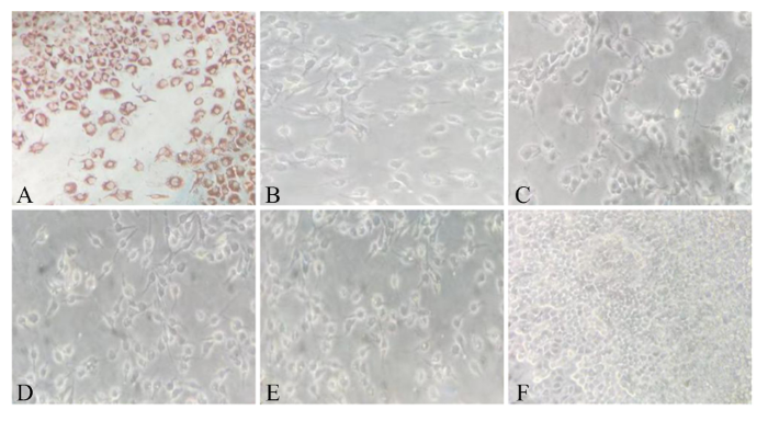 新窗口打开|下载原图ZIP|生成PPT
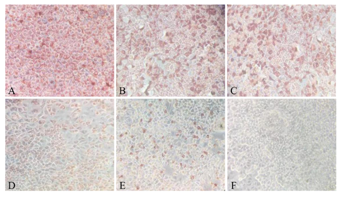 新窗口打开|下载原图ZIP|生成PPT