 ), 陈霓虹3,4(), 黄昌兵1,2
 1中国科学院心理研究所行为科学重点实验室, 北京 100101
2中国科学院大学心理学系, 北京 100049
3清华大学心理学系
4清华大学-IDG/麦戈文脑科学联合研究院, 北京 100084
收稿日期:2020-03-18出版日期:2021-01-15发布日期:2020-11-23通讯作者:席洁,陈霓虹E-mail:xij@psych.ac.cn;nihongch@mail.tsinghua.edu.cn基金资助:* 国家重点研发计划(2018YFC0705100);国家重点研发计划(2019YFC200108);国家自然科学基金(31470983);国家自然科学基金(31400877);国家自然科学基金(31971031);国家自然科学基金(31930053)Binocular disparity: Neural mechanisms and perceptual learning
WANG Getong1,2, XI Jie1,2(), CHEN Nihong3,4(), HUANG Changbing1,2 1CAS Key Laboratory of Behavioral Science, Institute of Psychology, Chinese Academy of Sciences, Beijing 100101, China
2Department of Psychology, University of Chinese Academy of Sciences, Beijing 100049, China
3Department of Psychology, School of Social Sciences, Tsinghua University, Beijing 100084, China
4IDG/McGovern Institute for Brain Research at Tsinghua University, Beijing 100084, China
Received:2020-03-18Online:2021-01-15Published:2020-11-23Contact:XI Jie,CHEN Nihong E-mail:xij@psych.ac.cn;nihongch@mail.tsinghua.edu.cn摘要/Abstract
摘要: 双眼瞳距使得空间某物体在左右眼视网膜的成像存在微小位置差异, 这种差异被称为双眼视差(binocular disparity), 是立体视知觉的重要信息来源。对双眼视差的心理物理学研究始于18世纪初, 迄今已有接近两百年的历史。近年来, 双眼视差研究主要集中在两方面。其一是用电生理、脑成像技术考察双眼视差在视觉背、腹侧通路的模块化表征, 其脑区表征反映出视觉系统的层级式、平行式加工规律。其二是应用知觉学习范式研究双眼视差的可塑性。未来研究应综合脑成像和神经调控技术考察双眼视差的神经机制及其学习效应, 包括双眼视差与多种深度线索间的信息整合和交互作用。应用方向上, 可结合虚拟现实等技术优化训练范式, 实现立体视力的康复和增强。
图/表 5
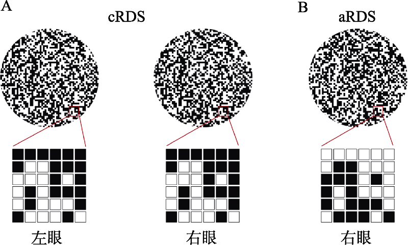
图1cRDS和aRDS示意图。A为相关随机点立体图(cRDS)示意图, 左右眼图片对应位置对比度相同, 经过双眼融合能够产生深度知觉; B为反相关随机点立体图(aRDS)示意图, 左右眼图片对应位置对比度取反, 虽然aRDS具有视差信息, 但由于打破了双眼之间的对应关系, 因此无法产生深度知觉。
图1cRDS和aRDS示意图。A为相关随机点立体图(cRDS)示意图, 左右眼图片对应位置对比度相同, 经过双眼融合能够产生深度知觉; B为反相关随机点立体图(aRDS)示意图, 左右眼图片对应位置对比度取反, 虽然aRDS具有视差信息, 但由于打破了双眼之间的对应关系, 因此无法产生深度知觉。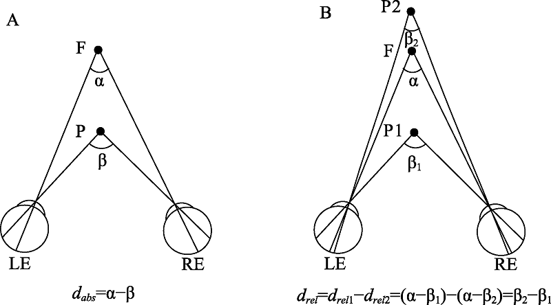
图2双眼视差示意图。双眼视差是空间中某一物体P在左眼(LE)和右眼(RE)视网膜上成像的水平差异。A为绝对视差示意图, F为注视点, 双眼视线在注视点F处夹角为α, 在物体P处夹角为β, 绝对视差dabs为(α-β), 其大小与注视点F的位置有关; B为相对视差示意图, F为注视点, 双眼视线在注视点F处夹角为α, 在物体P1和P2处夹角分别为β1和β2, 两物体之间的相对视差drel等于两者绝对视差的差值, 其值为(β2-β1), 其大小与注视点F的位置无关。
图2双眼视差示意图。双眼视差是空间中某一物体P在左眼(LE)和右眼(RE)视网膜上成像的水平差异。A为绝对视差示意图, F为注视点, 双眼视线在注视点F处夹角为α, 在物体P处夹角为β, 绝对视差dabs为(α-β), 其大小与注视点F的位置有关; B为相对视差示意图, F为注视点, 双眼视线在注视点F处夹角为α, 在物体P1和P2处夹角分别为β1和β2, 两物体之间的相对视差drel等于两者绝对视差的差值, 其值为(β2-β1), 其大小与注视点F的位置无关。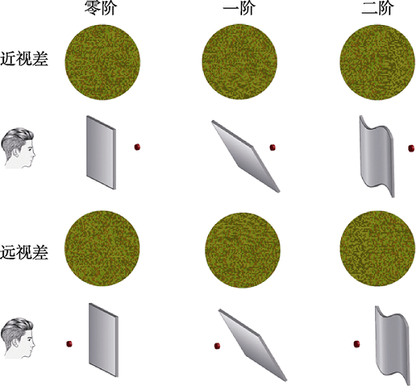
图3零阶、一阶和二阶视差RDS刺激示意图。佩戴红绿眼镜观看每幅红绿图像, 可形成图下方对应的深度知觉。零阶视差表征物体与注视点之间的远近关系, 一阶视差形成具有视差梯度的三维斜面, 二阶视差形成的则是具有视差梯度和曲率的三维形状。立体示意图中红色圆点代表注视点。
图3零阶、一阶和二阶视差RDS刺激示意图。佩戴红绿眼镜观看每幅红绿图像, 可形成图下方对应的深度知觉。零阶视差表征物体与注视点之间的远近关系, 一阶视差形成具有视差梯度的三维斜面, 二阶视差形成的则是具有视差梯度和曲率的三维形状。立体示意图中红色圆点代表注视点。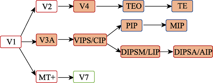
图4双眼视差的神经表征。箭头表示信息传递方向。图中白底标示脑区主要加工绝对视差, 橙底标示脑区主要加工相对视差。绿框标示现有研究发现的人类大脑皮层激活区域, 蓝框标示现有研究发现的猴大脑皮层激活区域, 红框标示人和猴共有的激活区域。MT+: middle temporal complex, 颞中回; VIPS: ventral IPS area, 顶内沟腹侧区; CIP: caudal intraparietal area, 顶内沟后部; DIPSM: the dorsal IPS medial area, 背侧顶内沟内侧区域; LIP: lateral intraparietal area, 顶内沟外侧区; DIPSA: the dorsal IPS anterior area, 背侧顶内沟前部; AIP: anterior intraparietal area, 顶内沟前部; PIP: posterior intraparietal area, 顶内沟后侧区; MIP: medial intraparietal area, 顶内沟内侧区; TE: the superior temporal sulcus, 颞上沟; TEO: temporal-occipital area。
图4双眼视差的神经表征。箭头表示信息传递方向。图中白底标示脑区主要加工绝对视差, 橙底标示脑区主要加工相对视差。绿框标示现有研究发现的人类大脑皮层激活区域, 蓝框标示现有研究发现的猴大脑皮层激活区域, 红框标示人和猴共有的激活区域。MT+: middle temporal complex, 颞中回; VIPS: ventral IPS area, 顶内沟腹侧区; CIP: caudal intraparietal area, 顶内沟后部; DIPSM: the dorsal IPS medial area, 背侧顶内沟内侧区域; LIP: lateral intraparietal area, 顶内沟外侧区; DIPSA: the dorsal IPS anterior area, 背侧顶内沟前部; AIP: anterior intraparietal area, 顶内沟前部; PIP: posterior intraparietal area, 顶内沟后侧区; MIP: medial intraparietal area, 顶内沟内侧区; TE: the superior temporal sulcus, 颞上沟; TEO: temporal-occipital area。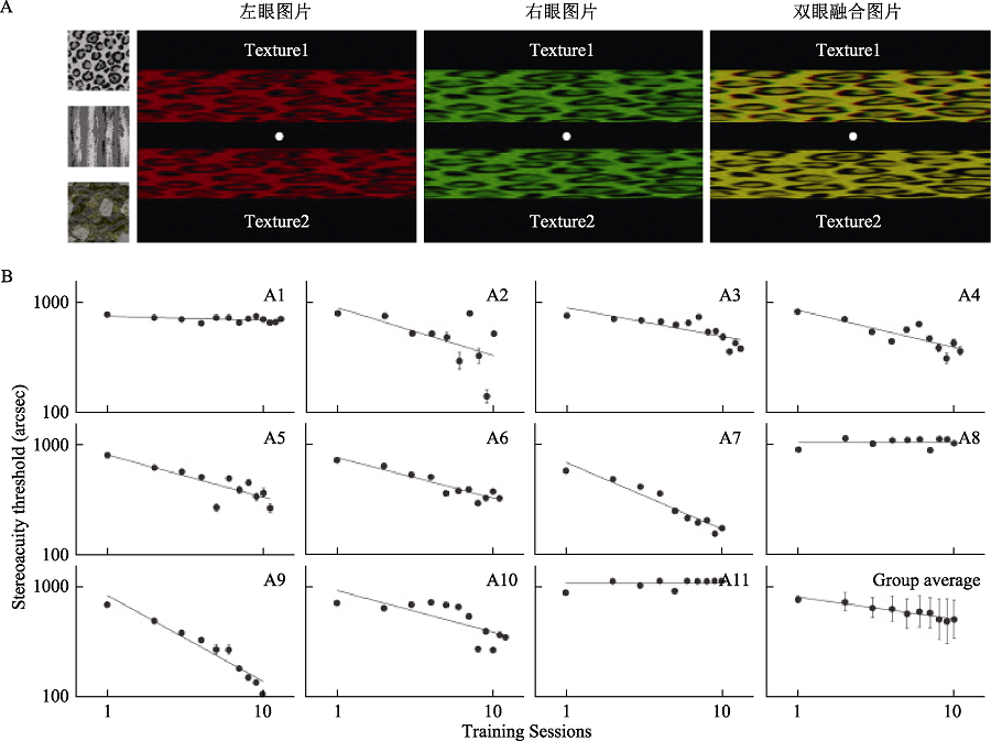
图5立体视训练(Xi et al., 2014)。A为训练使用的刺激, 第一行为刺激的三种纹理, 第二行以其中一种纹理图案为例, 从左至右依次为刺激的左眼、右眼及双眼融合后图片, 被试在实验中需佩戴红绿眼镜; B为11名弱视被试单人及平均学习曲线, 横坐标为训练次数, 纵坐标为立体视阈值, 研究发现训练后11名弱视被试中的9人立体视阈值显著降低。
图5立体视训练(Xi et al., 2014)。A为训练使用的刺激, 第一行为刺激的三种纹理, 第二行以其中一种纹理图案为例, 从左至右依次为刺激的左眼、右眼及双眼融合后图片, 被试在实验中需佩戴红绿眼镜; B为11名弱视被试单人及平均学习曲线, 横坐标为训练次数, 纵坐标为立体视阈值, 研究发现训练后11名弱视被试中的9人立体视阈值显著降低。参考文献 142
| [1] | 侯川. (1995). 立体视觉的发生机理与检测. 中国斜视与小儿眼科杂志, 3, 141-144. |
| [2] | 颜少明. (2006). 立体视觉检查图 (第3版). 北京: 人民卫生出版社. |
| [3] | Alexander, J. A. (1979). A new clinical test of stereopsis: Theoretical evaluation. The Australian Journal of Optometry, 62(5), 191-193. |
| [4] | Allouni, A. K., Thomas, O., Solomon, S. G., Krug, K., & Parker, A. J. (2005). Local and global binocular matching in V2 of the awake macaque. Society for Neuroscience Abstracts, 510, 8. |
| [5] | Andersen, R. A., & Buneo, C. A. (2002). Intentional maps in posterior parietal cortex. Annual Review of Neuroscience, 25(1), 189-220. doi: 10.1146/annurev.neuro.25.112701.142922URL |
| [6] | Anzai, A., Chowdhury, S. A., & DeAngelis, G. C. (2011). Coding of stereoscopic depth information in visual areas V3 and V3A. The Journal of Neuroscience, 31(28), 10270-10282. URLpmid: 21753004 |
| [7] | Anzai, A., & DeAngelis, G. C. (2010). Neural computations underlying depth perception. Current Opinion in Neurobiology, 20(3), 367-375. URLpmid: 20451369 |
| [8] | Astle, A. T., McGraw, P. V., & Webb, B. S. (2011). Recovery of stereo acuity in adults with amblyopia. BMJ Case Reports, 7-10. |
| [9] | Backus, B. T., Fleet, D. J., Parker, A. J., & Heeger, D. J. (2001). Human cortical activity correlates with stereoscopic depth perception. Journal of Neurophysiology, 86(4), 2054-2068. URLpmid: 11600661 |
| [10] | Ban, H., Preston, T. J., Meeson, A., & Welchman, A. E. (2012). The integration of motion and disparity cues to depth in dorsal visual cortex. Nature Neuroscience, 15(4), 636-643. doi: 10.1038/nn.3046URL |
| [11] | Barlow, H. B., Blakemore, C., & Pettigrew, J. D. (1967). The neural mechanism of binocular depth discrimination. The Journal of Physiology, 193(2), 327-342. URLpmid: 6065881 |
| [12] | Bohr, I., & Read, J. C. A. (2013). Stereoacuity with Frisby and revised FD2 stereo tests. PLoS One, 8(12), e82999. URLpmid: 24349416 |
| [13] | Born, R. T., & Bradley, D. C. (2005). Structure and function of visual area MT. Annual Review of Neuroscience, 28(1), 157-189. |
| [14] | Bosten, J. M., Goodbourn, P. T., Lawrance-Owen, A. J., Bargary, G., Hogg, R. E., & Mollon, J. D. (2015). A population study of binocular function. Vision Research, 110, 34-50. URLpmid: 25771401 |
| [15] | Bradshaw, M. F., & Glennerster, A. (2006). Stereoscopic acuity and observation distance. Spatial Vision, 19(1), 21-36. URLpmid: 16411481 |
| [16] | Bredfeldt, C. E., & Cumming, B. G. (2006). A simple account of cyclopean edge responses in macaque V2. The Journal of Neuroscience, 26(29), 7581-7596. URLpmid: 16855086 |
| [17] | Brookes, A., & Stevens, K. A. (1989). The analogy between stereo depth and brightness. Perception, 18(5), 601-614. URLpmid: 2602086 |
| [18] | Chang, D. H. F., Mevorach, C., Kourtzi, Z., & Welchman, A. E. (2014). Training transfers the limits on perception from parietal to ventral cortex. Current Biology, 24(20), 2445-2450. URLpmid: 25283780 |
| [19] | Chen, G., Lu, H. D., & Roe, A. W. (2008). A map for horizontal disparity in monkey V2. Neuron, 58(3), 442-450. URLpmid: 18466753 |
| [20] | Chino, Y. M., Smith, E. L., Hatta, S., & Cheng, H. (1997). Postnatal development of binocular disparity sensitivity in neurons of the primate visual cortex. The Journal of Neuroscience, 17(1), 296-307. URLpmid: 8987756 |
| [21] | Chopin, A., Bavelier, D., & Levi, D. M. (2019). The prevalence and diagnosis of 'stereoblindness' in adults less than 60 years of age: A best evidence synthesis. Ophthalmic and Physiological Optics, 39(2), 66-85. URLpmid: 30776852 |
| [22] | Ciner, E. B., Scheiman, M. M., Schanel-Klitsch, E., & Weil, L. (1989). Stereopsis testing in 18- to 35-month-old children using operant preferential looking. Optometry & Vision Science, 66(11), 782-787. URLpmid: 2616139 |
| [23] | Cooper, J., Feldman, J., & Medlin, D. (1979). Comparing stereoscopic performance of children using the Titmus, TNO, and randot stereo tests. Journal of the American Optometric Association, 50(7), 821-825. URLpmid: 500993 |
| [24] | Cottereau, B. R., McKee, S. P., Ales, J. M., & Norcia, A. M. (2011). Disparity-tuned population responses from human visual cortex. The Journal of Neuroscience, 31(3), 954-965. doi: 10.1523/JNEUROSCI.3795-10.2011URLpmid: 21248120 |
| [25] | Cowey, A., & Porter, J. (1979). Brain damage and global stereopsis. Proceedings of the Royal Society B: Biological Sciences, 204(1157), 399-407. |
| [26] | Cowey, A., & Wilkinson, F. (1991). The role of the corpus callosum and extra striate visual areas in stereoacuity in macaque monkeys. Neuropsychologia, 29(6), 465-479. URLpmid: 1944856 |
| [27] | Cumming, B. G., & Parker, A. J. (1997). Responses of primary visual cortical neurons to binocular disparity without depth perception. Nature, 389(6648), 280-283. URLpmid: 9305841 |
| [28] | Cumming, B. G., & Parker, A. J. (1999). Binocular neurons in V1 of awake monkeys are selective for absolute, not relative, disparity. The Journal of Neuroscience, 19(13), 5602-5618. URLpmid: 10377367 |
| [29] | DeAngelis, G. C., & Newsome, W. T. (1999). Organization of disparity-selective neurons in macaque area MT. The Journal of Neuroscience, 19(4), 1398-1415. URLpmid: 9952417 |
| [30] | DiCarlo, J. J., Zoccolan, D., & Rust, N. C. (2013). How does the brain solve visual object recognition?. Neuron, 73(3), 415-434. URLpmid: 22325196 |
| [31] | Ding, J., & Levi, D. M. (2011). Recovery of stereopsis through perceptual learning in human adults with abnormal binocular vision. Proceedings of the National Academy of Sciences of the United States of America, 108(37), E733-E741. doi: 10.1073/pnas.1105183108URL |
| [32] | Dodd, J. V., Krug, K., Cumming, B. G., & Parker, A. J. (2001). Perceptually bistable three-dimensional figures evoke high choice probabilities in cortical area MT. The Journal of Neuroscience, 21(13), 4809-4821. URLpmid: 11425908 |
| [33] | Do?Venciog?lu, D., Ban, H., Schofield, A. J., & Welchman, A. E. (2013). Perceptual integration for qualitatively different 3-D cues in the human brain. Journal of Cognitive Neuroscience, 25(9), 1527-1541. URLpmid: 23647559 |
| [34] | Durand, J.-B., Nelissen, K., Joly, O., Wardak, C., Todd, J. T., Norman, J. F., ... Orban, G. A. (2007). Anterior regions of monkey parietal cortex process visual 3D shape. Neuron, 55(3), 493-505. doi: 10.1016/j.neuron.2007.06.040URL |
| [35] | Durand, J.-B., Peeters, R., Norman, J. F., Todd, J. T., & Orban, G. A. (2009). Parietal regions processing visual 3D shape extracted from disparity. NeuroImage, 46(4), 1114-1126. URLpmid: 19303937 |
| [36] | Erkelens, C. J., & Collewijn, H. (1985). Motion perception during dichoptic viewing of moving random-dot stereograms. Vision Research, 25(4), 583-588. URLpmid: 4060612 |
| [37] | Feinberg, R., & Reuel, S. (1961). Device for testing visual acuity. US3011394A. |
| [38] | Fendick, M., & Westheimer, G. (1983). Effects of practice and the separation of test targets on foveal and peripheral stereoacuity. Vision Research, 23(2), 145-150. URLpmid: 6868389 |
| [39] | Finlay, D. C., Manning, M. L., Dunlop, D. P., & Dewis, S. A. M. (1989). Difficulties in the definition of 'stereoscotoma' using temporal detection of thresholds of dynamic random dot stereograms. Documenta Ophthalmologica, 72, 161-173. URLpmid: 2582997 |
| [40] | Fox, R., Patterson, R., & Francis, E. L. (1986). Stereoacuity in young children. Investigative Ophthalmology & Visual Science, 27(4), 598-600. URLpmid: 3957579 |
| [41] | Frisby, J. P., & Clatworthy, J. L. (1975). Learning to see complex random-dot stereograms. Perception, 4(2), 173-178. |
| [42] | Gallese, V., Murata, A., Kaseda, M., Niki, N., & Sakata, H. (1994). Deficit of hand preshaping after muscimol injection in monkey parietal cortex. Cognitive Neuroscience and Neuropsychology, 5(12), 1525-1529. |
| [43] | Gantz, L., Patel, S. S., Chung, S. T. L., & Harwerth, R. S. (2007). Mechanisms of perceptual learning of depth discrimination in random dot stereograms. Vision Research, 47(16), 2170-2178. URLpmid: 17588634 |
| [44] | Georgieva, S., Peeters, R., Kolster, H., Todd, J. T., & Orban, G. A. (2009). The processing of three-dimensional shape from disparity in the human brain. The Journal of Neuroscience, 29(3), 727-742. URLpmid: 19158299 |
| [45] | Giaschi, D., Narasimhan, S., Solski, A., Harrison, E., & Wilcox, L. M. (2013). On the typical development of stereopsis: fine and coarse processing. Vision Research, 89, 65-71. URLpmid: 23891704 |
| [46] | Goncalves, N. R., Ban, H., Sánchez-Panchuelo, R. M., Francis, S. T., Schluppeck, D., & Welchman, A. E. (2015). 7 Tesla fMRI reveals systematic functional organization for binocular disparity in dorsal visual cortex. The Journal of Neuroscience, 35(7), 3056-3072. URLpmid: 25698743 |
| [47] | Goodale, M. A., & Milner, A. D. (1992). Separate visual pathways for perception and action. Trends in Neuroscience, 15(1), 20-25. |
| [48] | Grefkes, C., & Fink, G. R. (2005). The functional organization of the intraparietal sulcus in humans and monkeys. Journal of Anatomy, 207(1), 3-17. URLpmid: 16011542 |
| [49] | Haefner, R. M., & Cumming, B. G. (2008). Adaptation to natural binocular disparities in primate V1 explained by a generalized energy model. Neuron, 57(1), 147-158. URLpmid: 18184571 |
| [50] | Hegdé, J., & van Essen, D. C. (2005). Role of primate visual area V4 in the processing of 3-D shape characteristics defined by disparity. Journal of Neurophysiology, 94(4), 2856-2866. URLpmid: 15987759 |
| [51] | Helmholtz, H. V. (1909). Handbuch der Physiologischen Optik. New York: Dover. |
| [52] | Hess, R. F., Mansouri, B., & Thompson, B. (2010). A new binocular approach to the treatment of amblyopia in adults well beyond the critical period of visual development. Restorative Neurology and Neuroscience, 28(6), 793-802. URLpmid: 21209494 |
| [53] | Hess, R. F., Thompson, B., Black, J. M., Machara, G., Zhang, P., Bobier, W. R., & Cooperstock, J. (2012). An iPod treatment of amblyopia: An updated binocular approach. Optometry (St. Louis, Mo.), 83(2), 87-94. |
| [54] | Hess, R. F., To, L., Zhou, J., Wang, G., & Cooperstock, J. R. (2015). Stereo vision: The haves and have-nots. i-Perception, 6(3), 2041669515593028. URLpmid: 27433314 |
| [55] | Hinkle, D. A., & Connor, C. E. (2002). Three-dimensional orientation tuning in macaque area V4. Nature Neuroscience, 5(7), 665-670. URLpmid: 12068303 |
| [56] | Howarth, P. A. (2008). The adverse health and safety effects of viewing visual images. Displays, 29(2), 45-46. doi: 10.1016/j.displa.2007.09.012URL |
| [57] | Hubel, D. H., & Wiesel, T. N. (1962). Receptive fields, binocular interaction and functional architecture in the cat's visual cortex. Journal of Physiology, 160(1), 106-154. |
| [58] | Jameson, D., & Hurvich, L. M. (1959). Note on factors influencing the relation between stereoscopic acuity and observation distance. Journal of the Optical Society of America, 49(6), 639. URLpmid: 13655159 |
| [59] | Janssen, P., Vogels, R., Liu, Y., & Orban, G. A. (2003). At least at the level of inferior temporal cortex, the stereo correspondence problem is solved. Neuron, 37(4), 693-701. URLpmid: 12597865 |
| [60] | Janssen, P., Vogels, R., & Orban, G. A. (1999). Macaque inferior temporal neurons are selective for disparity-defined three-dimensional shapes. Proceedings of the National Academy of Sciences of the United States of America, 96(14), 8217-8222. URLpmid: 10393975 |
| [61] | Janssen, P., Vogels, R., & Orban, G. A. (2000a). Selectivity for 3D shape that reveals distinct areas within macaque inferior temporal cortex. Science, 288(5473), 2054-2056. URLpmid: 10856221 |
| [62] | Janssen, P., Vogels, R., & Orban, G. A. (2000b). Three-dimensional shape coding in inferior temporal cortex. Neuron, 27(2), 385-397. URLpmid: 10985357 |
| [63] | Julesz, B. (1960). Binocular depth perception of computer-generated patterns. Bell System Technical Journal, 39(5), 1125-1162. |
| [64] | Julesz, B. (1971). Foundations of cyclopean perception. Boston: MIT Press. |
| [65] | Julesz, B. (1978). Global stereopsis: Cooperative phenomena in stereoscopic depth perception. In R. Held, H. W. Leibowitz, & H. L. Teuber (Eds.), Perception: Vol. 8: Handbook of Sensory Physiology(p. 215). Springer, Berlin, Heidelberg. |
| [66] | Julesz, B. (1986). Stereoscopic vision. Vision Research, 26(9), 1601-1612. URLpmid: 3303677 |
| [67] | Katsuyama, N., Yamashita, A., Sawada, K., Naganuma, T., Sakata, H., & Taira, M. (2010). Functional and histological properties of caudal intraparietal area of macaque monkey. Neuroscience, 167(1), 1-10. URLpmid: 20096334 |
| [68] | Leat, S. J., Pierre, J. S., Hasan-Abadi, S., & Faubert, J. (2001). The moving dynamic random dot stereosize test: Development, age norms, and comparison with the frisby, randot, and stereo smile tests. Journal of Pediatric Ophthalmology & Strabismus, 38(5), 284-294. URLpmid: 11587177 |
| [69] | Levi, D. M., Harwerth, R. S., & Smith, E. L. (1980). Binocular interactions in normal and anomalous binocular vision. Documenta Ophthalmologica, 49(2), 303-324. URLpmid: 7438987 |
| [70] | Liu, Y., Vogels, R., & Orban, G. A. (2004). Convergence of depth from texture and depth from disparity in macaque inferior temporal cortex. The Journal of Neuroscience, 24(15), 3795-3800. URLpmid: 15084660 |
| [71] | Long, N. R. (1982). Transfer of learning in transformed random-dot stereostimuli. Perception, 11(4), 409-414. URLpmid: 7182800 |
| [72] | Lu, Z.-L., Hua, T., Huang, C.-B., Zhou, Y., & Dosher, B. A. (2011). Visual perceptual learning. Neurobiology of Learning and Memory, 95(2), 145-151. doi: 10.1016/j.nlm.2010.09.010URLpmid: 20870024 |
| [73] | Manning, M. L., Finlay, D. C., Neill, R. A., & Frost, B. G. (1987). Detection threshold differences to crossed and uncrossed disparities. Vision Research, 27(9), 1683-1686. URLpmid: 3445498 |
| [74] | Marr, D. (1982). Vision. San Francisco: Freeman. URLpmid: 32575705 |
| [75] | Maruko, I., Zhang, B., Tao, X., Tong, J., Smith, E. L., & Chino, Y. M. (2008). Postnatal development of disparity sensitivity in visual area 2 (V2) of macaque monkeys. Journal of Neurophysiology, 100(5), 2486-2495. URLpmid: 18753321 |
| [76] | Mazziotti, R., Baroncelli, L., Ceglia, N., Chelini, G., Sala, G. D., Magnan, C., ... Pizzorusso, T. (2017). Mir-132/212 is required for maturation of binocular matching of orientation preference and depth perception. Nature Communication, 8, 15488. doi: 10.1038/ncomms15488URL |
| [77] | McKee, S. P., Levi, D. M., & Movshon, J. A. (2003). The pattern of visual deficits in amblyopia. Journal of Vision, 3(5), 380-405. URLpmid: 12875634 |
| [78] | Mendola, J. D., Dale, A. M., Fischl, B., Liu, A. K., & Tootell, R. B. H. (1999). The representation of illusory and real contours in human cortical visual areas revealed by functional magnetic resonance imaging. The Journal of Neuroscience, 19(19), 8560-8572. URLpmid: 10493756 |
| [79] | Murata, A., Gallese, V., Luppino, G., Kaseda, M., & Sakata, H. (2000). Selectivity for the shape, size, and orientation of objects for grasping in neurons of monkey parietal area AIP. Journal of Neurophysiology, 83(5), 2580-2601. URLpmid: 10805659 |
| [80] | Neri, P., Bridge, H., & Heeger, D. J. (2004). Stereoscopic processing of absolute and relative disparity in human visual cortex. Journal of Neurophysiology, 92(3), 1880-1891. URLpmid: 15331652 |
| [81] | Neri, P., Parker, A. J., & Blakemore, C. (1999). Probing the human stereoscopic system with reverse correlation. Nature, 401, 695-698. URLpmid: 10537107 |
| [82] | Nguyenkim, J. D., & DeAngelis, G. C. (2003). Disparity-based coding of three-dimensional surface orientation by macaque middle temporal neurons. The Journal of Neuroscience, 23(18), 7117-7128. URLpmid: 12904472 |
| [83] | Nienborg, H., & Cumming, B. G. (2006). Macaque V2 neurons, but not V1 neurons, show choice-related activity. The Journal of Neuroscience, 26(37), 9567-9578. doi: 10.1523/JNEUROSCI.2256-06.2006URL |
| [84] | Nikara, T., Bishop, P. O., & Pettigrew, J. D. (1968). Analysis of retinal correspondence by studying receptive fields of binocular single units in cat striate cortex. Experimental Brain Research, 6(4), 353-372. URLpmid: 5721765 |
| [85] | Nongpiur, M. E, & Sharma, P. (2010). Horizontal Lang two-pencil test as a screening test for stereopsis and binocularity. Indian Journal of Ophthalmology, 58(4), 287-290. URLpmid: 20534917 |
| [86] | Ogle, K. N. (1952). Disparity limits of stereopsis. Archives of Ophthalmology, 48(1), 50-60. URLpmid: 14932562 |
| [87] | Ogle, K. N. (1958). Note on stereoscopic acuity and observation distance. Journal of the optical society of America, 48(11), 794-798. URLpmid: 13588453 |
| [88] | Ohzawa, I., DeAngelis, G. C., & Freeman, R. D. (1990). Stereoscopic depth discrimination in the visual cortex: Neurons ideally suited as disparity detectors. Science, 249(4972), 1037-1041. URLpmid: 2396096 |
| [89] | Ohzawa, I., DeAngelis, G. C., & Freeman, R. D. (1997). Encoding of binocular disparity by complex cells in the cat's visual cortex. Journal of Neurophysiology, 77(6), 2879-2909. URLpmid: 9212245 |
| [90] | Orban, G. A. (2011). The extraction of 3D shape in the visual system of human and nonhuman primates. Annual Review of Neuroscience, 34(1), 361-388. |
| [91] | O'Toole, A. J., & Kersten, D. J. (1992). Learning to see random-dot stereograms. Perception, 21(2), 227-243. URLpmid: 1513672 |
| [92] | Panum, P. L. (1940). Physiological investigations concerning vision with two eyes (C. Hubscher, Trans.). Hanover, NH: Dartmouth Eye Institute. |
| [93] | Patterson, R., & Fox, R. (1984). The effect of testing method on stereoanomaly. Vision Research, 24(5), 403-408. URLpmid: 6740961 |
| [94] | Poggio, G. F., & Fischer, B. (1977). Binocular interaction and depth sensitivity in striate and prestriate cortex of behaving rhesus monkey. Journal of Neurophysiology, 40(6), 1392-1405. URLpmid: 411898 |
| [95] | Poggio, G. F., Gonzalez, F., & Krause, F. (1988). Stereoscopic mechanisms in monkey visual cortex: Binocular correlation and disparity selectivity. The Journal of Neuroscience, 8(12), 4531-4550. URLpmid: 3199191 |
| [96] | Poggio, G. F., Motter, B. C., Squatrito, S., & Trotter, Y. (1985). Responses of neurons in visual cortex (V1 and V2) of the alert macaque to dynamic random-dot stereograms. Vision Research, 25(3), 397-406. URLpmid: 4024459 |
| [97] | Portela-Camino, J. A., Martín-González, S., Ruiz-Alcocer, J., Illarramendi-Mendicute, I., & Garrido-Mercado, R. (2018). A random dot computer video game improves stereopsis. Optometry and Vision Science, 95(6), 523-535. URLpmid: 29787486 |
| [98] | Ramachandran, V. S. (1976). Learning-like phenomena in stereopsis. Nature, 262(5567), 382-384. URLpmid: 958387 |
| [99] | Ramachandran, V. S., & Braddick, O. (1973). Orientation-specific learning in stereopsis. Perception, 2(3), 371-376. URLpmid: 4794134 |
| [100] | Richards, W. (1970). Stereopsis and stereoblindness. Experimental Brain Research, 10(4), 380-388. URLpmid: 5422472 |
| [101] | Richards, W. (1971). Anomalous stereoscopic depth perception. Journal of the Optical Society of America, 61(3), 410-414. doi: 10.1364/josa.61.000410URLpmid: 5542548 |
| [102] | Rogers, B., & Graham, M. (1982). Similarities between motion parallax and stereopsis in human depth perception. Vision Research, 22(2), 261-270. URLpmid: 7101762 |
| [103] | Romano, P. E., Romano, J. A., & Puklin, J. E. (1975). Stereoacuity development in children with normal binocular single vision. American Journal of Ophthalmology, 79(6), 966-971. doi: 10.1016/0002-9394(75)90679-0URLpmid: 1137000 |
| [104] | Roy, J. P., Komatsu, H., & Wurtz, R. H. (1992). Disparity sensitivity of neurons in monkey extrastriate area MST. The Journal of Neuroscience, 12(7), 2478-2492. URLpmid: 1613542 |
| [105] | Sakata, H. (2003). The role of the parietal cortex in grasping. Advances in Neurology, 93, 121-139. URLpmid: 12894405 |
| [106] | Sakata, H., Taira, M., Murata, A., & Mine, S. (1995). Neural mechanisms of visual guidance of hand action in the parietal cortex of the monkey. Cerebral Cortex, 5(5), 429-438. URLpmid: 8547789 |
| [107] | Sasieni, L. S. (1978). The frisby stereotest. Optician, 176, 7-10. |
| [108] | Schmitt, C., Kromeier, M., Bach, M., & Kommerell, G. (2002). Interindividual variability of learning in stereoacuity. Graefe's Archive for Clinical and Experimental Ophthalmology, 240(9), 704-709. doi: 10.1007/s00417-002-0458-yURL |
| [109] | Schoemann, M. D., Lochmann, M., Paulus, J., & Michelson, G. (2017). Repetitive dynamic stereo test improved processing time in young athletes. Restorative Neurology and Neuroscience, 35(4), 413-421. URLpmid: 28671146 |
| [110] | Scholl, B., Burge, J., & Priebe, N. J. (2013). Binocular integration and disparity selectivity in mouse primary visual cortex. Journal of Neurophysiology, 109(12), 3013-3024. URLpmid: 23515794 |
| [111] | Sereno, M. E., Trinath, T., Augath, M., & Logothetis, N. K. (2002). Three-dimensional shape representation in monkey cortex. Neuron, 33(4), 635-652. URLpmid: 11856536 |
| [112] | Simons, K. (1981). Stereoacuity norms in young children. Archives of Ophthalmology, 99(3), 439-445. URLpmid: 7213162 |
| [113] | Snyder, L. H., Batista, A. P., & Andersen, R. A. (1997). Coding of intention in the posterior parietal cortex. Nature (London), 386(6621), 167-170. doi: 10.1038/386167a0URL |
| [114] | Solimini, A. G. (2013). Are there side effects to watching 3D movies? A prospective crossover observational study on visually induced motion sickness. PLoS ONE, 8(2), e56160. URLpmid: 23418530 |
| [115] | Sowden, P., Davies, I., Rose, D., & Kaye, M. (1996). Perceptual learning of stereoacuity. Perception, 25(9), 1043-1052. URLpmid: 8983044 |
| [116] | Srivastava, S., Orban, G. A., de Mazière, P. A., & Janssen, P. (2009). A distinct representation of three-dimensional shape in macaque anterior intraparietal area: Fast, metric, and coarse. The Journal of Neuroscience, 29(34), 10613-10626. doi: 10.1523/JNEUROSCI.6016-08.2009URLpmid: 19710314 |
| [117] | Taira, M., Mine, S., Georgopoulos, A. P., Murata, A., & Sakata, H. (1990). Parietal cortex neurons of the monkey related to the visual guidance of hand movement. Experimental Brain Research, 83(1), 29-36. URLpmid: 2073947 |
| [118] | Takemura, A., Inoue, Y., Kawano, K., Quaia, C., & Miles, F. A. (2001). Single-unit activity in cortical area MST associated with disparity-vergence eye movements: Evidence for population coding. Journal of Neurophysiology, 85(5), 2245-2266. URLpmid: 11353039 |
| [119] | Tanabe, S., Umeda, K., & Fujita, I. (2004). Rejection of false matches for binocular correspondence in macaque visual cortical area V4. The Journal of Neuroscience, 24(37), 8170-8180. URLpmid: 15371518 |
| [120] | Tanabe, S., Yasuoka, S., & Fujita, I. (2008). Disparity-energy signals in perceived stereoscopic depth. Journal of Vision, 8(3), 1-10. URLpmid: 18484820 |
| [121] | Tanaka, H., Uka, T., Yoshiyama, K., Kato, M., & Fujita, I. (2001). Processing of shape defined by disparity in monkey inferior temporal cortex. Journal of Neurophysiology, 85(2), 735-744. URLpmid: 11160508 |
| [122] | Thomas, O. M., Cumming, B. G., & Parker, A. J. (2002). A specialization for relative disparity in V2. Nature Neuroscience, 5(5), 472-478. URLpmid: 11967544 |
| [123] | Tomac, S., & Altay, Y. (2000). Near stereoacuity: Development in preschool children; Normative values and screening for binocular vision abnormalities; A study of 115 children. Binocular Vision Strabismus Quarterly, 15(3), 221-228. URLpmid: 10960225 |
| [124] | Tootell, R. B. H., & Nasr, S. (2017). Columnar segregation of magnocellular and parvocellular streams in human extrastriate cortex. The Journal of Neuroscience, 37(33), 8014-8032. URLpmid: 28724749 |
| [125] | Tsao, D. Y., Vanduffel, W., Sasaki, Y., Fize, D., Knutsen, T. A., Mandeville, J. B., ... Tootell, R. B. H. (2003). Stereopsis activates V3A and caudal intraparietal areas in macaques and humans. Neuron, 39(3), 555-568. URLpmid: 12895427 |
| [126] | Tsodyks, M., & Gilbert, C. (2004). Neural networks and perceptual learning. Nature, 431(7010), 775-781. URLpmid: 15483598 |
| [127] | Tsutsui, K. I., Jiang, M., Yara, K., Sakata, H., & Taira, M. (2001). Integration of perspective and disparity cues in surface-orientation-selective neurons of area CIP. Journal of Neurophysiology, 86(6), 2856-2867. URLpmid: 11731542 |
| [128] | Uka, T., & DeAngelis, G. C. (2006). Linking neural representation to function in stereoscopic depth perception: Roles of the middle temporal area in coarse versus fine disparity discrimination. The Journal of Neuroscience, 26(25), 6791-6802. URLpmid: 16793886 |
| [129] | Uka, T., Tanaka, H., Yoshiyama, K., Kato, M., & Fujita, I. (2000). Disparity selectivity of neurons in monkey inferior temporal cortex. Journal of Neurophysiology, 84(1), 120-132. URLpmid: 10899190 |
| [130] | Uka, T., Tanabe, S., Watanabe, M., & Fujita, I. (2005). Neural correlates of fine depth discrimination in monkey inferior temporal cortex. The Journal of Neuroscience, 25(46), 10796-10802. URLpmid: 16291953 |
| [131] | Ukai, K., & Howarth, P. A. (2008). Visual fatigue caused by viewing stereoscopic motion images: Background, theories, and observations. Displays, 29(2), 106-116. doi: 10.1016/j.displa.2007.09.004URL |
| [132] | Umeda, K., Tanabe, S., & Fujita, I. (2007). Representation of stereoscopic depth based on relative disparity in macaque area V4. Journal of Neurophysiology, 98(1), 241-252. doi: 10.1152/jn.01336.2006URLpmid: 17507498 |
| [133] | Verhoef, B.-E., Vogels, R., & Janssen, P. (2016). Binocular depth processing in the ventral visual pathway. Philosophical Transactions of the Royal Society B: Biological Sciences, 371(1697), 20150259. doi: 10.1098/rstb.2015.0259URL |
| [134] | von der Heydt, R., Zhou, H., & Friedman, H. S. (2000). Representation of stereoscopic edges in monkey visual cortex. Vision Research, 40(15), 1955-1967. URLpmid: 10828464 |
| [135] | Watanabe, M., Tanaka, H., Uka, T., & Fujita, I. (2002). Disparity-selective neurons in area V4 of macaque monkeys. Journal of Neurophysiology, 87(4), 1960-1973. doi: 10.1152/jn.00780.2000URLpmid: 11929915 |
| [136] | Westheimer, G. (1979). Cooperative neural processes involved in stereoscopic acuity. Experimental Brain Research, 36(3), 585-597. URLpmid: 477784 |
| [137] | Wheatstone, C. (1838). On some remarkable, and hitherto unobserved, phenomena of binocular vision. Philosophical Transactions - Royal Society, 53, 371-394. |
| [138] | Wilcox, L. M., & Allison, R. S. (2009). Coarse-fine dichotomies in human stereopsis. Vision Research, 49(22), 2653-2665. URLpmid: 19520102 |
| [139] | Wong, B. P. H., Woods, R. L., & Peli, E. (2002). Stereoacuity at distance and near. Optometry and Vision Science, 79(12), 771-778. URLpmid: 12512685 |
| [140] | Wright, L. A., & Wormald, R. P. (1992). Stereopsis and ageing. Eye, 6(5), 473-476. doi: 10.1038/eye.1992.100URL |
| [141] | Xi, J., Jia, W.-L., Feng, L.-X., Lu, Z.-L., & Huang, C.-B. (2014). Perceptual learning improves stereoacuity in amblyopia. Investigative Ophthalmology and Visual Science, 55(4), 2384-2391. doi: 10.1167/iovs.13-12627URLpmid: 24508791 |
| [142] | Yamane, Y., Carlson, E. T., Bowman, K. C., Wang, Z., & Connor, C. E. (2008). A neural code for three-dimensional object shape in macaque inferotemporal cortex. Nature Neuroscience, 11(11), 1352-1360. doi: 10.1038/nn.2202URLpmid: 18836443 |
相关文章 15
| [1] | 周爱保, 胡砚冰, 周滢鑫, 李玉, 李文一, 张号博, 郭彦麟, 胡国庆. 听而不“闻”?人声失认症的神经机制[J]. 心理科学进展, 2021, 29(3): 414-424. |
| [2] | 赵小红, 童薇, 陈桃林, 吴冬梅, 张蕾, 陈正举, 方晓义, 龚启勇, 唐小蓉. 敬畏的心理模型及其认知神经机制[J]. 心理科学进展, 2021, 29(3): 520-530. |
| [3] | 魏真瑜, 邓湘树, 赵治瀛. 亲社会行为中的从众效应[J]. 心理科学进展, 2021, 29(3): 531-539. |
| [4] | 岳童, 黄希庭, 傅安国. 人们何以能够“舍生取义”?基于保护性价值观认知神经机制的解释[J]. 心理科学进展, 2021, 29(3): 540-548. |
| [5] | 郭滢, 龚先旻, 王大华. 错误记忆产生的认知与神经机制:信息加工视角[J]. 心理科学进展, 2021, 29(1): 79-92. |
| [6] | 刘启鹏, 赵小云, 王翠艳, 徐艺雅, 王淑燕. 反刍思维与注意脱离损坏的关系及其神经机制[J]. 心理科学进展, 2021, 29(1): 102-111. |
| [7] | 翁纯纯, 王宁. 时距知觉的动物研究范式及相关神经机制[J]. 心理科学进展, 2020, 28(9): 1478-1492. |
| [8] | 杨晓梦, 王福兴, 王燕青, 赵婷婷, 高春颍, 胡祥恩. 瞳孔是心灵的窗口吗?——瞳孔在心理学研究中的应用及测量[J]. 心理科学进展, 2020, 28(7): 1029-1041. |
| [9] | 程士静, 何文广. 语义认知的习得、发展和老化及其神经机制[J]. 心理科学进展, 2020, 28(7): 1156-1163. |
| [10] | 张晶晶, 梁啸岳, 陈伊笛, 陈庆荣. 音乐句法加工的认知机制与音乐结构的影响模式[J]. 心理科学进展, 2020, 28(6): 883-892. |
| [11] | 张静, 陈巍. 身体拥有感及其可塑性:基于内外感受研究的视角[J]. 心理科学进展, 2020, 28(2): 305-315. |
| [12] | 杨国春, 伍海燕, 齐玥, 刘勋. 人类性别加工的认知神经机制[J]. 心理科学进展, 2020, 28(12): 2008-2017. |
| [13] | 李灵, 侯晓旭, 张亚, 隋雪. 食物线索注意偏向及其神经机制[J]. 心理科学进展, 2020, 28(12): 2040-2051. |
| [14] | 岳童, 黄希庭, 徐颖, 潘思存. 价值观的稳定性与可变性:基于认知神经科学的视角[J]. 心理科学进展, 2020, 28(12): 2091-2101. |
| [15] | 王鑫, 杭明丽, 梁丹丹. 动词论元结构复杂性加工的认知神经机制[J]. 心理科学进展, 2020, 28(1): 62-74. |
PDF全文下载地址:
http://journal.psych.ac.cn/xlkxjz/CN/article/downloadArticleFile.do?attachType=PDF&id=5293
