全文HTML
--> --> -->

单分子光谱[14]具有消除系综平均的独特优势[15-17], 已经在共轭聚合物光学特性的研究方面取得了长足的进展. Barbara研究组首先将单分子光谱技术应用于聚苯乙烯中的共轭聚合物poly-para-phenylene vinylene-poly-p-pyridyl vinylene(PPV-PPyV), 发现具有多发色团的共轭聚合物单分子表现出单个发射体性质[18]. 此后, 通过测量时间分辨的荧光轨迹获得共轭聚合物单分子的光物理特性得到广泛的应用[19,20]. 然而, 基于荧光轨迹测量无法观测共轭聚合物单分子发色团吸收和发射动力学过程. Schroeyers等[21]通过荧光寿命和散焦发射模式的同步探测区分了单个共轭聚合物链上的不同荧光发射发色团. Habuchi研究组[22]利用超分辨技术标记了共轭聚合物单分子中的发射位点. 2004年, Lupton研究组[23]在低温下通过偏振相关的激发和发射光谱测量, 发现共轭聚合物单分子吸收和发射偏振特性的转换, 研究了共轭聚合物的超快分子内能量转移. 另外, 利用计算机模拟对共轭聚合物分子进行荧光激发和发射偏振各向异性测量[24], 可以表征其结构各向异性[25,26]和能量转移特性[27]. 2008年, Lin等[28]在低温环境下利用荧光激发和发射各向异性测量研究了单个共轭聚合物链内吸收和发射偏振的相关性和其中的能量迁移过程. 2017年, Orrit研究组[29]通过光热显微技术和荧光成像获得了共轭聚合物单分子的吸收和荧光发射成像. 然而, 这些方法都不能用于实时跟踪共轭聚合物单分子吸收激发光和发射荧光的发色团的动态演变过程.
本文利用相对相位调制的超短脉冲对激发共轭聚合物单分子获得共轭聚合物单分子荧光时域成像和频域重构成像, 通过调制用于激发的超短脉冲对的相对相位, 并对共轭聚合物单分子荧光做傅里叶变换, 获得共轭聚合物单分子发色团的吸收特性; 通过测量共轭聚合物单分子散焦光斑探测其发射偶极取向; 进而实时观测共轭聚合物单分子发色团吸收和发射特性的动态演化过程.
2.1.样品制备
实验研究的共轭聚合物分子Poly[2,7-(9,9-dioctylfluorene)-alt-4,7-bis(thiophen-2-yl)benzo-2,1,3-thiadiazole](PFO-DBT)(最高已占分子轨道为3.53 eV, 最低未占分子轨道为5.4 eV, 重均分子量约为10000—50000, Sigma-Aldrich)是由宽带隙的9,9-dioctylfluorene (DOF)和窄带隙的4,7-bis(thiophen-2-yl)benzo-2,1,3-thiadiazole (DBT)通过Suzuki偶联反应形成的交替共轭聚合物[30], 窄带的DBT单元形成激子陷阱, DOF上的激子通过分子内能量转移到DBT单元并被局域. PFO-DBT样品用甲苯溶剂溶解并稀释, 并与0.5%的poly(methyl methacrylate)(PMMA, Sigma-Aldrich)宿主基质混合, 得到PFO-DBT分子浓度为1 × 10⁻⁸ mol/L的溶液. 混合液以2500 r/min的转速旋转涂覆于盖玻片上, 获得掺杂于PMMA聚合物薄膜中的分散的共轭聚合物分子样品. 共轭聚合物单分子样品的制备条件对聚合物分子链的构象有很大的影响[12]. 我们之前的研究发现, 利用甲苯溶剂制备的PFO-DBT共轭聚合物链呈现折叠构象, 发色团之间由于有效的链间能量转移导致PFO-DBT单分子表现出单个发射体发射特性[13]. PFO-DBT共轭聚合物单分子掺杂于基质PMMA聚合物薄膜中, 有效避免了由氧气导致的共轭聚合物单分子快速光漂白, 同时PMMA聚合物用来固定PFO-DBT, 阻止了共轭聚合物链主干的扭转运动.2
2.2.实验装置
基于散焦宽场显微系统的单分子荧光成像装置如图1(a)所示. 脉冲宽度为400 fs的脉冲激光器(FemtoFiber pro TVIS, Toptica, 中心波长532 nm, 重复频率20 MHz)发出的水平偏振超短脉冲光被50/50分束器(BS)分束后, 分别被两个反射镜反射, 并重新在BS处合为一束, 构成迈克耳孙干涉结构的超短脉冲对激发系统. 其中一束光路中放置四分之一波片(


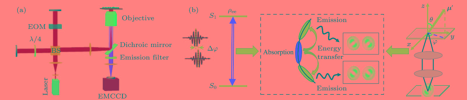 图 1 (a)实验装置示意图; (b)利用相对相位调制的脉冲对激发的共轭聚合物单分子散焦宽场成像原理示意图
Figure1. (a) Schematic of the experimental setup; (b) schematic diagram of defocused wide-field imaging of single conjugated polymer molecules excited with phase-modulated ultrashort laser pulse pairs.
2
2.3.实验原理
共轭聚合物分子被光激发后, 激发态局域于一个发色团, 然后通过能量转移到邻近的能量最低的发色团发出荧光, 如图1(b)所示. 发色团具有特定的吸收偶极矩和发射偶极矩. 因此, 可以通过测量共轭聚合物单分子吸收和发射跃迁偶极取向的变化实现对其吸收和发射特性动态演化的实时跟踪.首先考虑相对相位差为


将(3)和(4)式代入到(2)式中, 最终得到激发态布居概率


共轭聚合物单分子的发射跃迁偶极取向可以通过散焦宽场荧光成像测量. 我们在之前的研究中发现利用甲苯制备的PFO-DBT共轭聚合物单分子显示出单个发色团发射特性[13], 即每一时刻只有一个能量最低的发色团发射荧光. 在散焦宽场荧光成像中, 单个发色团的荧光在EMCCD上的散焦投影显示出特定的双瓣状强度分布模式, 这种强度分布模式明显区分于多个发色团同时发射形成的环形或圆形强度分布, 且其分布模式取决于单个发色团发射跃迁偶极取向. 基于散焦系统成像基本理论[31,32], 通过计算可以得到不同取向跃迁偶极子辐射对应的散焦模式. 实验观测到的散焦成像与最接近的理论模型匹配, 得到共轭聚合物单分子的发射偶极取向. 图1(b)右半部分为发射跃迁偶极矩为


利用傅里叶变换方法和散焦成像技术可以得到发色团吸收与发射偶极特性并且实时分析其动力学演化. 对于发射偶极取向动态演化的测量, 其时间分辨率主要受限于EMCCD的积分时间和共轭聚合物单分子荧光的信号背景比. 理论上, 如果共轭聚合物单分子具有足够高的信号背景比, 对发射偶极取向动态演化分析的时间分辨率可以达到EMCCD的分辨极限33 ms. 同时, 由于受到宽场成像EMCCD的最高时间分辨率33 ms的限制, 在通过对荧光信号傅里叶变换得到调制频谱信息时, 根据采样定律, 所能选取的最大调制频率约为15 Hz. 若要通过傅里叶变换得到调制频谱信息, 需要对至少一个周期的调制信号采样分析, 采样帧数至少需要3帧以上, 对应的吸收偶极取向动态演化最高时间分辨率约为100 ms. 而在实验过程中, 考虑到单分子荧光信号背景比对实验的影响, 我们选取调制频率为1 Hz, 散焦宽场成像积分时间为100 ms, 傅里叶变换帧数为50帧, 以获得足够的荧光信号实现对吸收和发射特性的分析. 如果样品的信号背景比足够高, 采用时间分辨率更高的探测装置并选取更大的调制频率, 对吸收和发射特性动态演化的测量可以获得更高的时间分辨率.
 图 2 共轭聚合物单分子散焦宽场荧光成像时域序列图与利用傅里叶变换频域信息重构的成像序列图 (a)—(c)上半部分为实验测得的散焦宽场荧光成像随时间变化序列, 下半部分为相应的拟合结果; (d)—(f)为与散焦宽场荧光成像同样区域分子的频域信息重构成像图, 不同颜色代表相位的差异, 其中红色代表正相位, 白色代表负相位, 上半部分为直接重构成像结果, 下半部分为拟合结果
Figure2. Schematic of the time-domain imaging sequence and reconstructed frequency-domain imaging by Fourier transform for single conjugated polymer molecules based on defocused wide-field fluorescence imaging. The upper part of (a), (b) and (c) gives the experimental results of defocused wide-field fluorescence imaging, while the lower part shows the simulation results. (d), (e) and (f) are the reconstructed frequency-domain imaging at the same area, where red color represents positive phase and white represents negative phase, the upper part gives the results of reconstructed imaging, while the lower part shows the simulation results.
图3所示为图2中黄色圆圈所标示的PFO-DBT单分子Ⅰ, Ⅱ和Ⅲ的荧光调制轨迹和发射偶极取向方位角随时间变化的轨迹. 图3(a)表示施加在EOM上的调制信号, 用于调制脉冲对之间的相对相位; 图3(b)—(d)对应分子的荧光调制轨迹. 由图3(b)可以看到, 分子Ⅰ的荧光调制信号与施加在EOM上的锯齿波信号保持反相, 表明分子Ⅰ参与光吸收的发色团保持不变. 同时, 共轭聚合物单分子发射偶极取向方位角角度几乎保持恒定不变. 我们对方位角角度进行统计分析, 得到其平均值为34.0° ± 0.1°, 如图3(e)所示. 在约11 s后没有显示相应的角度变化, 是由于分子荧光信号变弱, 难以提取有效的角度信息或对角度拟合不准确. 由于PMMA宿主基质阻止了聚合物链的扭转运动, 而分支结构的不同也会导致临近发色团的发射偶极取向有很明显的差异[23], 因此, 可以认为在成像过程中聚合物单分子发射荧光的发色团也没有改变. 对于分子Ⅱ, 初始时共轭聚合物单分子荧光调制轨迹与EOM的调制信号轨迹同相, 而在6 s后反相, 如图3(c) 所示. 但是共轭聚合物单分子方位角角度保持恒定, 其平均值为47.7° ± 1.9°, 如图3(f) 所示. 因此分子Ⅱ参与光吸收的发色团发生变化, 但始终由同一能量最低的发色团发射荧光. 对于分子Ⅲ, 荧光调制轨迹与EOM的调制信号保持同相(图3(d)), 而方位角角度在正、负值之间频繁变化, 其统计结果出现两个明显的峰值, 分别为70.9° ± 7.9°和–67.2° ± 0.8°(图3(g)). 也就是说, 分子Ⅲ参与光吸收的发色团没有明显变化, 但其发射偶极取向频繁变化, 表明发射荧光的发色团发生了变化. 从图3中可以发现, 分子Ⅰ和分子Ⅱ在实验时间内只有一个发色团发出荧光. 相比而言, 分子Ⅲ可能存在两个甚至三个能量最低的发色团依次发射荧光. 这与我们之前研究发现的PFO-DBT共轭聚合物单分子发射荧光的发色团数目分布一致[13].
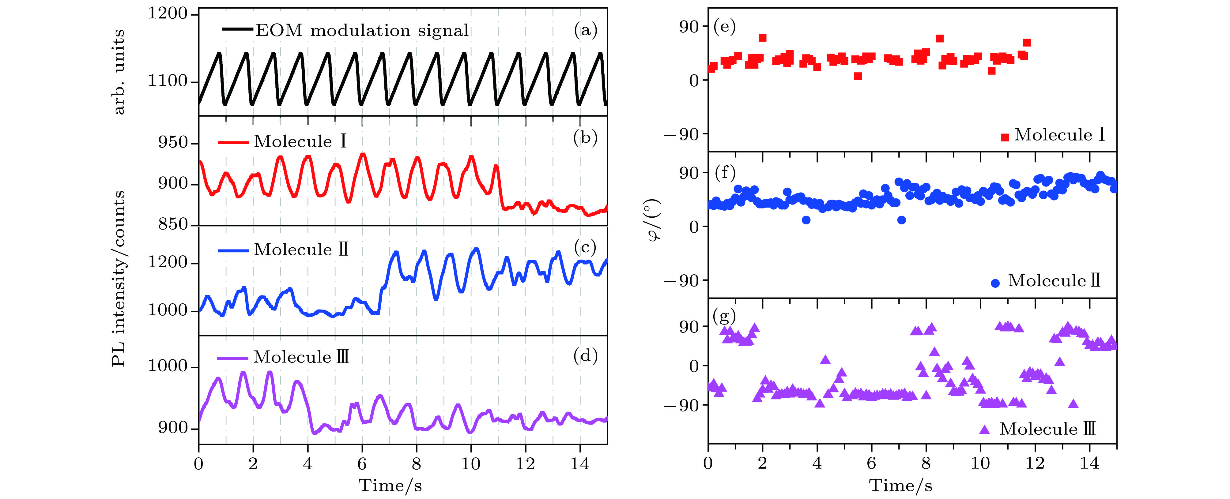 图 3 共轭聚合物单分子Ⅰ, Ⅱ和Ⅲ的荧光调制轨迹和相应发射偶极取向发射角的变化 (a)施加在EOM上的锯齿波信号; (b), (c), (d)每个分子的荧光调制轨迹; (e), (f), (g)角度的变化
Figure3. Modulated fluorescence trajectories and corresponding emission angle of single conjugated polymer molecules Ⅰ, Ⅱ and Ⅲ: (a) The sawtooth wave signal applied on EOM that used for phase modulation of pulse pairs; (b), (c), and (d) the modulated fluorescence trajectories of each molecule; (e), (f), and (g) the change of the angle for molecules Ⅰ, Ⅱ and Ⅲ, respectively.
PFO-DBT分子还存在吸收偶极矩和发射偶极矩同时变化的情况, 如图4所示. 在实验时间内分子1的散焦光斑模式和频域重构成像的相位未发生变化, 表明分子1有固定的吸收跃迁偶极矩和发射跃迁偶极矩. 从图4(f)和图4(h)中可以看到, 分子1的荧光调制轨迹与EOM上施加的锯齿波信号(图4(e))反相, 发射偶极取向的方位角角度总是负值(–81.2° ± 0.1°). 与之相比, 分子2的发射偶极取向方位角角度在实验时间内发生了明显变化, 并且分子的频域重构成像相位也发生了变化, 表明分子2的吸收跃迁偶极取向和发射跃迁偶极取向都发生了变化. 图4(g)和图4(i)所示荧光调制轨迹和角度随时间的轨迹同样显示了这一变化. 在约5 s之后, 荧光调制轨迹和锯齿波信号之间的相关性从反相变为同相, 而共轭聚合物单分子2的发射偶极取向方位角也显示出明显变化, 存在约三个明显的峰值位置(–76° ± 0.2°, –17.6° ± 0.2°和15.1° ± 2.7°). 吸收和发射偶极取向同时发生变化, 表明部分PFO-DBT共轭聚合物单分子存在多个发色团依次吸收激光且有多个发色团依次发射荧光, 显示出共轭聚合物单分子中复杂的能量转移过程.
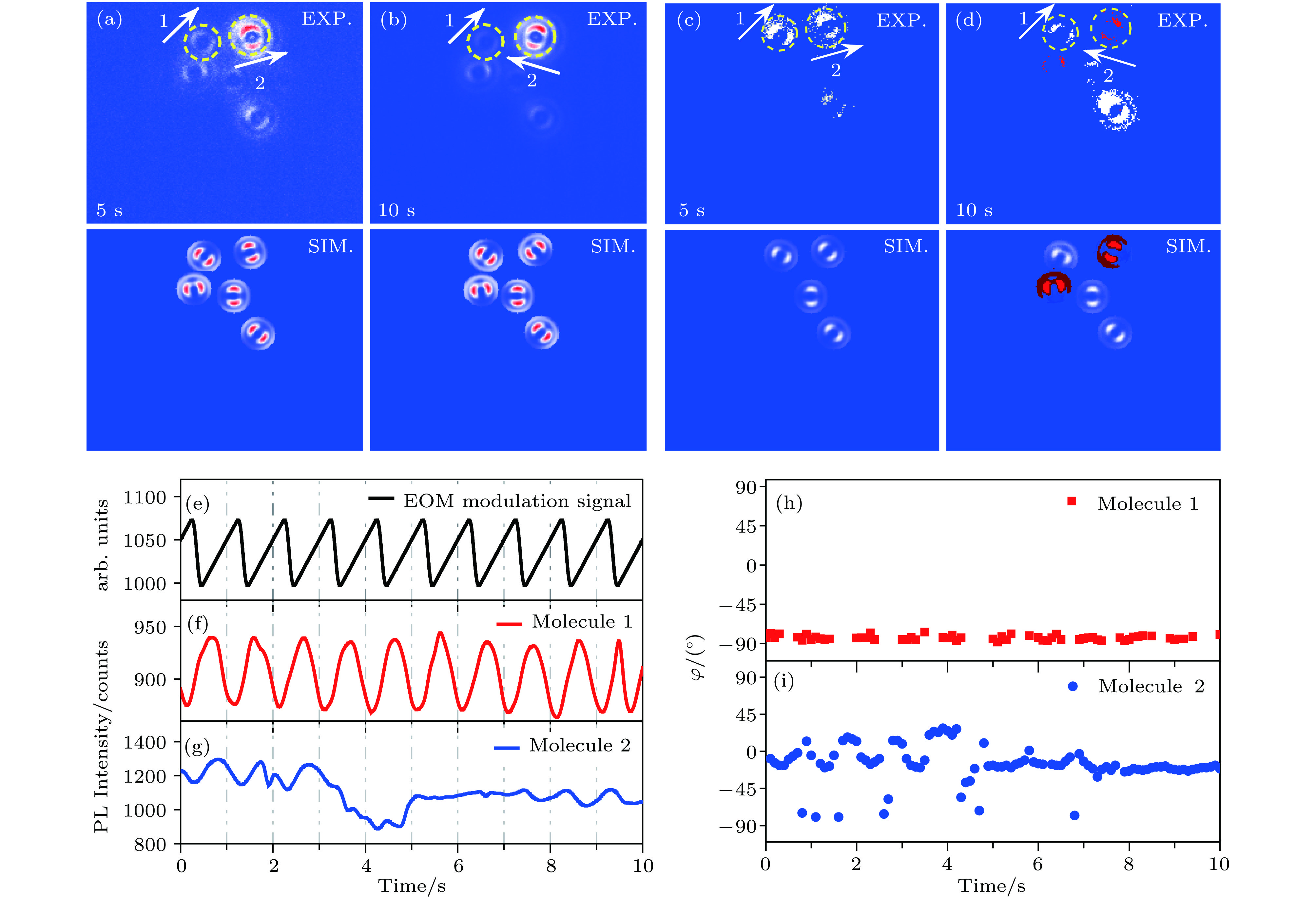 图 4 (a), (b)共轭聚合物单分子散焦宽场成像时域序列图和相应模式的拟合结果; (c), (d)频域信息重构成像和相应的拟合结果, 其中红色代表正相位, 白色代表负相位; (e)施加在EOM上的锯齿波信号; (f), (g)分别显示分子1和2的荧光调制轨迹; (h), (i)分别显示分子1和2的发射角的变化; 图中展示了共轭聚合物单分子吸收和发射偶极取向均保持恒定(分子1)以及吸收和发射偶极取向同时发生变化(分子2)的情况
Figure4. (a) and (b) are the snapshots of time-domain imaging based on defocused wide-field fluorescence imaging of single conjugated polymer molecules and corresponding simulation results; (c) and (d) show the reconstructed frequency-domain imaging and corresponding simulation results, where red color represents positive phase and white represents negative phase; (e) the sawtooth wave signal applied on EOM that used for phase modulation of pulse pairs; (f) and (g) show the fluorescence modulation trajectories of molecules 1 and 2, respectively; (h) and (i) show the change of emission angle of molecules 1 and 2, respectively. The absorption and emission dipole orientation of single conjugated polymer molecule 1 keep constant, while that of molecule 2 change simultaneously.
我们实验测得了PFO-DBT共轭聚合物单分子参与光吸收和荧光发射的发色团的三种动态过程. 共轭聚合物单分子吸收偶极取向和发射偶极取向均保持恒定, 说明PFO-DBT中参与光吸收和荧光发射的为同一个发色团; 或者参与光吸收和荧光发射的为不同发色团, 但是实验过程中没有发生改变. 吸收和发射偶极取向同时发生变化, 说明在这个过程中, 至少有两个发色团依次参与了光吸收并且依次发射. 也就是说, 初始由一个发色团吸收光子后, 发出荧光, 在发色团被淬灭后, 由另一个发色团吸收光子并发出荧光, 两个发色团的取向有明显差异. 而吸收或发射偶极取向其中之一发生变化, 说明在实验过程中由同一个发色团吸收, 至少两个发色团依次发射; 或者同一个发色团发射, 至少两个发色团依次参与光吸收.
