尹佳雯, 竺凯琪, 叶冰琪, 刘佳露, 余乾鹏, 徐孙, 孙晴, 左照江


浙江农林大学省部共建亚热带森林培育国家重点实验室, 浙江 杭州 311300
收稿日期:2020-11-24;修回日期:2021-02-19;网络出版日期:2021-06-11
基金项目:国家自然科学基金(31870585);浙江省基础公益研究计划(LGN19C150006);国家级大学生创新创业训练计划(201910341007,202010341007);浙江农林大学学生科研训练项目(102-2013200125,2020KX0031)
*通信作者:左照江, E-mail: zuozhaojiang@126.com.
摘要:[目的] 蓝藻挥发性有机化合物(VOCs)对其他藻类的化感作用可促进蓝藻成为富营养化水体优势种群,本研究旨在以VOCs主要成分α-紫罗酮为例揭示其化感致死机制。[方法] 采用α-紫罗酮处理莱茵衣藻,测定藻细胞生长以及致死浓度下藻细胞光合性能、caspase-likes活性和DNA ladders。[结果] 采用0.05和0.1 mmol/L α-紫罗酮处理24 h后,莱茵衣藻细胞生长均受到明显抑制,其中0.1 mmol/L处理时部分藻细胞发生死亡,死亡率为38.3%。采用0.2 mmol/L α-紫罗酮处理时,藻细胞全部死亡,同时光合色素逐渐降解、Fv/Fm逐渐降低并消失,这表明藻细胞死亡并非坏死。在藻细胞死亡过程中,caspase-9-like和caspase-3-like活性明显增强;DNA在处理1 h时出现ladders,并逐渐降解为100-250 bp片段。[结论] 这表明蓝藻VOCs可通过诱导细胞程序性死亡以发挥化感作用。
关键词:蓝藻莱茵衣藻α-紫罗酮细胞程序性死亡化感机制
Programmed cell death in Chlamydomonas reinhardtii induced by cyanobacterial volatile α-ionone
Jiawen Yin, Kaiqi Zhu, Bingqi Ye, Jialu Liu, Qianpeng Yu, Sun Xu, Qing Sun, Zhaojiang Zuo


State Key Laboratory of Subtropical Silviculture, Zhejiang A&F University, Hangzhou 311300, Zhejiang Province, China
Received: 24 November 2020; Revised: 19 February 2021; Published online: 11 June 2021
*Corresponding author: Zhaojiang Zuo, E-mail: zuozhaojiang@126.com.
Foundation item: Supported by the National Natural Science Foundation of China (31870585), by the Basic Public Welfare Research Project of Zhejiang Province (LGN19C150006), by the National Students' Innovation and Entrepreneurship Training Program (201910341007, 202010341007) and by the Student Research Training Program in Zhejiang A&F University (102-2013200125, 2020KX0031)
Abstract: [Objective] Allelopathic effects of cyanobacterial volatile organic compounds (VOCs) on other algae promote cyanobacteria becoming the dominant species in eutrophicated waters. The aim of the present study is to uncover the allelopathic lethal mechanism of cyanobacterial VOCs by using α-ionone. [Methods] In the treatment with α-ionone, the cell growth of Chlamydomonas reinhardtii was investigated, and the photosynthetic abilities, caspase-like activities and DNA ladders were investigated at lethal concentration. [Results] When C. reinhardtii cells were treated with α-ionone at 0.05 and 0.1 mmol/L, the cell growth was significantly inhibited, and 38.3% of the cells were killed by 0.1 mmol/L α-ionone. However, all the cells were killed by 0.2 mmol/L α-ionone. During the cell death, the photosynthetic pigments gradually degraded with prolonging the treatment time, and Fv/Fm gradually declined and even disappeared, indicating that the cell death is not a necrosis. Meanwhile, the activities of caspase-9-like and caspase-3-like increased remarkably. In the treatment with 0.2 mmol/L α-ionone for 1 h, DNA showed ladders and then gradually degraded to the fragments of 100-250 bp. [Conclusion] This suggests that cyanobacterial VOCs play the allelopathic role by inducing programmed cell death.
Keywords: cyanobacteriaChlamydomonas reinhardtiiα-iononeprogrammed cell deathallelopathic mechanism
蓝藻通过次生代谢途径释放大量的挥发性有机化合物(volatile organic compounds,VOCs),主要包括呋喃类、含硫类、烷烃类、萜烯类、苯类、醇类、醛类、酮类和酯类化合物,这些VOCs溶于水后不仅会降低水质并造成饮用水危机[1-2],还会抑制其他藻类生长以促使蓝藻成为富营养化水体优势种群[3-5]。
水华微囊藻(Microcystis flos-aquae)和铜绿微囊藻(M. aeruginosa)是形成蓝藻水华的两种典型藻种,无氮和无磷条件可促进其VOCs产生与释放,同时这些VOCs可抑制其他藻类生长[6-7]。在蓝藻VOCs主要成分中,桉树脑和柠檬烯可通过抑制细胞生长、诱导光合色素降解和降低光合性以抑制甚至杀死莱茵衣藻(Chlamydomonas reinhardtii)和小球藻(Chlorella vulgaris)[6-8];α-紫罗酮、β-紫罗酮、β-环柠檬醛和香叶基丙酮可抑制蛋白核小球藻(C. pyrenoidosa)生长[3];0.1–0.5 mg/mL β-环柠檬醛会使谷皮菱形藻(Nitzschia palea)细胞破裂[9]。由此可见,蓝藻VOCs在蓝藻成为富营养化水体优势种群中具有化感作用,然而其化感机制尚不清楚。
细胞程序性死亡(programmed cell death,PCD)是细胞感受到某种信号或受到某些因素刺激后,为维持内环境稳定而发生的一种主动性消亡过程。此过程不仅在高等植物和动物生长发育以及响应生物和非生物胁迫过程中发生[10-11],还在单细胞生物中发生,例如酸[12-13]、高温[14]、盐[15]、紫外线[15]、H2O2[16]、除藻剂[17]等胁迫条件可诱导单细胞藻类和酵母发生PCD。在此过程中,细胞会发生一系列变化,主要包括:细胞收缩与空泡化、细胞器退化、caspase (-like)活化、细胞核先浓缩后破裂、DNA降解成ladders等[14, 16-17]。
在蓝藻VOCs中,α-紫罗酮、β-紫罗酮和β-环柠檬醛主要通过类胡萝卜素降解形成,是富营养化水体中的主要嗅味物质[4, 18],并且在2–5 mg/mL (约10–26 mmol/L)时对蛋白核小球藻产生化感作用[3]。衣藻隶属于绿藻门,广泛存在于河流、湖泊、海洋等水体中。在富营养化水体藻种演替过程中,蓝藻大量繁殖生长后,衣藻等绿藻大量减少[19-20]。因此,本研究通过测定α-紫罗酮对莱茵衣藻细胞生长的影响以及致死浓度下藻细胞光合性能、caspase-likes活性和DNA ladders等变化,以期从PCD角度揭示蓝藻VOCs对其他藻类的化感致死机制,进而提升对蓝藻成为富营养化水体优势种群的认识以利于其有效防治。
1 材料和方法 1.1 实验材料 莱茵衣藻CC400株系(中国科学院遗传与发育生物学研究所刘翠敏研究员惠赠)隶属于绿藻门(Chlorophyta)团藻目(Volvocales)衣藻属(Chlamydomonas),采用TAP培养基[21]进行培养。培养条件为:温度25 ℃、光照(16 h)/黑暗(8 h)、光强50 μmol/(m2·s)、摇床转速100 r/min。待藻细胞达到对数生长期时,5000 r/min离心后转入新鲜培养基中,使藻细胞密度为5×106细胞/mL,并采用0.05、0.10、0.20 mmol/L α-紫罗酮(上海源叶生物科技有限公司)进行处理,以未处理作为对照。待α-紫罗酮处理莱茵衣藻24 h后,测定藻细胞密度;待0.2 mmol/L α-紫罗酮处理0、0.5 (caspase-likes活性)、1.0、2.0、6.0、12.0、24.0 h时测定藻细胞光合色素吸收光谱、光合性能、caspase-likes活性和DNA ladders。4次重复。
1.2 藻细胞密度与死亡率测定 采用中性红对莱茵衣藻进行染色以区分活细胞和死细胞(红色),利用血细胞计数板(25×16)计数两种藻细胞数量,并计算活细胞密度与藻细胞死亡率[17]。
1.3 光合色素吸收光谱测定 取3 mL莱茵衣藻培养液,5000 r/min离心5 min后,用3 mL 96%乙醇重悬藻细胞并置于黑暗处静提24 h。待光合色素提取完成后,采用TU-1900 UV-Vis分光光度计在400–700 nm处进行扫描以测定吸收光谱,并计算每106个细胞的吸收光谱[17]。
1.4 叶绿素荧光测定 取约1×10⁷个藻细胞,6000 r/min离心5 min,将藻细胞重悬于10 μL培养基中,用移液枪吸取后滴于3 cm2滤纸上以形成大小约0.5 cm2斑点,黑暗中放置15 min。采用YZQ-500非调制式叶绿素荧光仪测定其叶绿素荧光,并计算光系统Ⅱ (PSII)最大光量子产量(Fv/Fm)[7]。
1.5 Caspase-9-like和caspase-3-like活性测定 Caspase-9-like和caspase-3-like活性分别采用caspase-9和caspase-3活性检测试剂盒进行测定。取25 mL莱茵衣藻培养液,5000 r/min离心5 min后,加入100 μL裂解缓冲液冰浴裂解10 min。在4 ℃下离心后,取上清液并分别加入100 μmol/L caspase-3反应液(Ac-DEVD-pNA)和caspase-9反应液(IETD-pNA)。在37 ℃孵育2 h后,通过测定反应产物在405 nm处的吸光值,从而计算caspase-9-like和caspase-3-like活性。
1.6 DNA ladders检测 取10 mL莱茵衣藻培养液,5000 r/min离心5 min后,加入350 μL NET溶液[100 mmol/L NaCl、50 mmol/L EDTA、20 mmol/L Tris-HCl (pH 8.0)]重悬藻细胞,并加入25 μL SDS (200 g/L)和25 μL蛋白酶K (10 g/L),55 ℃水浴2 h后加入200 μL 5 mol/L KAC。在4 ℃下离心后,取上清加入等体积的酚: 氯仿: 异戊醇(25:24:1)抽提2次后,加入2倍体积的无水乙醇,在–70 ℃条件下沉淀DNA。将DNA溶于适量ddH2O中,加入RNase A消化30 min后,采用1.5%琼脂糖凝胶电泳检测DNA ladders[13]。
1.7 数据处理 采用Origin 8.0进行单因素方差分析并绘图。
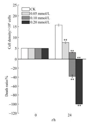
2 结果和分析 2.1 α-紫罗酮对藻细胞生长的影响 采用0.05和0.1 mmol/L α-紫罗酮处理莱茵衣藻24 h后,其藻细胞密度均极显著(P < 0.01)低于对照,同时0.1 mmol/L处理时藻细胞死亡率为38.3% (P < 0.01)。采用0.2 mmol/L α-紫罗酮处理24 h后,藻细胞全部死亡(图 1)。
 |
| 图 1 α-紫罗酮对莱茵衣藻细胞生长的影响 Figure 1 Effects of α-ionone on the growth of C. reinhardtii. CK: the control. **: compared to the control, the significant difference at P < 0.01 level. |
| 图选项 |
2.2 α-紫罗酮对光合色素吸收光谱的影响 未采用α-紫罗酮处理时,莱茵衣藻光合色素吸收光谱在藻细胞生长过程中未发生明显变化(图 2-A);采用0.2 mmol/L α-紫罗酮处理后,其光合色素吸收光谱在413、433、457、663 nm处的吸收峰均随处理时间延长而逐渐降低,这表明光合色素逐渐降解(图 2-B)。
 |
| 图 2 0.2 mmol/L α-紫罗酮对莱茵衣藻光合色素吸收光谱的影响 Figure 2 Effects of 0.2 mmol/L α-ionone on absorbance spectra of photosynthetic pigments in C. reinhardtii. A: the control, without α-ionone treatment; B: the treatment with α-ionone at 0.2 mmol/L. |
| 图选项 |
2.3 α-紫罗酮对Fv/Fm的影响 采用0.2 mmol/L α-紫罗酮处理莱茵衣藻1 h后,其Fv/Fm显著(P < 0.01)低于对照。随处理时间延长,Fv/Fm逐渐降低,并在处理24 h时消失(图 3)。
 |
| 图 3 0.2 mmol/L α-紫罗酮对莱茵衣藻Fv/Fm的影响 Figure 3 Effects of 0.2 mmol/L α-ionone on Fv/Fm in C. reinhardtii. **: compared to the control, significant difference at P < 0.01 level. |
| 图选项 |
2.4 α-紫罗酮对caspase-likes活性的影响 0.2 mmol/L α-紫罗酮处理莱茵衣藻1 h后,caspase-9-like和caspase-3-like活性均增至最高,与对照相比分别增加了4.5 (P < 0.01)和5.1 (P < 0.01)倍。随处理时间延长,caspase-9-like和caspase-3-like活性呈现逐渐降低趋势,但均显著(P < 0.01)高于对照(图 4)。
 |
| 图 4 0.2 mmol/L α-紫罗酮对莱茵衣藻caspase-likes活性的影响 Figure 4 Effects of 0.2 mmol/L α-ionone on the activities of caspase-likes in C. reinhardtii. A: Caspase-9-like; B: Caspase-3-like. **: compared to the control, significant difference at P < 0.01 level. |
| 图选项 |
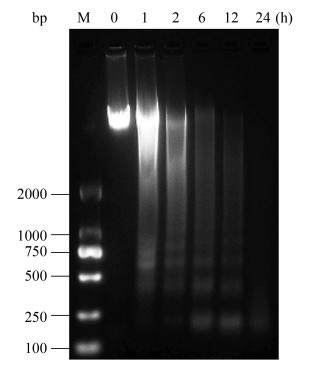
2.5 α-紫罗酮对DNA ladders的影响 采用0.2 mmol/L α-紫罗酮处理莱茵衣藻1 h后,其DNA出现ladders。随着处理时间延长,DNA降解逐渐增强,在处理24 h时降解为100–250 bp片段(图 5)。
 |
| 图 5 0.2 mmol/L α-紫罗酮对DNA ladders的影响 Figure 5 Effect of 0.2 mmol/L α-ionone on DNA ladders. M: DNA marker. |
| 图选项 |
3 讨论 细胞死亡可分为坏死和PCD,其中发生PCD时细胞生理活性逐渐消失,例如乙酸、甲萘醌、除藻化合物(芳樟醇和α-松油醇)诱导莱茵衣藻PCD[13, 17, 22];H2O2诱导杜氏藻(Dunaliella tertiolecta) PCD[23];以及H2O2、NaCl和KCl诱导Micrasterias denticulata PCD[24]时,藻细胞光合性能均逐渐消失。采用0.2 mmol/L α-紫罗酮处理莱茵衣藻后,其光合色素逐渐降解(图 2),Fv/Fm逐渐减弱至24 h时完全消失(图 3),这表明藻细胞死亡并非坏死,而可能是PCD。
Caspases活化与细胞PCD过程密切相关,其最早在哺乳动物细胞中发现,而植物和藻类细胞中与其功能相似的酶则被称为caspase-likes。在发生PCD时,细胞色素c通过线粒体膜上的渗透转移孔进入细胞质后与Apaf-1和caspase-9相结合,从而激活PCD途径中的caspase级联反应[25]。Caspase-9是内源途径启动分子,可激活多种下游caspases,包括引发DNA降解的caspase-3[26]。此外,外源途径的caspase-8也可激活caspase-3[27]。在藻细胞中,caspase-like活化是其PCD的关键特征[22, 28],抑制其活化可阻止细胞死亡[22]。α-紫罗酮处理可激活莱茵衣藻caspase-9-like和caspase-3-like,且两者活性变化趋势相一致,这表明caspase-3-like应该是由caspase-9-like通过内源途径激活(图 4),进而引发DNA降解(图 5)。
当细胞发生PCD时,DNA快速降解并呈现ladders是其典型特征[24],然而并非所有PCD过程都能产生ladders[13, 17]。例如,UV、H2O2和甲萘醌诱导莱茵衣藻PCD时会出现DNA ladders[22, 29-30],而乙酸、蜂毒素、芳樟醇和α-松油醇诱导则不出现DNA ladders[13, 17, 31],这可能是由于藻细胞株系和诱导因子不同所致。在本研究中,0.2 mmol/L α-紫罗酮处理莱茵衣藻1 h时出现PCD典型特征——DNA ladders,随着处理时间延长DNA降解逐渐增强,在24 h时降解为100–250 bp片段(图 5)。
蓝藻VOCs对其他藻类生长具有明显的化感作用[3-4, 6-7]。α-紫罗酮是蓝藻VOCs的主要化感成分之一[3],其可通过诱导PCD以杀死莱茵衣藻,这表明蓝藻VOCs可通过诱导PCD以发挥化感作用,从而促进蓝藻成为富营养化水体优势种群。
References
| [1] | Ye CL, Yang YY, Xu QH, Ying BB, Zhang MQ, Gao B, Ni BB, Yakefu Z, Bai Y, Zuo ZJ. Volatile organic compound emissions from Microcystis aeruginosa under different phosphorus sources and concentrations. Phycological Research, 2018, 66(1): 15-22. DOI:10.1111/pre.12201 |
| [2] | Zuo ZJ. The review of research advances in algal volatile organic compounds. Acta Hydrobiologica Sinica, 2017, 41(6): 1369-1379. (in Chinese) 左照江. 藻类挥发性有机化合物研究进展. 水生生物学报, 2017, 41(6): 1369-1379. |
| [3] | Ikawa M, Sasner JJ, Haney JF. Activity of cyanobacterial and algal odor compounds found in lake waters on green alga Chlorella pyrenoidosa growth. Hydrobiologia, 2001, 443(1/2/3): 19-22. DOI:10.1023/A:1017535801766 |
| [4] | Zuo ZJ. Why algae release volatile organic compounds-the emission and roles. Frontiers in Microbiology, 2019, 10: 491. DOI:10.3389/fmicb.2019.00491 |
| [5] | Li YX, Li DH. Competition between toxic Microcystis aeruginosa and nontoxic Microcystis wesenbergii with Anabaena PCC7120. Journal of Applied Phycology, 2012, 24(1): 69-78. DOI:10.1007/s10811-010-9648-x |
| [6] | Zuo ZJ, Yang YY, Xu QH, Yang WT, Zhao JX, Zhou L. Effects of phosphorus sources on volatile organic compound emissions from Microcystis flos-aquae and their toxic effects on Chlamydomonas reinhardtii. Environmental Geochemistry and Health, 2018, 40(4): 1283-1298. DOI:10.1007/s10653-017-0055-y |
| [7] | Xu QH, Yang L, Yang WT, Bai Y, Hou P, Zhao JX, Zhou L, Zuo ZJ. Volatile organic compounds released from Microcystis flos-aquae under nitrogen sources and their toxic effects on Chlorella vulgaris. Ecotoxicology and Environmental Safety, 2017, 135: 191-200. DOI:10.1016/j.ecoenv.2016.09.027 |
| [8] | Zhao JX, Yang L, Zhou L, Bai Y, Wang B, Hou P, Xu QH, Yang WT, Zuo ZJ. Inhibitory effects of eucalyptol and limonene on the photosynthetic abilities in Chlorella vulgaris (Chlorophyceae). Phycologia, 2016, 55(6): 696-702. DOI:10.2216/16-38.1 |
| [9] | Chang DW, Hsieh ML, Chen YM, Lin TF, Chang JS. Kinetics of cell Lysis for Microcystis aeruginosa and Nitzschia palea in the exposure to β-cyclocitral. Journal of Hazardous Materials, 2011, 185(2/3): 1214-1220. |
| [10] | Tan K, Wen CX, Feng HL, Chao XT, Su H. Nuclear dynamics and programmed cell death in Arabidopsis root hairs. Plant Science, 2016, 253: 77-85. DOI:10.1016/j.plantsci.2016.08.006 |
| [11] | Yekkour A, Tran D, Arbelet-Bonnin D, Briand J, Mathieu F, Lebrihi A, Errakhi R, Sabaou N, Bouteau F. Early events induced by the toxin deoxynivalenol lead to programmed cell death in Nicotiana tabacum cells. Plant Science, 2015, 238: 148-157. DOI:10.1016/j.plantsci.2015.06.004 |
| [12] | Guaragnella N, Antonacci L, Giannattasio S, Marra E, Passarella S. Catalase T and Cu, Zn-superoxide dismutase in the acetic acid-induced programmed cell death in Saccharomyces cerevisiae. FEBS Letters, 2008, 582(2): 210-214. DOI:10.1016/j.febslet.2007.12.007 |
| [13] | Zuo ZJ, Zhu YR, Bai YL, Wang Y. Acetic acid-induced programmed cell death and release of volatile organic compounds in Chlamydomonas reinhardtii. Plant Physiology and Biochemistry, 2012, 51: 175-184. DOI:10.1016/j.plaphy.2011.11.003 |
| [14] | Dingman JE, Lawrence JE. Heat-stress-induced programmed cell death in Heterosigma akashiwo (Raphidophyceae). Harmful Algae, 2012, 16: 108-116. DOI:10.1016/j.hal.2012.02.003 |
| [15] | Bidle KD. Programmed cell death in unicellular phytoplankton. Current Biology, 2016, 26(13): R594-R607. DOI:10.1016/j.cub.2016.05.056 |
| [16] | Zhou TR, Zheng J, Cao HS, Wang XJ, Lou K, Zhang XH, Tao Y. Growth suppression and apoptosis-like cell death in Microcystis aeruginosa by H2O2: a new insight into extracellular and intracellular damage pathways. Chemosphere, 2018, 211: 1098-1108. DOI:10.1016/j.chemosphere.2018.08.042 |
| [17] | Chen YT, Weng YY, Zhou M, Meng YY, Liu JL, Yang L, Zuo ZJ. Linalool- and α-terpineol-induced programmed cell death in Chlamydomonas reinhardtii. Ecotoxicology and Environmental Safety, 2019, 167: 435-440. DOI:10.1016/j.ecoenv.2018.10.062 |
| [18] | Liu XS, Shi CF, Xu XG, Li XJ, Xu Y, Huang HY, Zhao YP, Zhou YW, Shen HC, Chen C, Wang GX. Spatial distributions of β-cyclocitral and β-ionone in the sediment and overlying water of the west shore of Taihu Lake. Science of the Total Environment, 2017, 579: 430-438. DOI:10.1016/j.scitotenv.2016.11.079 |
| [19] | Yao XJ, Liu DF, Yang ZJ, Fang XF, Hu XL, Fang LJ, Tian ZB. Distribution characteristics of phytoplankton community structure in Xiangxi bay of Three Gorges reservoir during spring and summer. Journal of Sichuan University: Engineering Science Edition, 2012, 44(S2): 211-220. (in Chinese) 姚绪姣, 刘德富, 杨正健, 方小凤, 胡响铃, 方丽娟, 田泽斌. 三峡水库香溪河库湾水华高发期浮游植物群落结构分布特征. 四川大学学报: 工程科学版, 2012, 44(S2): 211-220. |
| [20] | Ma QL, Hu F, Guo QW, Xu ZC, Yao LA, Zhao XM. Eutrophication characteristics and distribution of phytoplankton communities in Liuduzhai reservoir in summer. Acta Scientiae Circumstantiae, 2014, 34(6): 1497-1504. (in Chinese) 马千里, 胡芳, 虢清伟, 许振成, 姚玲爱, 赵学敏. 六都寨水库夏季富营养化状况与浮游植物分布特征研究. 环境科学学报, 2014, 34(6): 1497-1504. |
| [21] | Gorman DS, Levine RP. Cytochrome f and plastocyanin: their sequence in the photosynthetic electron transport chain of Chlamydomonas reinhardi. Proceedings of the National Academy of Sciences of the United States of America, 1965, 54(6): 1665-1669. DOI:10.1073/pnas.54.6.1665 |
| [22] | Sirisha VL, Sinha M, D'Souza JS. Menadione-induced caspase-dependent programmed cell death in the green chlorophyte Chlamydomonas reinhardtii. Journal of Phycology, 2014, 50(3): 587-601. DOI:10.1111/jpy.12188 |
| [23] | Segovia M, Berges JA. Inhibition of caspase-like activities prevents the appearance of reactive oxygen species and dark-induced apoptosis in the unicellular chlorophyte Dunaliella tertiolecta1. Journal of Phycology, 2009, 45(5): 1116-1126. DOI:10.1111/j.1529-8817.2009.00733.x |
| [24] | Affenzeller MJ, Darehshouri A, Andosch A, Lütz C, Lütz-Meindl U. Salt stress-induced cell death in the unicellular green alga Micrasterias denticulata. Journal of Experimental Botany, 2009, 60(3): 939-954. DOI:10.1093/jxb/ern348 |
| [25] | Li P, Nijhawan D, Budihardjo I, Srinivasula SM, Ahmad M, Alnemri ES, Wang XD. Cytochrome c and dATP-dependent formation of apaf-1/caspase-9 complex initiates an apoptotic protease cascade. Cell, 1997, 91(4): 479-489. DOI:10.1016/S0092-8674(00)80434-1 |
| [26] | Murik O, Elboher A, Kaplan A. Dehydroascorbate: a possible surveillance molecule of oxidative stress and programmed cell death in the green alga Chlamydomonas reinhardtii. New Phytologist, 2014, 202(2): 471-484. DOI:10.1111/nph.12649 |
| [27] | Riedl SJ, Salvesen GS. The apoptosome: signalling platform of cell death. Nature Reviews Molecular Cell Biology, 2007, 8(5): 405-413. DOI:10.1038/nrm2153 |
| [28] | Ba?ares-Espa?a E, Kromkamp JC, López-Rodas V, Costas E, Flores-Moya A. Photoacclimation of cultured strains of the cyanobacterium Microcystis aeruginosa to high-light and low-light conditions. FEMS Microbiology Ecology, 2013, 83(3): 700-710. DOI:10.1111/1574-6941.12025 |
| [29] | Nedelcu AM. Evidence for p53-like-mediated stress responses in green algae. FEBS Letters, 2006, 580(13): 3013-3017. DOI:10.1016/j.febslet.2006.04.044 |
| [30] | Vavilala SL, Gawde KK, Sinha M, D'Souza JS. Programmed cell death is induced by hydrogen peroxide but not by excessive ionic stress of sodium chloride in the unicellular green alga Chlamydomonas reinhardtii. European Journal of Phycology, 2015, 50(4): 422-438. DOI:10.1080/09670262.2015.1070437 |
| [31] | Yordanova ZP, Woltering EJ, Kapchina-Toteva VM, Iakimova ET. Mastoparan-induced programmed cell death in the unicellular alga Chlamydomonas reinhardtii. Annals of Botany, 2013, 111(2): 191-205. DOI:10.1093/aob/mcs264 |
