 ,*,2), 杨刚*, 逄燕*, 钟希祥*, 李梦麒*, 薛贺波*, 齐轶鹏*, 史艺?*
 †
EXPERIMENTAL STUDY ON HEMODYNAMICS OF AORTIC VALVE UNDER VARIED CARDIAC OUTPUT USING PIV 1)
Liu Zhaomiao,*,2), Yang Gang*, Pang Yan*, Zhong Xixiang*, Li Mengqi*, Xue Hebo*, Qi Yipeng*, Shi Yi?* †
通讯作者: 2) 赵淼, 教授, 主要研究方向: 微尺度流体力学、血流动力学、流固耦合理论及工程应用. E-mail:lzm@bjut.edu.cn
收稿日期:2019-08-23接受日期:2019-10-21网络出版日期:2019-10-21
| 基金资助: |
Received:2019-08-23Accepted:2019-10-21Online:2019-10-21
作者简介 About authors

摘要
主动脉瓣发生病变时导致心排出量(cardiac output, CO)减少,而心排出量减少与主动脉瓣血流动力学耦合作用, 引发瓣膜继发性疾病.本文基于医学影像数据三维重构带有冠状动脉的主动脉根部,制备高度光滑和透明的主动脉根部实验模型, 构建体外脉动循环模拟系统,利用粒子图像测速技术(particle image velocimetry,PIV)研究冠状动脉存在时心排出量对主动脉瓣速度分布、黏性剪应力(viscous shear stress, VSS)和雷诺剪应力(Reynolds shear stress, RSS)等血流动力学的影响.研究结果表明: 冠状动脉的存在改变了主动脉窦中的涡旋运动和涡度,冠状动脉存在时流体经由冠状动脉流出, 主动脉窦中的涡旋运动逐渐消失,涡度较早开始减小. 峰值期, 中心对称流动两侧区域存在正、负高黏性剪切区域,存在冠状动脉一侧的升主动脉下游存在高雷诺剪应力区域.心排出量显著影响主动脉瓣的速度分布、VSS和RSS等血液流动和受力状况.随着心排出量增大, 冠状动脉存在时峰值期的最大速度、VSS和RSS增大, 即$CO=2.1$, 2.8, 3.5和4.2 l/min时, 最大速度分别为0.98, 1.13, 1.21和1.37 m/s, 最大VSS分别为0.87, 0.95, 0.96和1.02 N/m$^{2}$, 最大RSS分别为103.76, 116.25, 138.68和146.55 N/m$^{2}$. 心排出量较低时,主动脉瓣较低的跨瓣流动速度和黏性剪应力易导致血栓形成,研究结果可为主动脉瓣置换术提供理论参考.
关键词:
Abstract
Reduced cardiac output (CO) always occurs in aortic valve diseases. The hemodynamics of aortic valve is affected by reduced CO, causing secondary valvular diseases. In this paper, a three-dimensional reconstruction of aortic root geometric model with left coronary artery is complished based on medical imaging data, a highly smooth and transparent aortic root experimental model is casted, and an in vitro pulsating circulation system is constructed. Particle image velocimetry (PIV) is used to investigate the effect of CO on hemodynamics of aortic valve with or without the left coronary artery, such as the velocity, viscous shear stress (VSS), Reynolds shear stress (RSS), and so on. The results show that aortic sinus hemodynamics are influenced by left coronary artery that the presence of left coronary artery changes the vortex and vorticity in the sinus. In the case of the presence of left coronary artery, fluids in the aortic sinus flows out through the left coronary artery which leads to vortex gradually disappears and vorticity early decreases. At the peak systolic, regions of positive and negative VSS are exist in both sides of the centrosymmetric systolic jet and RSS is especially elevated in the ascending aorta on the side of left coronary artery. In addition, the hemodynamics of aortic valve, such as the velocity, VSS and RSS, are significantly affected by the CO. The maximum velocity, VSS and RSS increase with the increasing of CO, namely, the maximum velocity is 0.98, 1.13, 1.21 and 1.37 m/s, the maximum VSS is 0.87, 0.95, 0.96 and 1.02 N/m$^{2}$, and the maximum RSS is 103.76, 116.25, 138.68 and 146.55 N/m$^{2}$ when $CO=2.1$, 2.8, 3.5 and 4.2 l/min, respectively. At low CO, the values of transvalvular flow velocity and VSS of aortic valve are small, which may easily lead to thrombosis. The research findings can provide theoretical references for the aortic valve implantation.
Keywords:
PDF (9110KB)元数据多维度评价相关文章导出EndNote|Ris|Bibtex收藏本文
本文引用格式
刘赵淼, 杨刚, 逄燕, 钟希祥, 李梦麒, 薛贺波, 齐轶鹏, 史艺. 不同心排出量下主动脉瓣血流动力学的PIV实验研究1). 力学学报[J], 2019, 51(6): 1918-1926 DOI:10.6052/0459-1879-19-231
Liu Zhaomiao, Yang Gang, Pang Yan, Zhong Xixiang, Li Mengqi, Xue Hebo, Qi Yipeng, Shi Yi.
引言
心脏瓣膜疾病是瓣膜出现结构或功能改变时血液无法顺利保持正常单一方向的流动而引发的病症,是发病率和致死率较高的心血管疾病之一[1-4]主动脉瓣由于位于压力负荷较大的左心室和升主动脉之间,容易发生主动脉瓣狭窄和关闭不全等心脏瓣膜疾病, 导致瓣膜不能正常开启和关闭,改变跨瓣的血液流动, 引发心排出量减少[5-6].心排出量减少改变主动脉瓣的血流动力学, 而改变的血流动力学又作用于主动脉瓣,引发瓣膜继发性疾病.心排出量与主动脉瓣血液流动和瓣叶所受剪应力密切相关,心排出量减少显著影响主动脉瓣的血流动力学[7-9].Zhang等[10]建立理想主动脉根部模型, 搭建体外脉动循环模拟系统,并利用粒子图像测速技术(particle image velocimetry,PIV)研究不同心排出量下主动脉根部的血液流动和结构变形,发现心排出量在血流动力学和结构变形方面起决定性作用.Yap等[11]研究不同心排出量对主动脉瓣瓣叶表面流体剪应力的影响,发现随着每搏量增加流体剪应力增大, 随着心率增大, 剪应力降低.Vahidkhah等[12]建立理想化的主动脉瓣模型,利用流固耦合研究心排出量减少和瓣叶血栓形成的关系, 结果表明随着心排出量减少,血液在瓣叶停留面积增大, 可能导致血栓形成.脉动流动研究表征整个心动周期内主动脉瓣附近血液流动和瓣叶力学行为,而稳态流动研究则较好地评估瓣叶有效开口面积和跨瓣压差.Seaman等[13]利用PIV研究了6种稳态流速下正常瓣膜和钙化瓣膜的血流动力学,发现在6种稳态流速下钙化瓣膜具有正常瓣膜功能, 随着流速增大,主动脉瓣速度、跨瓣压差和有效开口面积增大.
上述研究局限于使用理想化主动脉根部和主动脉瓣模型,通过改变心排出量研究瓣叶血液流动和力学行为,未考虑冠状动脉存在对主动脉瓣血液流动的影响.而Querzoli等[14]研究冠状动脉存在对主动脉瓣血流动力学的影响,发现:与无冠脉相比,冠状动脉存在时主动脉窦中的涡流被冠状动脉血流产生的吸力吸走,瓣膜闭合延迟且速度更快.Moore等[15]则研究了冠状动脉对瓣叶力学行为和主动脉窦血流动力学的影响,结果表明冠状动脉血流的存在使主动脉窦涡旋运动位于主动脉窦更深处,增大了瓣叶基部附近的流速和瓣膜开口面积. 因此,研究心排出量时有必要考虑冠状动脉存在对主动脉瓣血流动力学的影响.
目前, 主动脉瓣心排出量的研究多使用未考虑冠状动脉的理想化模型,研究获取的主动脉瓣附近血流动力学与真实人体带冠脉的血流动力学尚有差距. 另外,心排出量减少与主动脉瓣和主动脉根部相互耦合作用,对不同心排出量下主动脉瓣附近的剪切环境研究有待深入.本文基于医学影像数据三维重构带有冠状动脉的真实主动脉根部,制备高度光滑、透明的实验模型, 建立体外脉动循环模拟系统,利用PIV研究不同心排出量对主动脉瓣附近速度、VSS和RSS等影响,从流体力学角度探索心排出量减少对主动脉瓣影响的血流动力学机理,为认识主动脉瓣疾病和瓣膜置换术提供理论参考.
1 材料与方法
1.1 实验模型
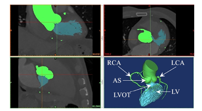
基于一例共228张、层厚0.5 mm的正常人体心脏CT影像数据(中国医学科学院阜外医院提供),利用Mimics 18.0 (Materialise, Leuven, Belgium)进行阈值分割和区域生长,三维重构左冠状动脉(left coronary artery, LCA)、右冠状动脉(right coronary artery, RCA)、升主动脉(ascending aorta, AA)、主动脉窦(aortic sinus, AS)、左心室流出道(left ventricle outflow tract, LVOT)和左心室(left ventricle, LV), 保存STL文件[16-17], 如图1所示.图1
 新窗口打开|下载原图ZIP|生成PPT
图1基于医学影像数据提取主动脉根部
Fig.1Aortic root obtained based on CT Data
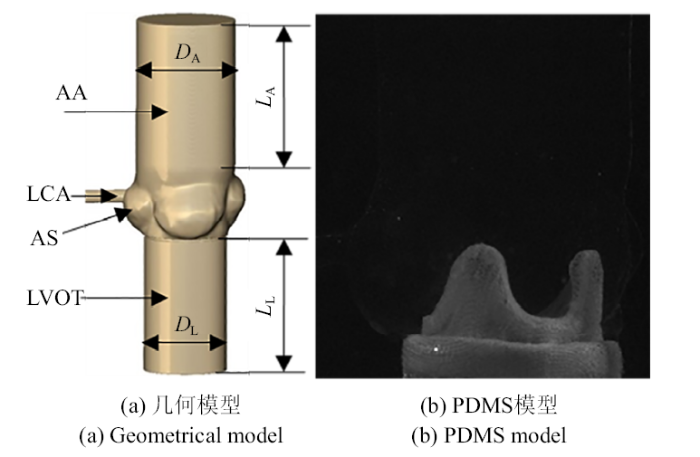
将STL文件输入触觉式设计系统Freeform (3D Systems, USA),对模型表面光滑和切割删除无用组织.左、右冠状动脉对主动脉根部流动的影响具有相似性,本文借鉴Querzoli等[14]的模型设计, 保留左冠状动脉而删除右冠状动脉,研究冠状动脉存在时主动脉根部的流动特性.主动脉根部几何模型包含LCA, AA, AS和LVOT,其中AA直径$D_{\rm A}=36$ mm、长度$L_{\rm A}=40$ mm, LVOT直径$D_{\rm L}=30$ mm,长度$L_{\rm L}=40$ mm, 如图2(a)所示. 利用3D打印技术,以水溶性材料聚乙烯醇(polyvinyl alcohol, PVA)打印主动脉根部,以聚二甲基硅氧烷(polydimethylsiloxane, PDMS; A胶:B胶=10:1)浇筑主动脉根部,水浴溶解PVA后得到高度光滑和透明的主动脉根部PDMS实验模型, 如图2(b)所示.
图2
 新窗口打开|下载原图ZIP|生成PPT
图2主动脉根部.
Fig.2Aortic root
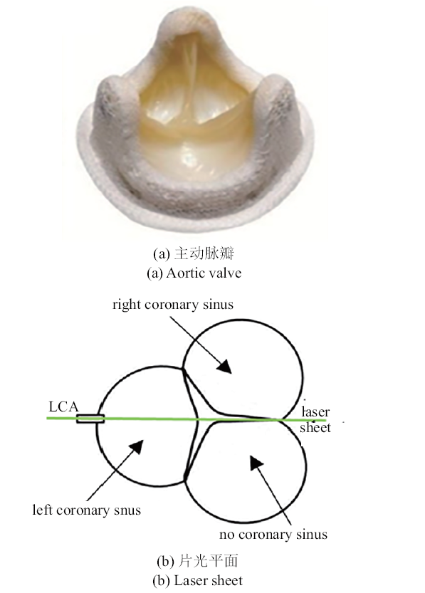
主动脉瓣为瓣环直径25 mm的美敦力Hancock II T510型人工生物主动脉瓣(Medtronic,Minneapolis, USA), 瓣孔直径22.5 mm, 缝合环直径33 mm, 瓣膜高度18 mm,主动脉伸入长度13.5 mm, 如图3(a)所示, 实验时经过主动脉瓣的片光平面如图3(b)所示
图3
 新窗口打开|下载原图ZIP|生成PPT
图3主动脉瓣及片光平面
Fig.3Aortic valve and laser plane
1.2 实验装置
粒子图像测速技术是一种非侵入式的全流场流动测量及显示技术,广泛应用于主动脉瓣的速度场和剪应力环境研究[18-20].PIV技术直接地展示主动脉根部中的流体速度,并通过后处理得到VSS和RSS等流体动力学特征,能够揭示心排出量减少可能对主动脉瓣瓣叶造成的影响.PIV技术原理是在待测流体中布撒示踪粒子, 使用脉冲激光片光源照亮目标流场区域,由CCD相机记录连续两次曝光时间间隔的两幅粒子图像,通过计算机处理获得示踪粒子的速度,并以该粒子速度代表粒子所在位置的流场速度[21].PIV系统主要由双腔Nd: YAG激光器(Dantec Dynamics, Denmark; 能量10 mJ, 波长532 Nm,脉冲时间4 Ns)、CCD相机(LaVision, Germany, Imager Pro;像素$1344\times 1024$)、同步控制器、片光元件、导光臂和计算机组成[22-23].双腔Nd:YAG激光器产生的激光经过导光臂、柱面镜和球面镜等片光元件引导后在实验模型中心平面形成厚度约为1 mm的片光,激发均匀布撒于工作流体中的示踪粒子. 示踪粒子为直径为10~$\mu$m的镀银中空玻璃微球(S-HGS-10, Dantec Dynamics, Denmark).CCD相机记录单位时间内示踪粒子的两帧图像, 计算机处理后获得目标流场.
循环系统由脉动式血液泵(Harvard Apparatus,USA)、储液池、压力计、节流阀和顺应腔等组成[24].脉动式血液泵用于模拟心脏功能, 可提供不同的心排出量.顺应腔和节流阀通过管路与升主动脉出口连接, 用于调节升主动脉出口的平均压力.节流阀控制进入顺应腔的工作流体, 压缩顺应腔中的可压缩空气,使升主动脉出口达到主动脉瓣最大张开幅度的平均压力[25-26].工作流体由脉动式血液泵从储液室中泵出进入主动脉根部,流经主动脉瓣后到达三通管, 一路进入顺应腔, 另一路经节流阀后回到储液池,形成循环.
左心室流出道(LVOT)由脉动式血液泵提供不同的心排出量,实验时调节脉动式血液泵的心率(heart rate, HR)与每搏量(stroke volume, SV),实现符合ISO 5840: 2015标准的不同心排出量[10-11], 如表1所示.升主动脉(AA)出口平均压力通过顺应腔和节流阀调节为1333.22~Pa.左冠状动脉(LCA)出口为自由出口, 因为冠状动脉顺应性远小于升主动脉顺应性,可忽略冠状动脉顺应性对整体顺应性的影响[27].
图4
 新窗口打开|下载原图ZIP|生成PPT
图4实验系统
Fig.4Experimental system
Table 1
表1
表1实验中的心排出量设置
Table 1
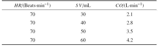 |
新窗口打开|下载CSV
工作流体选择体积比为40%/60%的丙三醇/去离子水混合溶液,该溶液物理性质类似于血液, 其密度$\rho =1100$ kg/m$^{3}$、常温时黏度$\mu =0.004\,0$~Pa$\cdot $s, 折射率($n=1.38$)与主动脉根部PDMS模型折射率相近($n=1.41$)[28].
1.3 物理量
涡度是速度场的旋度, 描述流体的旋转情况,高涡度区域表示流体中的高剪切区域[29], 涡度由式(1)定义式中, $U$为$X$方向上的瞬时速度, $V$为$Y$方向上的瞬时速度.
黏性剪应力(viscous shear stress, VSS)是表征流体相邻两层之间的剪切作用,与瓣膜血栓、血小板及红细胞溶血密切相关[30], 由式(2)定义
式中, $\mu $为工作流体的动力黏度, 单位N$\cdot$S/m$^2$.
雷诺剪应力(Reynolds shear stress,RSS)是由速度场的时间变化引起的流体流层之间剪应力的统计量[31-32],
由式(3)$\sim\!$式(5)定义
式中, $\rho $为流体的密度, $\overline U $为$X$方向平均速度, $\overline V $为$Y$方向的平均速度, $\overline{u'} $为$X$方向的平均瞬时速度波动, $\overline{v'} $为$Y$方向的平均瞬时速度波动.
2 结果与讨论
2.1 冠状动脉存在对主动脉瓣附近速度分布的影响
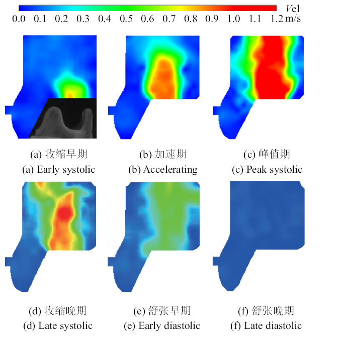
为了认识心动周期内主动脉瓣附近流体流动, 图5展示了$CO=4.2$ l/min时心动周期内主动脉瓣附近的速度分布云图. 收缩早期, 主动脉瓣瓣叶开始张开,瓣口处出现血液流动. 加速期, 主动脉瓣张开幅度增大,血液流过主动脉瓣时形成中心对称流动, 并且向主动脉瓣下游进一步流动.收缩峰值期, 主动脉瓣瓣叶完全打开, 血液中心对称流动加剧,血液跨瓣流动达到最大速度1.37 m/s. 收缩晚期, 主动脉瓣开始关闭,主动脉瓣附近的血液流动现象与峰值期现象相似, 但流动速度降低. 舒张早期,随着主动脉瓣的关闭, 跨瓣流动逐渐消失. 舒张晚期, 瓣膜完全关闭,主动脉瓣口血液流动现象停止.图5
 新窗口打开|下载原图ZIP|生成PPT
图5$CO=4.2$ l/min时主动脉瓣附近速度分布云图
Fig.5Velocity contours near the aortic valve at $CO=4.2$ l/min
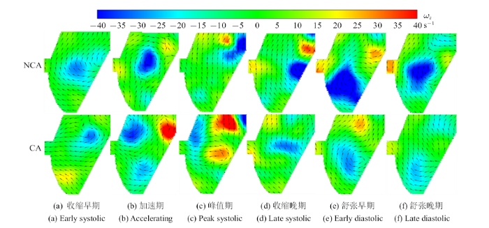
冠状动脉起源于主动脉窦, 其严重影响主动脉窦中的血液流动和受力状况,从而影响主动脉瓣的血流动力学[14-15]. 图6为$CO=4.2$ l/min,心动周期内冠状动脉存在与否时主动脉窦的速度矢量和涡度分布云图. 在涡度云图中,红色区域表示逆时针旋转(正)涡度, 而蓝色区域表示顺时针旋转(负)涡度.当冠状动脉不存在时, 从收缩早期开始, 流体在主动脉窦中开始形成涡旋运动,并且在整个心动周期内一直存在涡旋运动. 在收缩早期至收缩晚期,负涡度的强度逐渐增大; 收缩晚期后, 负涡度的强度逐渐减小,且流体由于无法从冠状动脉流出而在冠脉口处形成正涡度. 当冠状动脉存在时,从收缩晚期开始, 由于流体经由冠状动脉流出, 主动脉窦中的涡旋运动逐渐消失,该现象与文献[14]的研究结果一致.
图6
 新窗口打开|下载原图ZIP|生成PPT
图6$CO=4.2$ l/min时, 左冠状动脉是否存在时主动脉窦速度矢量和涡度分布
Fig.6Velocity vectors and vorticity contours of sinus in presence or absence of left coronary artery at $CO=4.2$ l/min (NCA: No coronary artery, CA: Coronary artery)
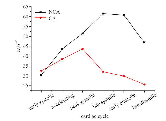
图7为$CO=4.2$ l/min, 心动周期内冠状动脉存在与否时主动脉窦负涡度变化曲线.冠状动脉不存在时, 在收缩晚期涡度达到最大为61.50 s$^{ - 1}$. 然而,与冠状动脉不存在时相比, 冠状动脉存在时负涡度在峰值期取得最大为43.63 s$^{-1}$, 涡度较早开始出现减少即从峰值期后负涡度逐渐较小,涡度小于冠状动脉不存在时. 研究表明[15],冠状动脉存在时流体从冠状动脉流出, 改善了主动脉窦中的血流动力学,且考虑冠状动脉所获得的血流动力学更接近真实状况. 因此,本文后续将在冠状动脉存在的情况下开展心排出量研究.
图7
 新窗口打开|下载原图ZIP|生成PPT
图7$CO=4.2$ l/min时, 心动周期内冠状动脉存在与否时主动脉窦负涡度变化曲线
Fig.7Curve of negative vorticity of sinus in the presence or absence of left coronary artery during cardiac cycle at $CO=4.2$ l/min
2.2 心排出量对主动脉瓣附近速度分布的影响
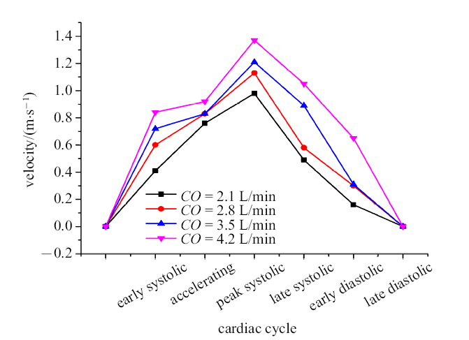
图8为心动周期内不同时刻不同心排出量下主动脉瓣附近的最大速度变化曲线.在整个心动周期内, 瓣叶张开后主动脉瓣出现流动, 随着瓣叶开口面积增大,跨瓣的血液流动速度增大, 并在收缩峰值期取得最大速度. 峰值期后,随着主动脉瓣逐渐关闭, 最大速度逐渐降低, 直至瓣膜关闭无流动.随着心排出量的增加, 心动周期内不同时刻的最大速度都增大, 且$CO=2.1$, 2.8, 3.5和4.2 l/min时, 在峰值期时中心对称流动取得最大速度,分别为0.98, 1.13, 1.21和1.37 m/s.图8
 新窗口打开|下载原图ZIP|生成PPT
图8心动周期内不同心排出量下的最大速度变化曲线
Fig.8Curves of peak velocity during cardiac cycle under varied $CO$
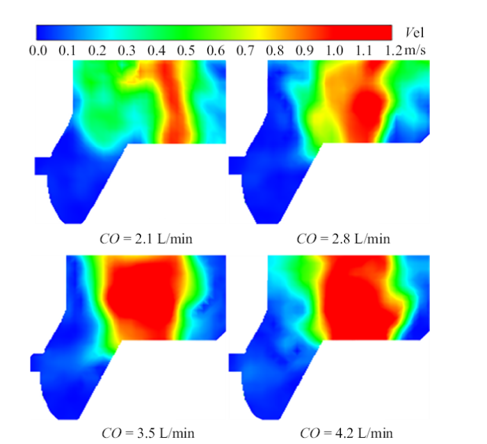
图9为峰值期时不同心排出量下主动脉瓣附近速度分布云图. 心排出量较低时,如$CO=2.1$ l/min, 中心对称流动速度较小、流动剧烈程度较低,主动脉瓣下游存在区域较大的低速区域. 随着心排出量的增加, 如$CO=4.2$ l/min,中心对称流动加剧, 流动速度和流动高速区域增大. 研究[33]表明,在血流速度较低的区域易产生低黏性剪应力, 最终导致血栓形成. 心排出量较低时,主动脉瓣瓣口中心对称流动速度较低, 瓣膜下游血液流动减弱, 形成较大的低速区域,为血栓形成提供有利环境. 同时, 心排出量较低时,较低的跨瓣流动可能导致供血不足.
图9
 新窗口打开|下载原图ZIP|生成PPT
图9峰值期不同心排出量下主动脉瓣附近的速度分布云图
Fig.9Velocity contours near the aortic valve during peak systolic under varied $CO$
2.3 心排出量对主动脉瓣附近黏性剪应力的影响
黏性剪应力是血细胞承受的真实物理剪切应力,其在血细胞损伤机制中扮演重要角色[34].图10为收缩峰值不同心排出量下主动脉瓣附近的黏性剪应力分布云图.由于中心对称流动两侧边缘存在较大的速度梯度(如图9所示),该区域存在与收缩射流边缘一致且彼此平行的正、负高黏性剪切区域,该现象与前人关于黏性剪应力分布的研究结果一致[29].
图10
 新窗口打开|下载原图ZIP|生成PPT
图10峰值期不同心排出量下主动脉瓣附近VSS分布云图
Fig.10VSS contours near the aortic valve during peak systolic under varied $CO$
心排出量较低时, 瓣叶开口面积较小,血液流动中心对称流动速度较低、高速区域较窄, 使得正负高黏性剪切区域距离较近.心排出量较高时, 主动脉瓣开口面积较大,血液流动中心对称流动速度和高速区域较大, 使得正负高黏性剪切区域距离较大.随着心排出量增大, 黏性剪应力增大, 即心排出量$CO=2.1$, 2.8, 3.5和4.2 l/min时, 峰值期的最大VSS分别为0.87, 0.95, 0.96和1.02 N/m$^{2}$. 研究[35-38]表明在低剪应力剪切环境时, 容易发生剪切诱导血小板活化, 导致血栓形成.在心排出量较低时, 黏性剪应力较低, 血栓形成的可能性较大.
2.4 心排出量对主动脉瓣附近雷诺剪应力的影响
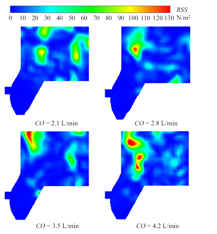
流体在改变方向时减速或加速产生雷诺剪应力, 其表征血流速度的湍流波动,与红细胞溶血密切相关[39].图11为收缩峰值期不同心排出量下主动脉瓣附近的雷诺剪应力分布云图. 峰值期,跨瓣的中心对 称流动达到最大速度,高速的跨瓣流动与升主动脉中相对缓慢流动的血液形成剪切层.在中心对称流动两侧由于速度波动较大, 存在雷诺剪应力较大的区域,特别是存在冠脉一侧的升主动脉下游雷诺剪应力最大. 随着心排出量增大,雷诺剪应力增大, 当$CO=2.1$, 2.8, 3.5和4.2 l/min时,最大$RSS$分别为103.76, 116.25, 138.68和146.55 N/m$^{2}$.
图11
 新窗口打开|下载原图ZIP|生成PPT
图11峰值期不同心排出量下主动脉瓣附近RSS分布云图
Fig.11RSS contours near the aortic valve during peak systolic under varied $CO$
研究[35,40]表明红细胞的溶血雷诺剪应力临界值为150 N/m$^{2}$,低于该剪应力值下的单位时间内红细胞溶血相对较低.尽管本文心排出量下的所有剪应力值都在红细胞安全剪切环境下,但随着心排出量的增大, 雷诺剪应力增大, 可能增大至红细胞溶血阈值范围内,因此$CO=4.2$ l/min时存在红细胞溶血的潜在危险.
在个体化病例中研究中, 冠状动脉的进出口条件通常与心肺循环系统相耦合,心排出量等进出口条件应通过建立集中参数模型等方法获取.但本文着重关注主动脉瓣病变引起的心排出量减少对主动脉瓣的影响,在PIV实验研究时采用符合ISO 5840: 2015标准的不同心排出量,通过主动脉根部的速度、VSS和RSS分布等血流动力学参数展示不同心排出量对主动脉瓣的影响.本文实验结果对主动脉瓣病变个体化病例研究具有借鉴意义,但个体化病例研究时应采用符合该病例的心排出量.
PIV流动显示技术是在待测流体中布撒示踪粒子,以示踪粒子的速度代表粒子所在位置的速度,并通过后处理得到VSS和RSS等流体动力学特征. 但当主动脉瓣运动时,主动脉瓣瓣叶的形状为随时间变化的不规则曲面,其容易遮挡拍摄平面中靠近瓣叶的示踪粒子,导致CCD相机无法记录瓣叶曲面表面的粒子运动. 因此,PIV实验研究时未能直接地获取主动脉瓣瓣叶表面的VSS和RSS随主动脉瓣张开闭合过程的变化.在未来研究中, 将在已有PIV实验的基础上, 结合计算流体力学方法,建立集中参数模型并开展主动脉瓣流固耦合分析,直接地展示瓣叶表面的应力分布随瓣叶张开闭合过程的变化.
3 结论
(1)冠状动脉存在时, 流体经由冠状动脉流出, 主动脉窦中的涡旋运动逐渐消失,涡度较早开始减小.(2)峰值期,中心对称流动两侧区域存在与流动边缘一致且彼此平行的正、负高黏性剪切区域,存在冠状动脉一侧的升主动脉下游存在高雷诺剪应力区域.
(3)随着心排出量增大,心动周期内不同时刻的最大速度、峰值期的黏性剪应力和雷诺剪应力增大.心排出量较低时, 主动脉瓣较低的跨瓣流动速度和黏性剪应力易导致血栓形成.
参考文献 原文顺序
文献年度倒序
文中引用次数倒序
被引期刊影响因子
DOIURL [本文引用: 1]

使用脉动工作站检测生物瓣膜在近似生理条件下的血流动力学特性,结合有限元分析研究了生物瓣膜在心动周期过程中的微观应力和应变情况,寻求一种快速评价人工心脏瓣膜结构—力学性能间关系的方法。结果表明,生物瓣膜(Edwards #2625)在体外脉动流检测条件下的平均跨瓣压差为10.8 mmHg、有效开口面积为2.0 cm2、返流百分比为8.4%,均符合ISO-5840国际检测标准。生物瓣膜有限元模拟结果揭示其在收缩期最大主应力达到425 kPa,应力集中在弯曲变形严重的腹部以及瓣叶缝合边;舒张期最大主应力为1.46 MPa,应力集中在瓣叶缝合边的两侧。在脉动检测的不同时间点,瓣膜有限元模型开口面积均与实验条件下样品的开口面积近似,证明了有限元模拟结果的可靠性。本文提出的体外脉动流检测实验与有限元仿真计算相结合的方法,为评价人工心脏瓣膜的结构—力学性能间关系提供一种高效可靠的途径。
DOIURL [本文引用: 1]
使用脉动工作站检测生物瓣膜在近似生理条件下的血流动力学特性,结合有限元分析研究了生物瓣膜在心动周期过程中的微观应力和应变情况,寻求一种快速评价人工心脏瓣膜结构—力学性能间关系的方法。结果表明,生物瓣膜(Edwards #2625)在体外脉动流检测条件下的平均跨瓣压差为10.8 mmHg、有效开口面积为2.0 cm2、返流百分比为8.4%,均符合ISO-5840国际检测标准。生物瓣膜有限元模拟结果揭示其在收缩期最大主应力达到425 kPa,应力集中在弯曲变形严重的腹部以及瓣叶缝合边;舒张期最大主应力为1.46 MPa,应力集中在瓣叶缝合边的两侧。在脉动检测的不同时间点,瓣膜有限元模型开口面积均与实验条件下样品的开口面积近似,证明了有限元模拟结果的可靠性。本文提出的体外脉动流检测实验与有限元仿真计算相结合的方法,为评价人工心脏瓣膜的结构—力学性能间关系提供一种高效可靠的途径。
DOIURLPMID
Prosthesis positioning in transcatheter aortic valve implantation procedures represents a crucial aspect for procedure success as demonstrated by many recent studies on this topic. Possible complications, device performance, and, consequently, also long-term durability are highly affected by the adopted prosthesis placement strategy. In the present work, we develop a computational finite element model able to predict device-specific and patient-specific replacement procedure outcomes, which may help medical operators to plan and choose the optimal implantation strategy. We focus in particular on the effects of prosthesis implantation depth and release angle. We start from a real clinical case undergoing Corevalve self-expanding device implantation. Our study confirms the crucial role of positioning in determining valve anchoring, replacement failure due to intra or para-valvular regurgitation, and post-operative device deformation.
DOIURLPMID [本文引用: 1]
[本文引用: 1]
[本文引用: 1]
DOIURLPMID [本文引用: 1]
In developed countries, aortic stenosis is the most prevalent of all valvular heart diseases. A manifestation of ageing, the disorder is becoming more frequent as the average age of the population increases. Symptomatic severe disease is universally fatal if left untreated yet is consistent with a typical lifespan when mechanical relief of the stenosis is provided in a timely fashion. Management of mild disease, severe asymptomatic disease, and far advanced disease, and the effect of new percutaneous treatments, provide both controversy and exciting promise to care of patients with aortic stenosis. We discuss these issues in this Review.
DOIURLPMID [本文引用: 1]
The aim of the present in vitro study was the evaluation of the fluid dynamical performance of the Carpentier-Edwards PERIMOUNT Magna Ease depending on the prosthetic size (21, 23, and 25?mm) and the cardiac output (3.6-6.4?L/min). A self-constructed flow channel in combination with particle image velocimetry (PIV) enabled precise results with high reproducibility, focus on maximal and local peek velocities, strain, and velocity gradients. These flow parameters allow insights into the generation of forces that act on blood cells and the aortic wall. The results showed that the 21 and 23?mm valves have a quite similar performance. Maximal velocities were 3.03 ± 0.1 and 2.87 ± 0.13?m/s; maximal strain E xx , 913.81 ± 173.25 and 896.15 ± 88.16?1/s; maximal velocity gradient E yx , 1203.14 ± 221.84?1/s and 1200.81 ± 61.83?1/s. The 25?mm size revealed significantly lower values: maximal velocity, 2.47 ± 0.15?m/s; maximal strain E xx , 592.98 ± 155.80?1/s; maximal velocity gradient E yx , 823.71 ± 38.64?1/s. In summary, the 25?mm Magna Ease was able to create a wider, more homogenous flow with lower peak velocities especially for higher flow rates. Despite the wider flow, the velocity values close to the aortic walls did not exceed the level of the smaller valves.
DOIURLPMID
The rationale of this paper is to investigate right ventricular (RV) hemodynamics in relation to changes in cardiac output, and in particular to study exercise-induced stresses at the RV outflow tract (RVOT), which is a common site of ventricular arrhythmias in the athlete's heart. We hypothesize that the thin-walled RVOT is exposed to high wall shear stresses (WSS) during physiological states associated with high cardiac output such as exercise, and therefore, may be particularly prone to substrate formation leading to ventricular tachyarrhythmias. 3D Particle Tracking Velocimetry (3D-PTV), an optical imaging method, has been performed in a novel anatomically accurate compliant silicone right heart model derived from a high resolution MRI heart scan of a healthy male proband. RV and RVOT flow patterns at resting conditions were obtained from two healthy athletic male proband's hearts and two patients with arrhythmogenic right ventricular cardiomyopathy/dysplasia (ARVC/D) via phase contrast magnetic resonance imaging (PC-MRI). The healthy case was used as a reference for validating the in vitro flow patterns of the silicone model, while the diseased cases were used to generalize our findings and investigate possible changes in hemodynamic stresses with RV morphological remodelling. Our results showed that both healthy and diseased geometries consistently displayed an increased WSS in the RVOT relative to the rest of the RV. We found that increases in cardiac output may lead to increases of mean kinetic energy (MKE), laminar viscous dissipation and WSS at the RVOT. Furthermore, higher peak WSS magnitudes were found for the diseased cases. The identified high WSS regions may correlate with the common site of RVOT ventricular tachycardia in athletes and patients with ARVC/D. Our results imply that exercise, as well as anatomical and functional remodeling might alter RV wall shear stress both in magnitude and spatial distribution, leading to increased hemodynamic stresses in the RVOT.
[本文引用: 1]
[本文引用: 1]
DOIURL [本文引用: 2]
DOIURL [本文引用: 2]
Aortic valve (AV) calcification is a highly prevalent disease with serious impact on mortality and morbidity. Although exact causes and mechanisms of AV calcification are unclear, previous studies suggest that mechanical forces play a role. Since calcium deposits occur almost exclusively on the aortic surfaces of AV leaflets, it has been hypothesized that adverse patterns of fluid shear stress on the aortic surface of AV leaflets promote calcification. The current study characterizes AV leaflet aortic surface fluid shear stresses using Laser Doppler velocimetry and an in vitro pulsatile flow loop. The valve model used was a native porcine valve mounted on a suturing ring and preserved using 0.15% glutaraldehyde solution. This valve model was inserted in a mounting chamber with sinus geometries, which is made of clear acrylic to provide optical access for measurements. To understand the effects of hemodynamics on fluid shear stress, shear stress was measured across a range of conditions: varying stroke volumes at the same heart rate and varying heart rates at the same stroke volume. Systolic shear stress magnitude was found to be much higher than diastolic shear stress magnitude due to the stronger flow in the sinuses during systole, reaching up to 20 dyn/cm(2) at mid-systole. Upon increasing stroke volume, fluid shear stresses increased due to stronger sinus fluid motion. Upon increasing heart rate, fluid shear stresses decreased due to reduced systolic duration that restricted the formation of strong sinus flow. Significant changes in the shear stress waveform were observed at 90 beats/min, most likely due to altered leaflet dynamics at this higher heart rate. Overall, this study represents the most well-resolved shear stress measurements to date across a range of conditions on the aortic side of the AV. The data presented can be used for further investigation to understand AV biological response to shear stresses.
DOIURLPMID [本文引用: 1]
There is an increasing awareness of leaflet thrombosis following transcatheter aortic valve implantation (TAVI) and valve-in-valve (ViV) procedures. Nevertheless, the predisposing factors affecting transcatheter aortic valve (TAV) thrombosis have remained unclear. This study aimed to quantify the effects of reduced cardiac output (CO) on blood stasis on the TAV leaflets as a permissive factor for valve thrombosis.
DOIURLPMID [本文引用: 1]
The bicuspid aortic valve (BAV), which forms with two leaflets instead of three as in the normal tricuspid aortic valve (TAV), is associated with a spectrum of secondary valvulopathies and aortopathies potentially triggered by hemodynamic abnormalities. While studies have demonstrated an intrinsic degree of stenosis and the existence of a skewed orifice jet in the BAV, the impact of those abnormalities on BAV hemodynamic performance and energy loss has not been examined. This steady-flow study presents the comparative in vitro assessment of the flow field and energy loss in a TAV and type-I BAV under normal and simulated calcified states. Particle-image velocimetry (PIV) measurements were performed to quantify velocity, vorticity, viscous, and Reynolds shear stress fields in normal and simulated calcified porcine TAV and BAV models at six flow rates spanning the systolic phase. The BAV model was created by suturing the two coronary leaflets of a porcine TAV. Calcification was simulated via deposition of glue beads in the base of the leaflets. Valvular performance was characterized in terms of geometric orifice area (GOA), pressure drop, effective orifice area (EOA), energy loss (EL), and energy loss index (ELI). The BAV generated an elliptical orifice and a jet skewed toward the noncoronary leaflet. In contrast, the TAV featured a circular orifice and a jet aligned along the valve long axis. While the BAV exhibited an intrinsic degree of stenosis (18% increase in maximum jet velocity and 7% decrease in EOA relative to the TAV at the maximum flow rate), it generated only a 3% increase in EL and its average ELI (2.10 cm2/m2) remained above the clinical threshold characterizing severe aortic stenosis. The presence of simulated calcific lesions normalized the alignment of the BAV jet and resulted in the loss of jet axisymmetry in the TAV. It also amplified the degree of stenosis in the TAV and BAV, as indicated by the 342% and 404% increase in EL, 70% and 51% reduction in ELI and 48% and 51% decrease in EOA, respectively, relative to the nontreated valve models at the maximum flow rate. This study indicates the ability of the BAV to function as a TAV despite its intrinsic degree of stenosis and suggests the weak dependence of pressure drop on orifice area in calcified valves.
DOIURL [本文引用: 4]
DOIURLPMID [本文引用: 3]
Mechanical stresses on aortic valve leaflets are well-known mediators for initiating processes leading to calcific aortic valve disease. Given that non-coronary leaflets calcify first, it may be hypothesized that coronary flow originating from the ostia significantly influences aortic leaflet mechanics and sinus hemodynamics. High resolution time-resolved particle image velocimetry (PIV) measurements were conducted to map the spatiotemporal characteristics of aortic sinus blood flow and leaflet motion with and without physiological coronary flow in a well-controlled in vitro setup. The in vitro setup consists of a porcine aortic valve mounted in a physiological aorta sinus chamber with dynamically controlled coronary resistance to emulate physiological coronary flow. Results were analyzed using qualitative streak plots illustrating the spatiotemporal complexity of blood flow patterns, and quantitative velocity vector and shear stress contour plots to show differences in the mechanical environments between the coronary and non-coronary sinuses. It is shown that the presence of coronary flow pulls the classical sinus vorticity deeper into the sinus and increases flow velocity near the leaflet base. This creates a beneficial increase in shear stress and washout near the leaflet that is not seen in the non-coronary sinus. Further, leaflet opens approximately 10% farther into the sinus with coronary flow case indicating superior valve opening area. The presence of coronary flow significantly improves leaflet mechanics and sinus hemodynamics in a manner that would reduce low wall shear stress conditions while improving washout at the base of the leaflet.
DOIURLPMID [本文引用: 1]
This work investigates the effect of arterial bifurcation angulation on atherosclerosis development through in-silico simulations of coupled cell dynamics. The computational model presented here combines cellular pathways, fluid dynamics, and physiologically-realistic vessel geometries as observed in the human vasculature. The coupled cells model includes endothelial cells (ECs) and smooth muscle cells (SMCs) with ion dynamics, hetero and homotypic coupling, as well as electro-diffusive coupling. Three arterial bifurcation surface models were used in the coupled cells simulations. All three simulations showed propagating waves of Ca2+ in both the SMC and EC layers, following the introduction of a luminal agonist, in this case ATP. Immediately following the introduction of ATP concentration Ca2+ waves propagate from the area of high ATP toward the areas of low ATP concentration, forming complex patterns where waves interact with eachother, collide and fade. These dynamic phenomena are repeated with a series of waves of slower velocity. The underlying motivation of this research was to examine the macro-scale phenomena, given that the characteristic length scales of atherosclerotic plaques are much larger than a single cell. The micro-scale dynamics were modeled on macro-scale arterial bifurcation surfaces containing over one million cells. The results of the simulations presented here suggest that susceptibility to atherosclerosis development depends on the bifurcation angulation. In conjunction with findings reported in the literature, the simulation results demonstrate that arterial bifurcations containing wider angles have a more prominent influence on the coupled cells pathways associated with the development of atherosclerosis, by means of disturbed flow and lower SMC Ca2+ concentrations. The discussion of the results considers the findings of this research within the context of the potential link between information transport through frequency encoding of Ca2+ wave dynamics and development of atheroprone conditions.
[本文引用: 1]
[本文引用: 1]
[本文引用: 1]
[本文引用: 1]
DOIURLPMID [本文引用: 1]
Aortic valve (AV) calcification is a highly prevalent disease with serious impact on mortality and morbidity. The exact cause and mechanism of the progression of AV calcification is unknown, although mechanical forces have been known to play a role. It is thus important to characterize the mechanical environment of the AV. In the current study, we establish a methodology of measuring shear stresses experienced by the aortic surface of the AV leaflets using an in vitro valve model and adapting the laser Doppler velocimetry (LDV) technique. The valve model was constructed from a fresh porcine aortic valve, which was trimmed and sutured onto a plastic stented ring, and inserted into an idealized three-lobed sinus acrylic chamber. Valve leaflet location was measured by obtaining the location of highest back-scattered LDV laser light intensity. The technique of performing LDV measurements near to biological surfaces as well as the leaflet locating technique was first validated in two phantom flow systems: (1) steady flow within a straight tube with AV leaflet adhered to the wall, and (2) steady flow within the actual valve model. Dynamic shear stresses were then obtained by applying the techniques on the valve model in a physiologic pulsatile flow loop. Results show that aortic surface shear stresses are low during early systole (<5 dyn/cm2) but elevated to its peak during mid to late systole at about 18-20 dyn/cm2. Low magnitude shear stress (<5 dyn/cm2) was observed during early diastole and dissipated to zero over the diastolic duration. Systolic shear stress was observed to elevate only with the formation of sinus vortex flow. The presented technique can also be used on other in vitro valve models such as congenitally geometrically malformed valves, or to investigate effects of hemodynamics on valve shear stress. Shear stress data can be used for further experiments investigating effects of fluid shear stress on valve biology, for conditioning tissue engineered AV, and to validate numerical simulations.
[本文引用: 1]
[本文引用: 1]
[本文引用: 1]
[本文引用: 1]
[本文引用: 1]
[本文引用: 1]
DOIURLPMID [本文引用: 1]
Experimental flow field characterization is a critical component of the assessment of the hemolytic and thrombogenic potential of heart valve substitutes, thus it is important to identify best practices for these experimental techniques. This paper presents a brief review of commonly used flow assessment techniques such as Particle image velocimetry (PIV), Laser doppler velocimetry, and Phase contrast magnetic resonance imaging and a comparison of these methodologies. In particular, recommendations for setting up planar PIV experiments such as recommended imaging instrumentation, acquisition and data processing are discussed in the context of heart valve flows. Multiple metrics such as residence time, local velocity and shear stress that have been identified in the literature as being relevant to hemolysis and thrombosis in heart valves are discussed. Additionally, a framework for uncertainty analysis and data reporting for PIV studies of heart valves is presented in this paper. It is anticipated that this paper will provide useful information for heart valve device manufacturers and researchers to assess heart valve flow fields for the potential for hemolysis and thrombosis.
DOIURLPMID [本文引用: 1]
Transcatheter aortic valve replacement (TAVR) is a life-saving alternative to surgical intervention. However, the identification of features associated with poor outcomes, including residual paravalvular leakage (PVL), leaflet calcification, and subclinical leaflet thrombosis, are cause to be concerned about valve durablilty (Mylotte and Piazza, 2015a, 2015b; Dasi et al., 2017; Makkar et al., 2015; Kheradvar et al., 2015a). The aim of this study is to optimize the potential of a hyaluronan (HA) enhanced polymeric transcatheter aortic valve (HA-TAV) that has promised to reduce blood damage causing-turbulent flow while maintaining durability. HA-enhanced linear low-density polyethylene (LLDPE) leaflets were sutured to novel cobalt chromium stents, size 26?mm balloon expandable stents. Hemodynamic performance was assessed in a left heart simulator under physiological pressure and flow conditions and compared to a 26?mm Medtronic Evolut and 26?mm Edwards SAPIEN 3. High-speed imaging and particle image velocimetry (PIV) were performed. The HA-TAV demonstrated an effective orifice area (EOA) within one standard deviation of the leading valve, SAPIEN 3.The regurgitant fraction (RF) of the HA-TAV (11.23?±?0.55%) is decreased in comparison the Evolut (15.74?±?0.73%) and slightly higher than the SAPIEN 3 (10.92?±?0.11%), which is considered trace regurgitation according to valve standards. A decreased number of higher principal Reynolds shear stresses were shown for the HA-TAV at each cardiac phase. The HA-TAV is directly comparable and in some cases superior to the leading commercially available prosthetic heart valves in in-vitro hemodynamic testing.
DOIURLPMID [本文引用: 1]
The aorta with its compliance plays a major role in hemodynamics as it saves a portion of ejected blood during systole which is then released in diastole. The aortic compliance decreases with increasing age, which is related to several cardiovascular imparities and diseases. Changes in flow patterns and pressure curves, due to varying aortic compliance, are difficult to investigate in vivo. As a result, the aim of the present work was to develop an in vitro setup enabling standardized investigations on the effect of compliance changes on flow patterns and pressure curves. Therefore an experimental setup with an anatomically correct silicone phantom of the aortic arch was developed, suitable for optical flow measurements under pulsatile inflow conditions. The setup was developed for precise adjustments of different compliances and optical flow measurements. Particle image velocimetry measurements were carried out downstream of the aortic valve in the center plane perpendicular to the valve with compliance adjusted between 0.62?×?10-3 to 1.82?×?10-3?mmHg-1. Preliminary results of the in vitro investigations showed that decreases in compliance results in significant increases in pressure changes with respect to time (dp/dt) and altered pressure curves in the aortic arch. In terms of flow, an increased aortic stiffness lead to higher mean velocities and decreased vortex development in the aortic sinuses. As in vivo validation and translation remains difficult, the results have to be considered as preliminary in vitro insights into the mechanisms of (age-related) compliance changes.
DOIURLPMID [本文引用: 1]
Potential applications of genome editing in assisted reproductive technology (ART) raise a vast array of strong opinions, emotional reactions and divergent perceptions. Acknowledging the need for caution and respecting such reactions, we observe that at least some are based on either a misunderstanding of the science or misconceptions about the content and flexibility of the existing legal frameworks. Combining medical, legal and ethical expertise, we present and discuss regulatory responses at the national, European and international levels. The discussion has an EU starting point and is meant as a contribution to the general international regulatory debate. Overall, this paper concludes that gene editing technologies should not be regulated autonomously. Rather, potential uses should be regulated under general, existing frameworks and where applicable by reference to sufficiently equivalent technologies and techniques already subject to specific regulation. To be clear, we do not argue for the hasty introduction of gene editing as a reproductive treatment option in the immediate future. We call for caution with regard to overreaching moratoria and prohibitions that will also affect basic research. We recommend flexible regulations that allow for further responsible research into the potential development of the technology. We call for an open and inclusive debate and argue that scientific communication should claim a more prominent role to counter the danger of widespread misinformation. A high level of transparency and accuracy should guide scientific communication while simultaneously global-scale responsibility and governance should be fostered by promoting cross-disciplinary thinking and multi-level stakeholder involvement in legal and regulatory processes.
DOIURL [本文引用: 1]
DOIURLPMID [本文引用: 2]
Leaflet thrombosis is a complication associated with transcatheter aortic valve (TAV) replacement (TAVR) correlated with sinus flow stasis. Sinus hemodynamics are important because they dictate shear stress and washout necessary to avoid stasis on TAV leaflets. Sinus flow is controlled by TAV axial deployment position but little is known regarding TAV axis misalignment effect. This study aims to elucidate TAV angular misalignment with respect to aortic root axis effect on sinus flow stasis potentially leading to leaflet thrombosis. Sinus hemodynamics were assessed in vitro using particle-image velocimetry in three different angular misalignments with respect to aorta axis: untilted, tilted away from the sinus and tilted towards sinus. A 26?mm Edwards SAPIEN3 was implanted in a 3D printed model of an anatomically realistic aortic root. TAV hemodynamics, sinus vortex tracking, leaflet shear stress probability density functions, and sinus blood time to washout were calculated. While pressure gradients differed insignificantly, blood velocity and vorticity decreased significantly in both tilted cases sinuses. Shear stress probability near the leaflet decreases with tilt indicating stasis. TAV tilted away from the sinus is the most unfavorable scenario with poor washout. TAV axial misalignment adds to factors list that could influence leaflet thrombosis risk through modifying sinus hemodynamics and washout.
DOIURL [本文引用: 1]
Patients with aortic stenosis present with calcium deposits on the native aortic valve, which can result in non-concentric expansion of Transcatheter Aortic Valve Replacement (TAVR) stents. The objective of this study is to evaluate whether eccentric deployment of TAVRs lead to turbulent blood flow and blood cell damage. Particle Image Velocimetry was used to quantitatively characterize fluid velocity fields, shear stress and turbulent kinetic energy downstream of TAVRs deployed in circular and eccentric orifices representative of deployed TAVRs in vivo. Effective orifice area (EOA) and mean transvalvular pressure gradient (TVG) values did not differ substantially in circular and eccentric deployed valves, with only a minor decrease in EOA observed in the eccentric valve (2.0 cm(2) for circular, 1.9 cm(2) for eccentric). Eccentric deployed TAVR lead to asymmetric systolic jet formation, with increased shear stresses (circular = 97 N/m(2) vs. eccentric = 119 N/m(2)) and regions of turbulence intensity (circular = 180 N/m(2) vs. eccentric = 230 N/m(2)) downstream that was not present in the circular deployed TAVR. The results of this study indicate that eccentric deployment of TAVRs can lead to altered flow characteristics and may potentially increase the hemolytic potential of the valve, which were not captured through hemodynamic evaluation alone.
DOIURL [本文引用: 1]
The congenital bicuspid aortic valve (BAV) is associated with increased leaflet calcification, ascending aortic dilatation, aortic stenosis (AS) and regurgitation (AR). Although underlying genetic factors have been primarily implicated for these complications, the altered mechanical environment of BAVs could potentially accelerate these pathologies. The objective of the current study is to characterize BAV hemodynamics in an in vitro system. Two BAV models of varying stenosis and jet eccentricity and a trileaflet AV (TAV) were constructed from excised porcine AVs. Particle Image Velocimetry (PIV) experiments were conducted at physiological flow and pressure conditions to characterize fluid velocity fields in the aorta and sinus regions, and ensemble averaged Reynolds shear stress and 2D turbulent kinetic energy were calculated for all models. The dynamics of the BAV and TAV models matched the characteristics of these valves which are observed clinically. The eccentric and stenotic BAV showed the strongest systolic jet (V = 4.2 m/s), which impinged on the aortic wall on the non-fused leaflet side, causing a strong vortex in the non-fused leaflet sinus. The magnitudes of TKE and Reynolds stresses in both BAV models were almost twice as large as comparable values for TAV, and these maximum values were primarily concentrated around the central jet through the valve orifice. The in vitro model described here enables detailed characterization of BAV flow characteristics, which is currently challenging in clinical practice. This model can prove to be useful in studying the effects of altered BAV geometry on fluid dynamics in the valve and ascending aorta. These altered flows can be potentially linked to increased calcific responses from the valve endothelium in stenotic and eccentric BAVs, independent of concomitant genetic factors.
DOIURL [本文引用: 1]
DOIURLPMID [本文引用: 1]
Transcatheter aortic valves (TAVs) represent the latest advances in prosthetic heart valve technology. TAVs are truly transformational as they bring the benefit of heart valve replacement to patients that would otherwise not be operated on. Nevertheless, like any new device technology, the high expectations are dampened with growing concerns arising from frequent complications that develop in patients, indicating that the technology is far from being mature. Some of the most common complications that plague current TAV devices include malpositioning, crimp-induced leaflet damage, paravalvular leak, thrombosis, conduction abnormalities and prosthesis-patient mismatch. In this article, we provide an in-depth review of the current state-of-the-art pertaining the mechanics of TAVs while highlighting various studies guiding clinicians, regulatory agencies, and next-generation device designers.
DOIURL [本文引用: 1]
Bileaflet mechanical heart valves (BMHV) are widely used to replace diseased heart valves. Implantation of BMHV, however, has been linked with major complications, which are generally considered to be caused by mechanically induced damage of blood cells resulting from the non-physiological hemodynamics environment induced by BMHV, including regions of recirculating flow and elevated Reynolds (turbulence) shear stress levels. In this article, we analyze the results of 2D high-resolution velocity measurements and full 3D numerical simulation for pulsatile flow through a BMHV mounted in a model axisymmetric aorta to investigate the mechanical environment experienced by blood elements under physiologic conditions. We show that the so-called Reynolds shear stresses neither directly contribute to the mechanical load on blood cells nor is a proper measurement of the mechanical load experienced by blood cells. We also show that the overall levels of the viscous stresses, which comprise the actual flow environment experienced by cells, are apparently too low to induce damage to red blood cells, but could potentially damage platelets. The maximum instantaneous viscous shear stress observed throughout a cardiac cycle is <15N/m2. Our analysis is restricted to the flow downstream of the valve leaflets and thus does not address other areas within the BMHV where potentially hemodynamically hazardous levels of viscous stresses could still occur (such as in the hinge gaps and leakage jets).
DOIURLPMID [本文引用: 2]
Valvular hemolysis and thrombosis are common complications associated with stenotic heart valves. This study aims to determine the extent to which hemodynamics induce such traumatic events. The viscous shear stress downstream of a severely calcified bioprosthetic valve was evaluated via in vitro 2D particle image velocimetry measurements. The blood cell membrane response to the measured stresses was then quantified using 3D immersed-boundary computational simulations. The shear stress level at the boundary layer of the jet flow formed downstream of the valve orifice was observed to reach a maximum of 1000-1700?dyn/cm(2), which was beyond the threshold values reported for platelet activation (100-1000?dyn/cm(2)) and within the range of thresholds reported for red blood cell (RBC) damage (1000-2000?dyn/cm(2)). Computational simulations demonstrated that the resultant tensions at the RBC membrane surface were unlikely to cause instant rupture, but likely to lead to membrane plastic failure. The resultant tensions at the platelet surface were also calculated and the potential damage was discussed. It was concluded that although shear-induced thrombotic trauma is very likely in stenotic heart valves, instant hemolysis is unlikely and the shear-induced damage to RBCs is mostly subhemolytic.
DOIURLPMID
Transcatheter aortic valves provide superior systolic hemodynamic performance in terms of valvular pressure gradient and effective orifice area compared with equivalent size surgical bioprostheses. However, in depth investigation of the flow field structures is of interest to examine the flow field characteristics and provide experimental evidence necessary for validation of computational models. The goal of this study was to compare flow field characteristics of the three most commonly used transcatheter and surgical valves using phase-locked particle image velocimetry (PIV). 26-mm Edwards SAPIEN 3, 26-mm Medtronic CoreValve, and 25-mm Carpentier-Edwards PERIMOUNT Magna were examined in a pulse duplicator with input parameters matching ISO-5840, that is, heart rate of 70 beats/min, cardiac output of 5 L/min, and mean aortic pressure of 100 mm Hg. A 2D PIV system was used to obtain flow velocity and viscous shear stress fields during the entire cardiac cycle. In vitro testing showed that the mean transvalvular pressure gradient was lowest for SAPIEN 3, followed by CoreValve, and PERIMOUNT Magna surgical bioprosthesis. In addition, the viscous shear stress magnitude within the jet boundary layer was higher in PERIMOUNT Magna than CoreValve and SAPIEN 3 at the peak of the flow. However, the measured shear stress values were below the known threshold for platelet activation and red blood damage. Therefore, shear-induced platelet activation is unlikely to take place during systole in the three bioprosthetic heart valves. The PIV measurements can be used for verification and validation of computational simulations.
[本文引用: 1]
[本文引用: 1]
DOIURLPMID [本文引用: 1]
This study aimed at assessment of post-transcatheter aortic valve (TAV) replacement hemodynamics and turbulence when a same-size SAPIEN 3 (Edwards Lifesciences Corp, Irvine, Calif) and Medtronic?Evolut (Minneapolis, Minn) were implanted in a rigid aortic root with physiological dimensions and in a representative root with calcific leaflets obtained from patient computed tomography scans.
DOIURLPMID [本文引用: 1]
A series of careful studies has been made on blood damage in a rotational viscometer. Specific attention has been focused on the effects of solid surface interaction, centrifugal force, air interface interaction, mixing of sheared and unsheared layers, cell-cell interaction, and viscous heating. The results show that there is a threshold shear stress, 1500 dynes/cm(2), above which extensive cell damage is directly due to shear stress, and the various secondary effects listed above are negligible. By analysis of these results and those of prior workers it is shown that the exposure time-shear stress plane is divided into two distinct regimes. In the regime of relatively low stresses and exposure times there is relatively little damage, and the damage is dominated by solid surface interaction effects. In the other regime, at high stresses and exposure times, stress effects alone dominate and very high rates of hemolysis occur. The experimental findings of all prior workers are shown to be consistent when interpreted in this way.
