 ), 侯璐璐1,2, 邱江3, 李长燃1, 王焕贞1
 1 西南大学心理学部, 心理健康教育中心, 重庆 400715
2 南京大学社会学院心理学系, 南京 210023
3 西南大学心理学部, 认知与人格教育部重点实验室, 重庆 400715
收稿日期:2016-07-23出版日期:2018-06-01发布日期:2018-04-28通讯作者:江琦E-mail:jiangqi@swu.edu.cn作者简介:江琦为共同第一作者。|侯璐璐为共同第一作者。The relationship between the caudate nucleus-orbitomedial prefrontal cortex connectivity and reactive aggression: A resting-state fMRI study
JIANG Qi1(), HOU Lulu1,2, QIU Jiang3, LI Changran1, WANG Huanzhen1 1 Mental Health Research Center of Southwest University, Faculty of Psychology, Southwest University, Chongqing 400715, China
2 Department of Psychology, School of Social and Behavior Sciences, Nanjing University, Nanjing 210023, China
3 Key Laboratory of Cognition and Personality of Southwest University, Faculty of Psychology, Southwest University, Chongqing 400715, China
Received:2016-07-23Online:2018-06-01Published:2018-04-28Contact:JIANG Qi E-mail:jiangqi@swu.edu.cn摘要/Abstract
摘要: 采用修改后的Taylor攻击范式, 将被试为虚拟对手选择的白噪音的惩罚强度作为反应性攻击的指标, 选取眶部内侧前额叶(Orbitomedial Prefrontal Cortex, OMPFC)作为种子点, 考察静息状态下正常人群OMPFC与其他脑区的连接及其与反应性攻击之间的关系。功能连接结果表明, 左侧OMPFC与右侧角回(Angular gyrus)、左侧OMPFC与双侧尾状核(Caudate nucleus)、右侧OMPFC与右侧尾状核的功能连接与反应性攻击显著负相关。格兰杰因果分析的结果进一步表明, 右侧尾状核到右侧OMPFC的效应连接与反应性攻击呈显著负相关, 尤其是与激发条件下的反应性攻击呈显著负相关。这表明, 静息状态下OMPFC与尾状核的连接与反应性攻击有着密切的关系。
图/表 9

图1实验流程图
图1实验流程图表1双侧OMPFC功能连接结果
| 脑区 | 半球 | MNI坐标 | 体素数量 | t |
|---|---|---|---|---|
| 种子点:左侧OMPFC | ||||
| 角回 | 右 | 48, -63, 51 | 51 | 5.58 |
| 尾状核 | 左 | -12, 15, 3 | 30 | 4.85 |
| 右 | 18, -15, 21 | 27 | 4.37 | |
| 内侧前额叶 | 右 | 33, 48, -6 | 74 | 5.66 |
| 右 | 51, 30, 33 | 32 | 4.95 | |
| 左 | -42, 48, 3 | 88 | 4.55 | |
| 种子点:右侧OMPFC | ||||
| 尾状核 | 右 | 12, 0, 15 | 54 | 4.41 |
| 内侧前额叶 | 右 | 33, 51, -6 | 69 | 5.92 |
| 右 | 42, 33, 39 | 48 | 4.75 | |
| 左 | -36, 45, 3 | 45 | 4.32 | |
表1双侧OMPFC功能连接结果
| 脑区 | 半球 | MNI坐标 | 体素数量 | t |
|---|---|---|---|---|
| 种子点:左侧OMPFC | ||||
| 角回 | 右 | 48, -63, 51 | 51 | 5.58 |
| 尾状核 | 左 | -12, 15, 3 | 30 | 4.85 |
| 右 | 18, -15, 21 | 27 | 4.37 | |
| 内侧前额叶 | 右 | 33, 48, -6 | 74 | 5.66 |
| 右 | 51, 30, 33 | 32 | 4.95 | |
| 左 | -42, 48, 3 | 88 | 4.55 | |
| 种子点:右侧OMPFC | ||||
| 尾状核 | 右 | 12, 0, 15 | 54 | 4.41 |
| 内侧前额叶 | 右 | 33, 51, -6 | 69 | 5.92 |
| 右 | 42, 33, 39 | 48 | 4.75 | |
| 左 | -36, 45, 3 | 45 | 4.32 | |
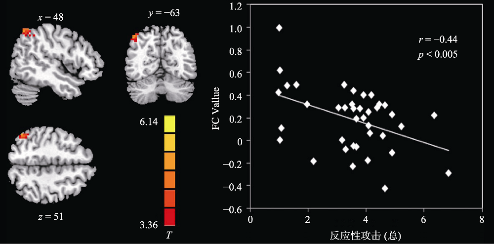
图2左侧OMPFC-右侧角回的功能连接与反应性攻击相关显著(FC值使用z转化之后的值注:彩图见电子版, 下同
图2左侧OMPFC-右侧角回的功能连接与反应性攻击相关显著(FC值使用z转化之后的值注:彩图见电子版, 下同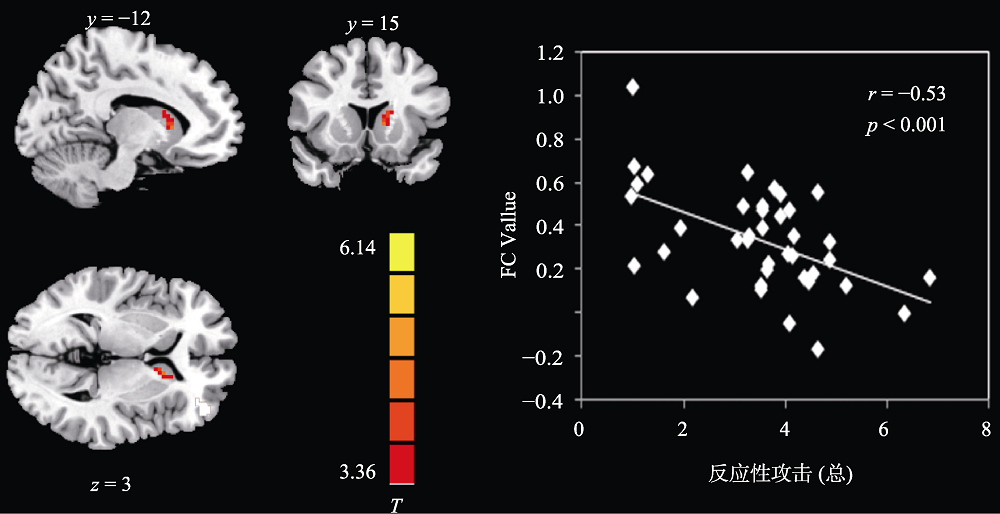
图3左侧OMPFC-左侧尾状核的功能连接与反应性攻击相关显著(FC值使用z转化之后的值)
图3左侧OMPFC-左侧尾状核的功能连接与反应性攻击相关显著(FC值使用z转化之后的值)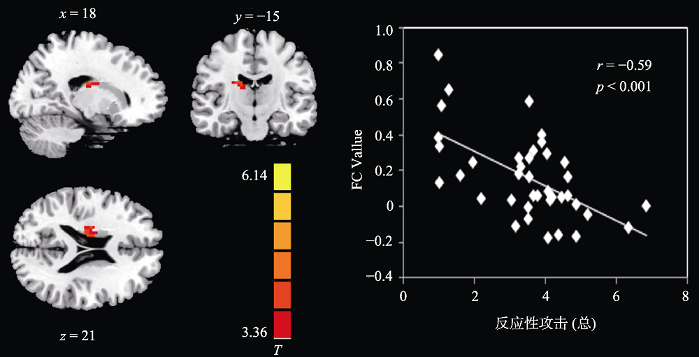
图4左侧OMPFC-右侧尾状核的功能连接与反应性攻击相关显著(FC值使用z转化之后的值)
图4左侧OMPFC-右侧尾状核的功能连接与反应性攻击相关显著(FC值使用z转化之后的值)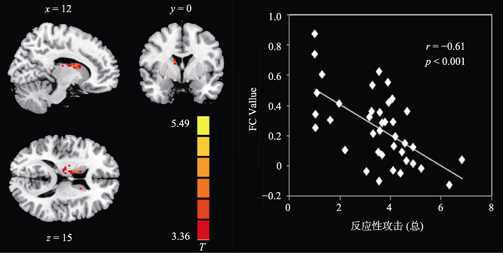
图5右侧OMPFC-右侧尾状核的功能连接与反应性攻击相关显著(FC值使用z转化之后的值)
图5右侧OMPFC-右侧尾状核的功能连接与反应性攻击相关显著(FC值使用z转化之后的值)表2功能连接值与反应性攻击的相关矩阵
| 功能连接 | 反应性攻击 (非激发条件) | 反应性攻击 (激发条件) | 反应性 攻击(总) |
|---|---|---|---|
| 左侧OMPFC- 右侧角回. | -0.43** | -0.36* | -0.44*** |
| 左侧OMPFC- 左侧尾状核 | -0.50** | -0.46** | -0.53*** |
| 左侧OMPFC- 右侧尾状核 | -0.51** | -0.54*** | -0.59*** |
| 右侧OMPFC- 右侧尾状核 | -0.54** | -0.55*** | -0.61*** |
表2功能连接值与反应性攻击的相关矩阵
| 功能连接 | 反应性攻击 (非激发条件) | 反应性攻击 (激发条件) | 反应性 攻击(总) |
|---|---|---|---|
| 左侧OMPFC- 右侧角回. | -0.43** | -0.36* | -0.44*** |
| 左侧OMPFC- 左侧尾状核 | -0.50** | -0.46** | -0.53*** |
| 左侧OMPFC- 右侧尾状核 | -0.51** | -0.54*** | -0.59*** |
| 右侧OMPFC- 右侧尾状核 | -0.54** | -0.55*** | -0.61*** |
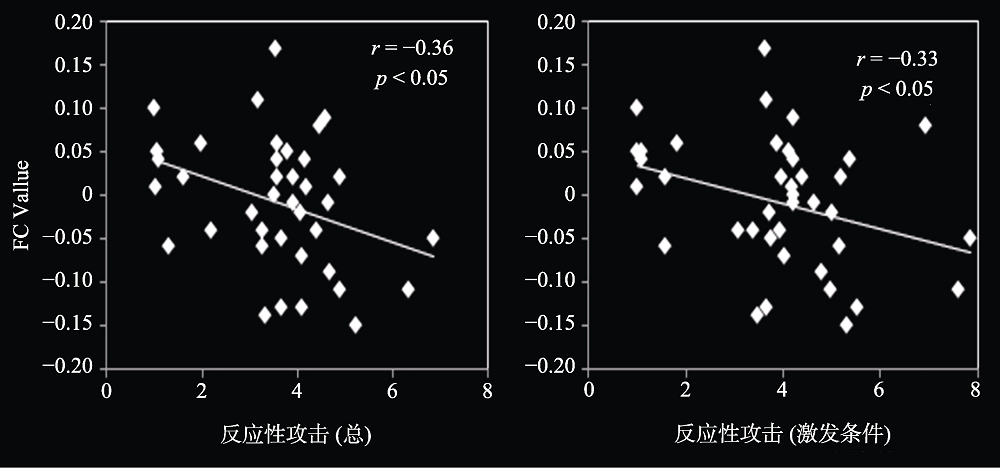
图6右侧尾状核→右侧OMPFC的效应连接与反应性攻击相关显著(左为反应性攻击、右为激发条件下的反应性攻击)
图6右侧尾状核→右侧OMPFC的效应连接与反应性攻击相关显著(左为反应性攻击、右为激发条件下的反应性攻击)表3效应连接值与反应性攻击的相关性统计
| 效应连接 | 反应性攻击 (非激发条件) | 反应性攻击 (激发条件) | 反应 性攻击(总) |
|---|---|---|---|
| 左侧OMPFC →右侧角回 | -0.05 | 0.02 | -0.01 |
| 左侧OMPFC →左侧尾状核 | 0.02 | -0.09 | -0.04 |
| 左侧OMPFC →右侧尾状核 | 0.21 | -0.01 | 0.11 |
| 右侧角回→ 左侧OMPFC | 0.08 | 0.02 | 0.05 |
| 左侧尾状核→ 左侧OMPFC | -0.21 | -0.16 | -0.20 |
| 右侧尾状核→ 左侧OMPFC | -0.19 | -0.17 | -0.20 |
| 右侧OMPFC →右侧尾状核 | 0.24 | 0.10 | 0.19 |
| 右侧尾状核→ 右侧OMPFC | -0.31 | -0.33* | -0.36* |
表3效应连接值与反应性攻击的相关性统计
| 效应连接 | 反应性攻击 (非激发条件) | 反应性攻击 (激发条件) | 反应 性攻击(总) |
|---|---|---|---|
| 左侧OMPFC →右侧角回 | -0.05 | 0.02 | -0.01 |
| 左侧OMPFC →左侧尾状核 | 0.02 | -0.09 | -0.04 |
| 左侧OMPFC →右侧尾状核 | 0.21 | -0.01 | 0.11 |
| 右侧角回→ 左侧OMPFC | 0.08 | 0.02 | 0.05 |
| 左侧尾状核→ 左侧OMPFC | -0.21 | -0.16 | -0.20 |
| 右侧尾状核→ 左侧OMPFC | -0.19 | -0.17 | -0.20 |
| 右侧OMPFC →右侧尾状核 | 0.24 | 0.10 | 0.19 |
| 右侧尾状核→ 右侧OMPFC | -0.31 | -0.33* | -0.36* |
参考文献 65
| [1] | Anderson C. A., & Bushman B. J . ( 2002). Human aggression. Annual Review of Psychology, 53(1), 27-51. doi: 10.1146/annurev.psych.53.100901.135231URL |
| [2] | Bettencourt B. A., Talley A., Benjamin A. J., & Valentine J . ( 2006). Personality and aggressive behavior under provoking and neutral conditions: A meta-analytic review. Psychological Bulletin, 132(5), 751-777. doi: 10.1037/0033-2909.132.5.751URLpmid: 16910753 |
| [3] | Beyer F., Münte T. F., Erdmann C., & Kr?mer U. M . ( 2014). Emotional reactivity to threat modulates activity in mentalizing network during aggression. Social Cognitive and Affective Neuroscience, 9(10), 1552-1560. doi: 10.1093/scan/nst146URLpmid: 23986265 |
| [4] | Beyer F., Münte T. F., G?ttlich M., & Kr?mer U. M . ( 2015). Orbitofrontal cortex reactivity to angry facial expression in a social interaction correlates with aggressive behavior. Cerebral Cortex, 25(9), 3057-3063. doi: 10.1093/cercor/bhu101URLpmid: 24842782 |
| [5] | Bj?rklund A., & Dunnett S. B . ( 2007). Dopamine neuron systems in the brain: An update. Trends in Neurosciences, 30(5), 194-202. doi: 10.1016/j.tins.2007.03.006URLpmid: 17408759 |
| [6] | Blair, R. J. R . ( 2012). Considering anger from a cognitive neuroscience perspective. Wiley Interdisciplinary Reviews: Cognitive Science, 3(1), 65-74. doi: 10.1002/wcs.154URLpmid: 22267973 |
| [7] | Brett M., Anton J. L., Valabregue R., & Poline J. B . ( 2002). Region of interest analysis using the MarsBar toolbox for SPM 99. NeuroImage, 16(2), S497. |
| [8] | Chen G., Hamilton J. P., Thomason M. E., Gotlib I. H., Saad Z. S., & Cox R. W . ( 2009). Granger causality via vector auto-regression tuned for fMRI data analysis. In Proceedings of the 17th annual scientific meeting and exhibition (Vol. 17, p. 1718). Honolulu, Hawaii. |
| [9] | Coccaro E. F., McCloskey M. S., Fitzgerald D. A., & Phan K. L . ( 2007). Amygdala and orbitofrontal reactivity to social threat in individuals with impulsive aggression. Biological Psychiatry, 62(2), 168-178. doi: 10.1016/j.biopsych.2006.08.024URLpmid: 17210136 |
| [10] | Coccaro E. F., Sripada C. S., Yanowitch R. N., & Phan K. L . ( 2011). Corticolimbic function in impulsive aggressive behavior. Biological Psychiatry, 69(12), 1153-1159. doi: 10.1016/j.biopsych.2011.02.032URLpmid: 21531387 |
| [11] | da Cunha-Bang S., Fisher P. M., Hjordt L. V., Perfalk E., Skibsted A. P., Bock C., .. Knudsen G. M . ( 2017). Violent offenders respond to provocations with high amygdala and striatal reactivity. Social Cognitive and Affective Neuroscience, 12(5), 802-810. doi: 10.1093/scan/nsx006URLpmid: 28338916 |
| [12] | Damasio H., Grabowski T., Frank R., Galaburda A. M., & Damasio A. R . ( 1994). The return of Phineas Gage: Clues about the brain from the skull of a famous patient. Science, 264(5162), 1102-1105. doi: 10.1126/science.8178168URLpmid: 8178168 |
| [13] | Davis M., & Whalen P. J . ( 2001). The amygdala: Vigilance and emotion. Molecular Psychiatry, 6(1), 13-34. doi: 10.1038/sj.mp.4000812URLpmid: 11244481 |
| [14] | D?biec, J. ( 2005). Peptides of love and fear: Vasopressin and oxytocin modulate the integration of information in the amygdala. BioEssays, 27(9), 869-873. doi: 10.1002/bies.20301URLpmid: 16108061 |
| [15] | Finger E. C., Marsh A. A., Mitchell D. G., Reid M. E., Sims C., Budhani S., .. Blair J. R . ( 2008). Abnormal ventromedial prefrontal cortex function in children with psychopathic traits during reversal learning. Archives of General Psychiatry, 65(5), 586-594. doi: 10.1001/archpsyc.65.5.586URL |
| [16] | Fite P. J., Rubens S. L., Preddy T. M., Raine A., & Pardini D. A . ( 2014). Reactive/proactive aggression and the development of internalizing problems in males: The moderating effect of parent and peer relationships. Aggressive Behavior, 40(1), 69-78. doi: 10.1002/ab.21498URLpmid: 23868672 |
| [17] | Fulwiler C. E., King J. A., & Zhang N. Y . ( 2012). Amygdala- orbitofrontal resting state functional connectivity is associated with trait anger. Neuroreport, 23(10), 606-610. doi: 10.1097/WNR.0b013e3283551cfcURLpmid: 22617448 |
| [18] | Gatzke-Kopp L. M., & Beauchaine T. P . ( 2007). Central nervous system substrates of impulsivity: Implications for the development of attention-deficit/hyperactivity disorder and conduct disorder. In D. Coch, G. Dawson, & K. W. Fischer (Eds.), Human behavior, Learning, and the developing brain: Atypical development (pp. 239-263). New York: Guilford. |
| [19] | Gatzke-Kopp L. M., Beauchaine T. P., Shannon K. E., Chipman J., Fleming A. P., Crowell S. E., .. Aylward E . ( 2009). Neurological correlates of reward responding in adolescents with and without externalizing behavior disorders. Journal of Abnormal Psychology, 118(1), 203-213. doi: 10.1037/a0014378URLpmid: 19222326 |
| [20] | Giancola P. R., & Parrott D. J . ( 2008). Further evidence for the validity of the Taylor aggression paradigm. Aggressive Behavior, 34(2), 214-229. doi: 10.1002/ab.20235URLpmid: 17894385 |
| [21] | Glenn A. L., & Yang Y. L . ( 2012). The potential role of the striatum in antisocial behavior and psychopathy. Biological Psychiatry, 72(10), 817-822. doi: 10.1016/j.biopsych.2012.04.027URLpmid: 22672927 |
| [22] | Gopal A., Clark E., Allgair A., D'Amato C., Furman M., Gansler D. A., & Fulwiler C . ( 2013). Dorsal/ventral parcellation of the amygdala: Relevance to impulsivity and aggression. Psychiatry Research: Neuroimaging, 211(1), 24-30. doi: 10.1016/j.pscychresns.2012.10.010URLpmid: 23352275 |
| [23] | Grahn J. A., Parkinson J. A., & Owen A. M . ( 2009). The role of the basal ganglia in learning and memory: Neuropsychological studies. Behavioural Brain Research, 199(1), 53-60. doi: 10.1016/j.bbr.2008.11.020URLpmid: 19059285 |
| [24] | Greicius M. D., Flores B. H., Menon V., Glover G. H., Solvason H. B., Kenna H., .. Schatzberg A. F . ( 2007). Resting-state functional connectivity in major depression: Abnormally increased contributions from subgenual cingulate cortex and thalamus. Biological Psychiatry, 62(5), 429-437. doi: 10.1016/j.biopsych.2006.09.020URLpmid: 17210143 |
| [25] | Hahn A., Stein P., Windischberger C., Weissenbacher A., Spindelegger C., Moser E., .. Lanzenberger R . ( 2011). Reduced resting-state functional connectivity between amygdala and orbitofrontal cortex in social anxiety disorder. NeuroImage, 56(3), 881-889. doi: 10.1016/j.neuroimage.2011.02.064URL |
| [26] | Hamilton J. P., Chen G., Thomason M. E., Schwartz M. E., & Gotlib I. H . ( 2011). Investigating neural primacy in major depressive disorder: Multivariate Granger causality analysis of resting-state fMRI time-series data. Molecular Psychiatry, 16(7), 763-772. doi: 10.1038/mp.2010.46URLpmid: 2925061 |
| [27] | Herpertz S. C., Nagy K., Ueltzh?ffer K., Schmitt R., Mancke F., Schmahl C., & Bertsch K . ( 2017). Brain mechanisms underlying reactive aggression in borderline personality disorder—Sex matters. Biological Psychiatry, 82(4), 257-266. doi: 10.1016/j.biopsych.2017.02.1175URLpmid: 28388995 |
| [28] | Hoptman M. J., D'Angelo D., Catalano D., Mauro C. J., Shehzad Z. E., Kelly A. M. C., .. Milham M. P . ( 2010). Amygdalofrontal functional disconnectivity and aggression in schizophrenia. Schizophrenia Bulletin, 36(5), 1020-1028. doi: 10.1093/schbul/sbp012URLpmid: 2930349 |
| [29] | Huber D., Veinante P., & Stoop R . ( 2005). Vasopressin and oxytocin excite distinct neuronal populations in the central amygdala. Science, 308(5719), 245-248. doi: 10.1126/science.1105636URLpmid: 15821089 |
| [30] | Koenigs M., & Tranel D . ( 2007). Irrational economic decision-making after ventromedial prefrontal damage: Evidence from the ultimatum game. Journal of Neuroscience, 27(4), 951-956. doi: 10.1523/JNEUROSCI.4606-06.2007URL |
| [31] | Kr?mer U. M., Jansma H., Tempelmann C., & Münte T. F . ( 2007). Tit-for-tat: The neural basis of reactive aggression. NeuroImage, 38(1), 203-211. doi: 10.1016/j.neuroimage.2007.07.029URLpmid: 17765572 |
| [32] | Kr?mer U. M., Riba J., Richter S., & Münte T. F . ( 2011). An fMRI study on the role of serotonin in reactive aggression. PLoS One, 6(11), e27668. doi: 10.1371/journal.pone.0027668URL |
| [33] | LeDoux, J. ( 1998). Fear and the brain: Where have we been, and where are we going? Biological Psychiatry, 44(12), 1229-1238. doi: 10.1016/S0006-3223(98)00282-0URLpmid: 9861466 |
| [34] | LeDoux, J. E . ( 2000). Emotion circuits in the brain. Annual Review of Neuroscience, 23, 155-184. doi: 10.1146/annurev.neuro.23.1.155URL |
| [35] | Lee G. P., Bechara A., Adolphs R., Arena J., Meador K. J., Loring D. W., & Smith J. R . ( 1998). Clinical and physiological effects of stereotaxic bilateral amygdalotomy for intractable aggression. The Journal of Neuropsychiatry and Clinical Neurosciences, 10(4), 413-420. doi: 10.1176/jnp.10.4.413URLpmid: 9813786 |
| [36] | Liu Y. L., Teng Z. J., Lan H. Y., Zhang X., & Yao D. Z . ( 2015). Short-term effects of prosocial video games on aggression: An event-related potential study. Frontiers in Behavioral Neuroscience, 9, 193. doi: 10.3389/fnbeh.2015.00193URLpmid: 4513560 |
| [37] | Lotze M., Veit R., Anders S., & Birbaumer N . ( 2007). Evidence for a different role of the ventral and dorsal medial prefrontal cortex for social reactive aggression: An interactive fMRI study. NeuroImage, 34(1), 470-478. doi: 10.1016/j.neuroimage.2006.09.028URLpmid: 17071110 |
| [38] | Maren, S. ( 2001). Neurobiology of Pavlovian fear conditioning. Annual Review of Neuroscience, 24, 897-931. doi: 10.1146/annurev.neuro.24.1.897URLpmid: 11520922 |
| [39] | Mark V. H., Sweet W., & Ervin F . ( 1975). Deep temporal lobe stimulation and destructive lesions in episodically violent temporal lobe epileptics. In W. Fields & W. Sweet (Eds.), Neural bases of violence and aggression (pp. 379-400). St. Louis: Warren H. Greem, Inc. |
| [40] | McCloskey M. S., Phan K. L., Angstadt M., Fettich K. C., Keedy S., & Coccaro E. F . ( 2016). Amygdala hyperactivation to angry faces in intermittent explosive disorder. Journal of Psychiatric Research, 79, 34-41. doi: 10.1016/j.jpsychires.2016.04.006URLpmid: 27145325 |
| [41] | McEwen C. A., & McEwen B. S . ( 2017). Social structure, adversity, toxic stress, and intergenerational poverty: An early childhood model. Annual Review of Sociology, 43, 445-472. doi: 10.1146/annurev-soc-060116-053252URL |
| [42] | Motzkin J. C., Newman J. P., Kiehl K. A., & Koenigs M . ( 2011). Reduced prefrontal connectivity in psychopathy. Journal of Neuroscience, 31(48), 17348-17357. doi: 10.1523/JNEUROSCI.4215-11.2011URLpmid: 3311922 |
| [43] | Narabayashi H., Nagao T., Saito Y., Yoshida M., & Nagahata M . ( 1963). Stereotaxic amygdalotomy for behavior disorders. Archives of Neurology, 9(1), 1-16. doi: 10.1001/archneur.1963.00460070011001URLpmid: 13937583 |
| [44] | Nelson R. J., & Trainor B. C . ( 2007). Neural mechanisms of aggression. Nature Reviews Neuroscience, 8(7), 536-546. doi: 10.1038/nrn2174URL |
| [45] | New A. S., Hazlett E. A., Buchsbaum M. S., Goodman M., Mitelman S. A., Newmark R., .. Siever L. J . ( 2007). Amygdala-prefrontal disconnection in borderline personality disorder. Neuropsychopharmacology, 32(7), 1629-1640. doi: 10.1038/sj.npp.1301283URL |
| [46] | Pietrini P., Guazzelli M., Basso G., Jaffe K., & Grafman J . ( 2000). Neural correlates of imaginal aggressive behavior assessed by positron emission tomography in healthy subjects. American Journal of Psychiatry, 157(11), 1772-1781. doi: 10.1176/appi.ajp.157.11.1772URL |
| [47] | Ramirez J. M., & Andreu J. M . ( 2006). Aggression, and some related psychological constructs (anger, hostility, and impulsivity) Some comments from a research project. Neuroscience and Biobehavioral Reviews, 30(3), 276-291. doi: 10.1016/j.neubiorev.2005.04.015URLpmid: 16081158 |
| [48] | Riva P., Gabbiadini A., Lauro L. J. R., Andrighetto L., Volpato C., & Bushman B. J . ( 2017). Neuromodulation can reduce aggressive behavior elicited by violent video games. Cognitive, Affective, and Behavioral Neuroscience, 17(2), 452-459. |
| [49] | Rosell D. R., & Siever L. J . ( 2015). The neurobiology of aggression and violence. CNS Spectrums, 20(3), 254-279. doi: 10.1017/S109285291500019XURLpmid: 25936249 |
| [50] | Rudebeck P. H., Bannerman D. M., & Rushworth M. F. S . ( 2008). The contribution of distinct subregions of the ventromedial frontal cortex to emotion, social behavior, and decision making. Cognitive, Affective, and Behavioral Neuroscience, 8(4), 485-497. doi: 10.3758/CABN.8.4.485URLpmid: 19033243 |
| [51] | Sagvolden T., Johansen E. B., Aase H., & Russell V. A . ( 2005). A dynamic developmental theory of attention- deficit/hyperactivity disorder (ADHD) predominantly hyperactive/impulsive and combined subtypes. Behavioral and Brain Sciences, 28(3), 397-419. doi: 10.1017/S0140525X05000075URLpmid: 16209748 |
| [52] | Sah P., Faber E. S. L., Lopez de Lopez M., & Power J. P . ( 2003). The amygdaloid complex: Anatomy and physiology. Physiological Reviews, 83(3), 803-834. doi: 10.1152/physrev.00002.2003URLpmid: 12843409 |
| [53] | Shannon K. E., Sauder C., Beauchaine T. P., & Gatzke-Kopp L. M . ( 2009). Disrupted effective connectivity between the medial frontal cortex and the caudate in adolescent boys with externalizing behavior disorders. Criminal Justice and Behavior, 36(11), 1141-1157. doi: 10.1177/0093854809342856URL |
| [54] | Siever, L. J . ( 2008). Neurobiology of aggression and violence. American Journal of Psychiatry, 165(4), 429-442. doi: 10.1017/S109285291500019XURLpmid: 18346997 |
| [55] | Song X. W., Dong Z. Y., Long X. Y., Li S. F., Zuo X. N., Zhu C. Z., .. Zang Y. F . ( 2011). REST: A toolkit for resting-state functional magnetic resonance imaging data processing. PLoS One, 6(9), e25031. doi: 10.1371/journal.pone.0025031URLpmid: 3176805 |
| [56] | Takeuchi H., Taki Y., Hashizume H., Sassa Y., Nagase T., Nouchi R., & Kawashima R . ( 2012). The association between resting functional connectivity and creativity. Cerebral Cortex, 22(12), 2921-2929. doi: 10.1093/cercor/bhr371URLpmid: 22235031 |
| [57] | Taylor, S. P . ( 1967). Aggressive behavior and physiological arousal as a function of provocation and the tendency to inhibit aggression. Journal of Personality, 35(2), 297-310. doi: 10.1111/j.1467-6494.1967.tb01430.xURLpmid: 6059850 |
| [58] | Tzourio-Mazoyer N., Landeau B., Papathanassiou D., Crivello F., Etard O., Delcroix N., .. Joliot M . ( 2002). Automated anatomical labeling of activations in SPM using a macroscopic anatomical parcellation of the MNI MRI single-subject brain. NeuroImage, 15(1), 273-289. doi: 10.1006/nimg.2001.0978URLpmid: 1177199511771995 |
| [59] | V?llm B., Richardson P., McKie S., Elliott R., Dolan M., & Deakin B . ( 2007). Neuronal correlates of reward and loss in cluster B personality disorders: A functional magnetic resonance imaging study. Psychiatry Research: Neuroimaging, 156(2), 151-167. doi: 10.1016/j.pscychresns.2007.04.008URLpmid: 17920821 |
| [60] | Wu Q. Z., Li D. M., Kuang W. H., Zhang T. J., Lui S., Huang X. Q., .. Gong Q. Y . ( 2011). Abnormal regional spontaneous neural activity in treatment-refractory depression revealed by resting-state fMRI. Human Brain Mapping, 32(8), 1290-1299. doi: 10.1002/hbm.21108URLpmid: 20665717 |
| [61] | Yan C. G., & Zang Y. F . ( 2010). DPARSF: A MATLAB toolbox for “pipeline” data analysis of resting-state fMRI. Frontiers in Systems Neuroscience, 413. doi: 10.3389/fnsys.2010.00013URLpmid: 2889691 |
| [62] | Zeng L. L., Shen H., Liu L., Wang L. B., Li B. J., Fang P., .. Hu D. W . ( 2012). Identifying major depression using whole-brain functional connectivity: A multivariate pattern analysis. Brain, 135(5), 1498-1507. doi: 10.1093/brain/aws059URLpmid: 22418737 |
| [63] | Zink C. F., Pagnoni G., Martin M. E., Dhamala M., & Berns G. S . ( 2003). Human striatal response to salient nonrewarding stimuli. Journal of Neuroscience, 23(22), 8092-8097. doi: 10.1016/S0304-3940(03)00712-2URLpmid: 12954871 |
| [64] | Zink C. F., Pagnoni G., Martin-Skurski M. E., Chappelow J. C., & Berns G. S . ( 2004). Human striatal responses to monetary reward depend on saliency. Neuron, 42(3), 509-517. doi: 10.1016/S0896-6273(04)00183-7URLpmid: 15134646 |
| [65] | Zuo X. N., Di Martino A., Kelly C., Shehzad Z. E., Gee D. G., Klein D. F., .. Milham M. P . ( 2010). The oscillating brain: Complex and reliable. NeuroImage, 49(2), 1432-1445. doi: 10.1016/j.neuroimage.2009.09.037URLpmid: 2856476 |
相关文章 7
| [1] | 蔡惠燕, 苗心, 王鹏飞, 林志为, 王孟成, 杨文登, 麻彦坤, 曾红. 长期戒断海洛因成瘾者冲动性相关脑区的结构及功能特征[J]. 心理学报, 2021, 53(8): 861-874. |
| [2] | 李芮, 夏凌翔. 攻击动机对特质愤怒与反应性攻击关系的中介作用:一项纵向研究[J]. 心理学报, 2021, 53(7): 788-797. |
| [3] | 金花, 梁紫平, 朱子良, 严世振, 林琳, 艾克旦·艾斯卡尔, 尹建忠, 姜云鹏, 田鑫. 整体运动知觉老化伴随颞中回静息态功能改变[J]. 心理学报, 2021, 53(1): 38-54. |
| [4] | 崔芳, 杨佳苗, 古若雷, 刘洁. 右侧颞顶联合区及道德加工脑网络的功能连接预测社会性框架效应:来自静息态功能磁共振的证据[J]. 心理学报, 2021, 53(1): 55-66. |
| [5] | 周衡, 何华, 于薇, 王爱君, 张明. 老年人声音诱发闪光错觉的大脑静息态低频振幅[J]. 心理学报, 2020, 52(7): 823-834. |
| [6] | 童丹丹, 李文福, 禄鹏, 杨文静, 杨东, 张庆林, 邱江. 科学发明情境中问题提出的脑机制再探[J]. 心理学报, 2020, 52(11): 1253-1265. |
| [7] | 孙岩,薄思雨,吕娇娇. 认知重评和表达抑制情绪调节策略的脑网络分析:来自EEG和ERP的证据[J]. 心理学报, 2020, 52(1): 12-25. |
PDF全文下载地址:
http://journal.psych.ac.cn/xlxb/CN/article/downloadArticleFile.do?attachType=PDF&id=4195
