 , 彭国平, 闵洁, 林晶
 武汉市中医医院 超声诊断科, 湖北 武汉 430020
2020-07-10 收稿, 2020-08-27 录用
*通讯作者: 陈艳
摘要: 探讨剪切波弹性成像定量参数对乳腺癌诊断和预后预测的作用。以乳腺癌患者60例作为观察组,乳腺良性结节患者60例作为对照组,利用剪切波弹性成像行超声检查,收集最大弹性模量值(Emax)。t检验分析两组Emax差异,并使用ROC曲线分析Emax诊断乳腺癌的价值。结果显示,与对照组比较,观察组AE-max、Shell1Emax、Shell2Emax、Shell3Emax均增高。AE-max、Shell1Emax、Shell2Emax和Shell3Emax对诊断乳腺癌均有一定价值,曲线下面积分别为0.723、0.893、0.818和0.791。AE-max、Shell1Emax、Shell2Emax和Shell3Emax对诊断乳腺癌患者术后复发均有一定价值,曲线下面积分别为0.754、0.884、0.824和0.782。说明剪切波弹性成像定量分析对诊断乳腺癌和预测预后具有一定价值。
关键词: 乳腺癌剪切波弹性成像最大弹性模量值诊断
The Value of Shear Wave Elastography Quantitative Parameters in the Differentiation of Benign and Malignant Breast Masses and the Prediction of Post-operative Recurrence of Breast Cancer
CHEN Yan
 , PENG Guoping, MIN Jie, LIN Jing
 Department of Ultrasound Diagnosis, Wuhan Hospital of Traditional Chinese Medicine, Wuhan 430020, Hubei, P. R. China
*Corresponding author: CHEN Yan
Abstract: This paper discusses the effect of shear wave elastography quantitative parameters on the diagnosis and prognosis of breast cancer.Sixty cases of breast cancer patients were taken as the observation group, and 60 cases of benign breast nodules were taken as the control group. The maximum elastic modulus (Emax) were collected for ultrasonic examination using shear wave elastography.T test was used to analyze the difference of Emax between the two groups, and ROC curve was used to analyze the value of Emax in the diagnosis of breast cancer. The results showed that compared with the control group, AE max, Shell1emax, shell2emax and Shell3emax in the observation group were increased. AE max, Shell1emax, Shell2emax and Shell3emax had certain value in the diagnosis of breast cancer. The area under the curve was 0.723, 0.893, 0.818 and 0.791, respectively. AE max, Shell1emax, Shell2emax and Shell3emax had certain value in the diagnosis of postoperative recurrence of breast cancer. The area under the curve was 0.754, 0.884, 0.824 and 0.782, respectively. It is concluded that the quantitative analysis of shear wave elastography has certain value in the diagnosis of breast cancer and prediction of prognosis.
Key words: breast cancershear wave elastographymaximum elastic modulusdiagnosis
乳腺癌是女性最常见的恶性肿瘤,近些年其死亡率明显下降,主要得益于早期诊断和早期治疗,其5年生存率可以达90%,但乳腺癌发病率呈上升和年轻化趋势[1-3],因此, 乳腺癌仍是威胁女性身心健康的重大疾病[4-6]。超声、X线、MRI和穿刺活检等均是诊治乳腺癌的重要检查手段,目前诊断的金标准仍是病理活检。超声检查虽然具有价格便宜、无创、简便、可反复检查等优势,但在临床上目前只能作为筛查[7-9],这也是超声诊断乳腺癌的瓶颈,而剪切波弹性成像(shear wave elastography)有望解决超声诊断乳腺癌的这个不足,有研究显示,三阴性乳腺癌患者剪切波弹性成像定量参数与乳腺良性结节患者明显不同[10]。本研究旨在探讨剪切波弹性成像定量参数对乳腺肿块良恶性鉴别及乳腺癌患者预后的预测价值。
1 资料与方法1.1 一般资料2017年1月~2018年6月,前瞻性收集我院收治的乳腺癌患者60例作为观察组,纳入标准:(1)乳腺癌,存在明确的结节,乳腺癌的诊断依据病理诊断;(2)年龄18~80;(3)同意参与本研究,已经签署知情同意书。排除标准:(1)复发性乳腺癌;(2)乳腺转移性癌;(3)检查前已接受手术、分子生物、放化疗等特殊治疗;(4)乳腺炎;(5)精神异常,不能配合检查。同期收集60例乳腺良性结节患者作为对照组。两组年龄、结节直径、患侧等差异均无统计学意义(P>0.05)。见表 1。
表1
| 表 1 两组患者一般资料比较 | ||||||||||||||||||||||||||||||||||||||
本研究获得我院伦理委员会批准。60例乳腺癌患者中,TNM分期为Ⅰ期、Ⅱ期、Ⅲ期的分别23、27、10例,肿瘤细胞分化程度为高、中、低分化的分别为42、12、6例。术后对患者随访1年,观察患者有无复发。
1.2 超声检查方法利用剪切波弹性成像行超声检查,收集最大弹性模量值(maximum elastic modulus, Emax)。采用Mindray Resona 7实时剪切波弹性成像超声诊断仪,探头频率3~11 MHz。常规超声检查记录结节声像图特征后转换为剪切波弹性成像模式,杨氏模量的量程设为0~180 kPa,打开质控图,探头缓慢移至病灶(距离结节边缘3 mm以上),屏气,图像稳定超过3 s冻结图像,利用Q-Box测量软件测量剪切波弹性成像定量参数,同时利用描迹法、Shell功能测量病灶1 mm、2 mm、3 mm范围内Emax,分别记录为AE-max、Shell1Emax、Shell2Emax和Shell3Emax。以上数据均由一名主任医师测量,同一患者分别测量3次取均值,同时收集常规形态。
1.3 观察指标AE-max、Shell1Emax、Shell2Emax、Shell3Emax、形态是否规则、边缘是否规整、有无包膜、回声是否均匀、是否有钙化、1年复发率。
1.4 统计分析采用SPSS 22.0软件进行统计学处理数据。计量资料以(均数±标准差)表示,两组间均数比较采用t检验;计数资料以率(%)表示,率的比较应用卡方检验。以病理结果为金标准,构建剪切波弹性成像各定量参数(AE-max、Shell1Emax、Shell2Emax和Shell3Emax)受试者工作曲线(ROC),计算曲线下面积(AUC),确定各参数最佳诊断界值,应用Z检验对ROC曲线进行比较。P<0.05为差异有统计学意义。
2 结果2.1 两组患者常规超声结果比较常规超声下,与对照组比较,观察组更多表现为形态不规则、边缘不规整、无包膜、回声不均匀和钙化。见表 2。
表2
| 表 2 两组患者常规超声结果比较 |
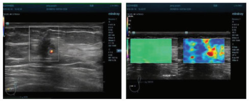
2.2 两组患者AE-max、Shell1Emax、Shell2Emax和Shell3Emax观察组AE-max、Shell1Emax、Shell2Emax、Shell3Emax均显著高于对照组(P=0.000)。数据见表 3,图 1为患者的剪切波弹性成像图。
表3
| 表 3 两组患者AE-max、Shell1Emax、Shell2Emax和Shell3Emax(kPa) |
图 1
 | 图 1 患者剪切波弹性成像图剪切波弹性成像蓝色为主,提示质地不硬,经测量,AE-max、Shell1Emax、Shell2Emax和Shell3Emax分别为134、187、165、174 kPa,最终病理证实为乳腺癌 |
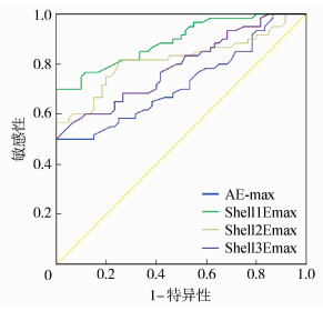
2.3 AE-max、Shell1Emax、Shell2Emax和Shell3Emax诊断乳腺结节患者乳腺癌的价值由ROC曲线(图 2)和表 4可知, AE-max、Shell1Emax、Shell2Emax和Shell3Emax对诊断乳腺癌均有一定价值。
图 2
 | 图 2 AE-max、Shell1Emax、Shell2Emax和Shell3Emax受试者工作曲线 |
表4
| 表 4 AE-max、Shell1Emax、Shell2Emax和Shell3Emax诊断乳腺癌的价值 |
2.4 剪切波弹性成像定量参数对乳腺癌患者术后复发的预测价值观察组的60例乳腺癌患者均行手术治疗,术后对患者随访1年,共有11例患者术后复发。剪切波弹性成像定量参数对术后复发均有一定的预测价值,与术后未复发的49例患者比较,复发患者的AE-max、Shell1Emax、Shell2Emax和Shell3Emax均显著增高(P < 0.05)。见表 5和表 6。
表5
| 表 5 剪切波弹性成像定量参数对乳腺癌患者术后复发的预测价值 |
表6
| 表 6 AE-max、Shell1Emax、Shell2Emax和Shell3Emax与术后复发的关联(kPa) |
3 讨论乳腺癌是女性最常见的恶性肿瘤,超声检查在乳腺癌的诊治中具有不可替代的价值,但常规超声检查仅能提供结节大小、形态学检查,不能进一步准确区分结节的良恶性,进一步检查依赖X线、MRI或穿刺活检。但穿刺活检是有创检查,且费用昂贵,X线、MRI具有辐射性。超声检查具有简便、价格便宜、可反复检查、用时短等优势,如可进一步明确乳腺结节性质,则具有重要意义。
剪切波弹性成像技术主要是测量机体组织的硬度从而评估组织的性质,具有实时、可视化、定量的特点,主要通过声辐射脉冲叩击组织施加激励,根据“马赫圆锥”原理,在不同组织中产生剪切波,并获取图像进行分析。虽然选择的感兴趣区不同,剪切波超声定量参数不同,但对最大弹性模量值,即Emax影响最小,故而Emax也是研究最多的一个参数[11-13]。癌组织一般质地较硬,因此可以用剪切波弹性成像技术诊断实体瘤,乳腺癌是实体瘤的一种,肿块质地较硬,目前已有研究采用剪切波技术诊断乳腺癌[14-16],具有较高的特异性和敏感性,支持本研究的结论。但需注意的是,少部分乳腺癌患者,富于细胞的髓样癌可稍软,个别也可呈囊性,如囊性乳头状癌,少数肿块周围有较多脂肪组织包裹,触诊时有柔韧感。因此,剪切波弹性技术尚不能作为诊断乳腺癌的金标准,但是可以作为常规超声检查的补充,减少不必要的病理活检,提高乳腺癌的早期诊断率。且超声弹性成像是一种无创伤性的诊断方法,能为良恶性乳腺结节的诊断提供一种有效的诊断信息,并对乳腺癌患者预后进行预测。但是剪切波弹性成像技术也存在不足,主要是对于较大的肿块,只能随机选取几个兴趣区,可能会存在选择误差,如本研究选择的测量值AE-max、Shell1Emax、Shell2Emax和Shell3Emax,虽然对乳腺癌的诊断具有较大价值,但是不同区域诊断价值不同,因此,在临床实践中需要注意取一个固定的感兴趣区作为检测点。此外,AE-max、Shell1Emax、Shell2Emax和Shell3Emax增大,说明乳腺癌组织中胶原纤维增加,乳腺癌组织中胶原纤维的增加不仅可以对组织起到支撑作用,对促进癌细胞的生长和侵袭也有重要影响,AE-max、Shell1Emax、Shell2Emax和Shell3Emax同样可以作为乳腺癌患者术后复发的预测因子。本研究发现,AE-max、Shell1Emax、Shell2Emax,以及Shell3Emax对预测乳腺癌患者术后1年复发的曲线下面积分别高达0.754、0.884、0.824和0.782,具有一定价值。但本研究的不足是样本量较少,对患者随访时间不够长,因此,对患者预后评估的价值尚有待进一步深入探讨。
综上所述,剪切波弹性成像定量分析对诊断乳腺癌和预测预后具有一定价值。
参考文献
| [1] | Huang M H, Blackwood J, Godoshian M, et al. Factors associated with self-reported falls, balance or walking difficulty in older survivors of breast, colorectal, lung, or prostate cancer:results from surveillance, epidemiology, and end results-medicare health outcomes survey linkage[J]. PLoS One, 2018, 13(12): e8573-e8578. |
| [2] | Figueira P V G, Haddad C A S, De Almeida R S, et al. Diagnosis of axillary web syndrome in patients after breast cancer surgery:epidemiology, risk factors, and clinical aspects:a prospective study[J]. American Journal of Clinical Oncology, 2018, 41(10): 992-996. DOI:10.1097/COC.0000000000000411 |
| [3] | Fairweather M, Jiang W, Keating N L, et al. Morbidity of local therapy for locally advanced metastatic breast cancer:an analysis of the surveillance, epidemiology, and end results (SEER)-medicare registry[J]. Breast Cancer Research Treat, 2018, 169(2): 287-293. DOI:10.1007/s10549-018-4689-y |
| [4] | Mailhot Vega R B, Balogun O D, Ishaq O F, et al. Estimating child mortality associated with maternal mortality from breast and cervical cancer[J]. Cancer, 2019, 125(1): 109-117. DOI:10.1002/cncr.31780 |
| [5] | Moller M H, Lousdal M L, Kristiansen I S, et al. Effect of organized mammography screening on breast cancer mortality:a population-based cohort study in Norway[J]. International Journal of Cancer, 2019, 144(4): 697-706. DOI:10.1002/ijc.31832 |
| [6] | Zheng J, Tabung F K, Zhang J, et al. Association between post-cancer diagnosis dietary inflammatory potential and mortality among invasive breast cancer survivors in the women's health initiative[J]. Cancer Epidemiology, Biomarkers and Prevention, 2018, 27(4): 454-463. DOI:10.1158/1055-9965.EPI-17-0569 |
| [7] | Badu-peprah A, Adu-sarkodie Y. Accuracy of clinical diagnosis, mammography and ultrasonography in preoperative assessment of breast cancer[J]. Ghana Medical Journal, 2018, 52(3): 133-139. |
| [8] | Choi E J, Lee E H, Kim Y M, et al. Interobserver agreement in breast ultrasound categorization in the mammography and ultrasonography study for breast cancer screening effectiveness (MUST-BE) trial:results of a preliminary study[J]. Ultrasonography, 2018, 12(4): 894-899. |
| [9] | Jales R M, Doria M T, Serra K P, et al. Power Doppler ultrasonography and shear wave elastography as complementary imaging methods for suspected local breast cancer recurrence[J]. Journal of Clinical Ultrasound, 2018, 37(6): 1493-1501. |
| [10] | 李菲.三阴性乳腺癌常规超声及声触诊组织成像定量剪切波弹性成像特征[D].济南: 山东大学, 2018. |
| [11] | Liu B, Zheng Y, Huang G, et al. Breast lesions:quantitative diagnosis using ultrasound shear wave elastography-a systematic review and meta-analysis[J]. Ultrasound in Medicine and Biology, 2016, 42(4): 835-847. DOI:10.1016/j.ultrasmedbio.2015.10.024 |
| [12] | Cong R, Li J, Guo S. A new qualitative pattern classification of shear wave elastograghy for solid breast mass evaluation[J]. European Journal of Radiology, 2017, 87(21): 111-119. |
| [13] | Au F W, Ghai S, Moshonov H, et al. Diagnostic performance of quantitative shear wave elastography in the evaluation of solid breast masses:determination of the most discriminatory parameter[J]. American Journal of Roentgenology, 2014, 203(3): W328-336. DOI:10.2214/AJR.13.11693 |
| [14] | Au F W, Ghai S, Lu F I, et al. Quantitative shear wave elastography:correlation with prognostic histologic features and immunohistochemical biomarkers of breast cancer[J]. Academic Radiology, 2015, 22(3): 269-277. DOI:10.1016/j.acra.2014.10.007 |
| [15] | Song E J, Sohn Y M, Seo M. Tumor stiffness measured by quantitative and qualitative shear wave elastography of breast cancer[J]. The British Journal of Radiology, 2018, 91(1086): 20170830. |
| [16] | Youk J H, Gweon H M, Son E J, et al. Shear-wave elastography of invasive breast cancer:correlation between quantitative mean elasticity value and immunohistochemical profile[J]. Breast Cancer Research Treat, 2013, 138(1): 119-126. DOI:10.1007/s10549-013-2407-3 |
