 ,1,2,*, 阙介民1,2, 张文定3, 孙翠丽1,2, 舒岩峰1,2, 侯叶茂3, 张久昶1,2, 史戎坚1,2, 魏龙1,2
Development and applications of paleontological computed tomography
WANG Yan-Fang1,2, WEI Cun-Feng,1,2,*, QUE Jie-Min1,2, ZHANG Wen-Ding3, SUN Cui-Li1,2, SHU Yan-Feng1,2, HOU Ye-Mao3, ZHANG Jiu-Chang1,2, SHI Rong-Jian1,2, WEI Long1,2收稿日期:2017-08-8网络出版日期:2019-01-20
| 基金资助: |
Corresponding authors: *weicf@ihep.ac.cn
Received:2017-08-8Online:2019-01-20

摘要
古生物化石研究传统的磨片法耗时,且具有破坏性,研究者不可能对珍贵的化石标本进行这样的操作,因此研究只能停留在化石的外表面。而古生物CT装置的出现使研究者可以对化石内部结构进行无损检测,得到上千层化石的图像数据。主要论述国内首套古生物CT装置的研制及典型应用,其中225-3D-μCT显微CT系统具有三维成像能力,最高分辨率达5 μm, 可以检测直径100 mm, 高度100 mm尺度的化石;450-TY-ICT通用型CT可对大尺寸化石高信噪比成像,检测范围是直径800 mm, 高度1000 mm, 分辨率达200 μm。这两台古生物CT以高成像性能满足了研究者对不同尺度化石的不同检测需求,成为中国古生物化石研究中非常重要的技术手段。
关键词:
Abstract
The traditional serial grinding method used to investigate the internal structure of fossils cannot be readily applied to valuable fossil specimens due to its destructive and time-consuming nature. Computed tomography (CT) is an ideal non-destructive technique for investigating the internal structure of fossils, in which thousands of serial images are obtained and used to produce an accurate reconstruction of the internal morphology. This paper reviews the design, development and applications of the first CT system in China dedicated exclusively to scanning fossils. The 225 kV three-dimensional (3D) fossil micro-CT (225-3D-μCT) is capable of high-resolution volumetric imaging, with a resolution up to 5 μm, and can accommodate specimens measuring up to 100 mm in diameter and 100 mm in length. The 450 kV ordinary fossil CT (450-TY-ICT) can produce high signal-to-noise ratio (SNR) images of specimens ranging up to 800 mm in diameter and 1000 mm in length, with a resolution up to 200 μm. Two paleontological CT facilities represent a high-performance platform offering the functional diversity needed to meet the demands of studying fossils at a variety of different scales. The two machines have become indispensable for paleontological research in China.
Keywords:
PDF (3834KB)元数据多维度评价相关文章导出EndNote|Ris|Bibtex收藏本文
本文引用格式
王燕芳, 魏存峰, 阙介民, 张文定, 孙翠丽, 舒岩峰, 侯叶茂, 张久昶, 史戎坚, 魏龙. 古生物CT装置的研制及应用. 古脊椎动物学报[J], 2019, 57(1): 84-92 DOI:10.19615/j.cnki.1000-3118.170921
WANG Yan-Fang, WEI Cun-Feng, QUE Jie-Min, ZHANG Wen-Ding, SUN Cui-Li, SHU Yan-Feng, HOU Ye-Mao, ZHANG Jiu-Chang, SHI Rong-Jian, WEI Long.
1 Introduction
The study of fossils is an essential part of efforts to unveil the secrets of the origin and evolution of life. In the past, serial grinding (Keyes, 1962; Mark, 2008) was the only method available to scientists who wished to investigate the internal structure of fossils. The fact that this technique destroys the fossil and is extremely time-consuming makes it difficult to apply, especially to particularly valuable fossil specimens such as human skulls. Computed tomography (CT) can be used to investigate various objects internally and is non-destructive, making it ideal as a paleontological method for extracting information on the internal structure of fossils.Medical CT is unsuitable for fossil scanning, because of its poor resolution. The voltage used to generate the scanning beam in the X-ray tube of a medical CT device is generally 160 kV or less, a level that is appropriate for harmlessly scanning the tissues of living humans but results in only sub-millimeter scale resolution. In industrial CT devices the X-ray tube voltage can be more variable, ranging from dozens of kV up to 15 MeV, and the resolution can be as good as 5 μm or even better.
Since 1996, paleontologists have been using high-resolution CT (HRCT) to study fossils (Beall et al., 1996). Paleontological papers based on studies using HRCT have been published in Nature, Science and other high-impact journals (e.g. Mickler et al., 2004; Clarke et al., 2005; Kearney et al., 2005; Kyle et al., 2005). However, before 2009 the only established industrial CT facilities readily accessible to paleontologists, like those at the University of Texas at Austin (a national shared multi-user facility since 1999) and the Australian National University (where the National Laboratory for X-ray Micro Computed Tomography (CTLab) maintains a number of X-ray micro-computed tomography (micro CT) instruments), were located outside China. As a result, they were of limited value to Chinese paleontologists, because many valuable specimens (such as fossil human skulls) are not allowed to leave China.
Industrial CT is a cutting-edge technology in paleontology, and is valuable as a non-destructive technique for investigating the internal structure of fossils. In order to promote paleontological research and protect intellectual property rights pertaining to the results of research on valuable Chinese specimens, it was deemed essential to develop a CT facility in China to be used exclusively for fossil scanning.
2 The development of paleontological CT
2.1 General concept
Paleontological CT is essentially a type of industrial CT, and paleontological CT as a method for scanning fossils has significant advantages over medical CT in terms of radiation intensity, penetration of the sample, and resolution. Compared with conventional industrial CT, our paleontological CT facility has the following special characteristics: 1) more suitable for objects (fossil specimens of invertebrates, fishes, amphibians, non-avian reptiles, birds, humans and other mammals, plants, etc.) that vary widely in size (5-800 mm in diameter) and composition; 2) able to provide superior spatial and density resolution. The second characteristic is essential for accurate imaging of the detailed internal structure of fossils, as this requires clear discrimination between the fossil and the surrounding matrix of rock or sediment even if the two are close in density.We have developed two paleontological CT devices, namely an ordinary CT device for relatively large fossil specimens and a micro-CT device for small ones. The 225 kV 3D fossil micro-CT (225-3D-μCT) is capable of high-resolution volumetric imaging, with a resolution up to 5 μm, and can accommodate specimens measuring up to 100 mm in diameter and 100 mm in length. The 450 kV ordinary fossil CT (450-TY-ICT) can produce high SNR (signal-to-noise ratio) images of specimens ranging up to 800 mm in diameter and 1000 mm in length, with a resolution up to 200 μm.
2.2 Design
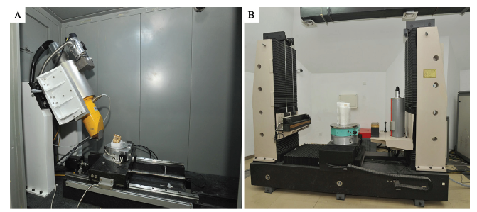
The system has been optimized using a high-performance X-ray tube with a small focal length to ensure good spatial resolution, and the micro-CT scanner also has a high-contrast detector with high pixel pitch to ensure good density resolution. To ensure stability, the platform on which specimens are placed for scanning is a single piece of high-quality natural marble in both CT scanners. The marble has been processed using a combination of automated and manual approaches, in order to minimize potential imprecision caused by fluctuations in temperature (which can cause small changes in the size of the piece of marble) and other environmental parameters, as well as smaller amounts of deformation caused by internal stress arising from the marble’s material properties. The fossil micro-CT is equipped with a high precision electronic control rotary table (HUBER410, Germany) that maintains its exact position when loaded with a fossil, ensuring that no errors are introduced as a result of specimen misalignment. In both devices, the system takes a series of X-ray images (also called “projections” or “frames”) at small angular intervals as the specimen rotates through a full 360°. Multiple frames are taken at each point, and averaged to produce a less noisy composite image. The composite images are then combined by software to produce the final CT data set for the specimen. Both paleontological CT devices are shown in Fig.1.Fig. 1
 新窗口打开|下载原图ZIP|生成PPT
Fig. 1Two paleontological CT devices
A. 225-3D-μCT; B. 450-TY-ICT
Control system In the 225-3D-μCT device, the rotary table on which fossils are placed is installed on a slide rail which runs parallel to a line drawn from the X-ray tube to the X-ray detector. The precision of the movement of the rotary table along the slide rail is guaranteed by a linear optical encoder, comprising a readhead paired with a scale. The readhead reads the scale in order to encode the current position of the rotary table, which is converted into a digital pulse signal and transmitted to the motion control system. This system ensures that the position of the rotary table is precisely known at all times. A positive limit point and a negative limit point, which respectively define the minimum allowable distance from the rotary table to the X-ray tube (which emits X-rays) and to the detector (which receives X-rays after they have passed through the specimen), constrain the movement of the rotary table. The boundaries represented by the positive limit point and negative limit point prevent fossil specimens from colliding with either the tube or the detector. A home point, considered to represent a zero point for the system, is set between the positive and negative limit points. Furthermore, a safety light curtain extending between a transmitting device and a receiving device is installed between the X-ray tube and the fossil sample, and is connected to the motion control system. If the fossil or another object interrupts the light curtain, motion of the system will immediately cease. This design ensures that the motion control system will be stopped by feedback before making contact with the fossil sample, preventing the sample from suffering damage under any circumstances.
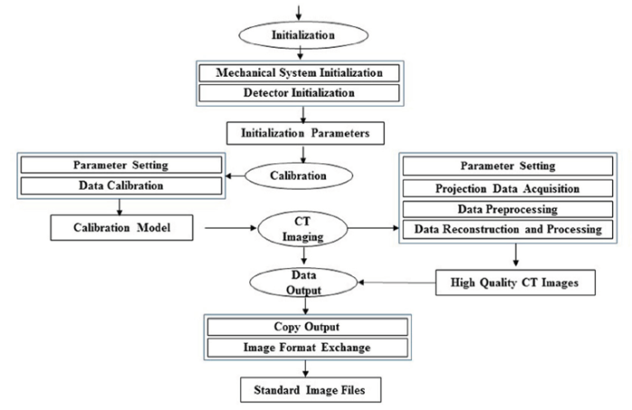
Data acquisition and image reconstruction software Custom-designed software with a Graphical User Interface (GUI ) allows users to synchronize the motion control system, the X-ray tube and the detector, utilizing protocols that make it possible to collect scanning data using different parameters. After data processing and reconstruction, the images can be 3D displayed, analyzed and outputted as image files to meet different needs. The software workflow is shown in Fig. 2.
Fig. 2
 新窗口打开|下载原图ZIP|生成PPT
Fig. 2Software workflow
2.3 Research innovations
Multi-scale and high precision CT equipment for paleontological research The machines described in this paper are the first two CT devices in China that have been designed exclusively for nondestructive investigation of fossils. Innovative features of these devices include their X-ray energy spectrum optimization, double collimating beam modulation, nonlinear structure tensor denoising technology and 3D tomographic imaging hardening correction techniques (Zhao et al., 2011). They provide CT images characterized by high spatial resolution and high contrast sensitivity, allowing accurate segmentation of fossil specimens. CT scanning surpasses traditional methods used to study the 3D internal structure of fossil specimens, such as serial grinding, in being non-destructive as well as accurate. The highest spatial resolution the equipment can provide is 5 μm, while the relative density resolution is 1‰. The maximum specimen diameter the equipment can accommodate is 800 mm, and specimens as small as 5 mm can be scanned in adequate detail. Accordingly, the CT facility as a whole is suitable for scanning fossils of most types (fishes, non-avian dinosaurs, birds, humans, non-human mammals, plants, etc.).A standardized procedure for 3D tomography of fossils We have developed a standardized procedure for scanning fossils and reconstructing them in 3D. Information about each fossil specimen, including specimen size, scanning parameters, calibration data, projection data, reconstructed images, and 3D visualization data, is recorded in order to create digital documents similar to databases. This procedure represents a non-destructive method for investigating fossil specimens at micron-grade resolution. A CT study typically includes calibration, selection of parameters to provide optimal quality, data acquisition and reconstruction, segmentation of fossil material to distinguish it from the surrounding rock, 3D restoration (i.e. correction for any deformation of the specimen), and accurate 3D measurement of volume and other geometric parameters. The quality of the final results is among the highest achieved by any group in China doing work of this type, and comparable to that achieved by leading facilities worldwide.
Quantitative evaluation of resolution in 3D tomography Through painstaking design, calibration and testing, we have developed a quantitative method to estimate the spatial resolution and density resolution needed to capture the morphology of a given specimen. In 3D tomography, it is necessary to obtain a 3D PSF (point spread function; describes the response of an imaging system to radiation from a point source) and a 3D CDF (contrast discrimination function) in order to accurately determine the spatial resolution and density resolution required for the specimen. Methods for calculating the 3D PSF and 3D CDF were presented by Wang et al. (2013) . This approach helps to improve the reliability of the tomography data collected during the scanning process, as well as the accuracy of the subsequent image processing (Wang et al., 2013). Our experience in this domain potentially provides a basis for the future establishment of general 3D micro-CT standards.
3 Applications of paleontological CT
First brought into service in 2009, our CT facilities have since logged over 6000 working hours and scanned more than 2000 fossils of various types. Our methods of scanning and subsequent data processing have proven to be highly effective, and have led to a number of major research breakthroughs (relating to, for example, the origin of vertebrate jaws, the evolution and development of mineralized hard tissue in vertebrates, and vertebrate brain evolution).Some configuration and scanning parameters for our two CT devices are shown in Table 1.
Table 1
Table 1Paleontological CT configuration and detection parameters
| CT | Model | Specimen diameter | Scanning projections | Fossil type |
|---|---|---|---|---|
| 225-3D-μCT | X-ray tube: Phoenix XS|225D, focus spot size: 5 μm Detector: Varian 4030CB, pixel size: 194 μm Rotary table: HUBER 410 | 5-100 mm | 720 projections used to reconstruct CT images 4 projections averaged at each angular interval | Fish Amphibians Reptiles Birds Mammals Humans Plants |
| 450-TY-ICT | X-ray tube: COMET XRS-450, focus spot size: 0.4 mm Detector: DT X-SCAN 0.4- 614HE, pixel size 0.4 mm Rotary table: HUBER 430 | 100-800 mm | 1440 projections used to reconstruct CT images 4 projections averaged at each angular interval |
新窗口打开|下载CSV
Our CT devices have been used to investigate many kinds of fossils (including fishes, non-avian dinosaurs, birds, mammals and plants) with high levels of precision and at a variety of scales. Research results have been published in Nature (Zhu et al., 2013), PNAS (Wu et al., 2011, 2014), PloS One (Wu et al., 2013; Xing et al., 2014), Journal of Human Evolution (Liu et al., 2013), Anthropological Science (Wu et al., 2012), Chinese Science Bulletin (Wu et al., 2008) and other important journals (Lu et al., 2012; Zhu et al., 2012; Zhang et al., 2013; Xing et al., 2015).
3.1 Reconstruction of an ancient human brain using CT
The Liujiang fossil skull was found in the Tongtianyan Cave in Liuzhou district, Guangxi Zhuang Autonomous Region by a local farmer in 1958. Its age has been variously estimated as 68-40 ka, based on uranium dating of several non-human mammalian teeth found together with the skull (Yuan et al., 1986), or older than 153 ka (Shen et al., 2002). Because the endocranial cavity is filled with hard matrix, earlier studies were restricted to the exterior morphology of the skull.Using the 450 kV paleontological CT device, researchers from the Institute of Vertebrate Paleontology and Paleoanthropology (IVPP) scanned the Liujiang skull and documented a difference in density between the bone wall of the skull and the associated matrix. They also obtained data on the morphology of the brain cavity in the Liujiang skull, and this new information has considerable importance for studies of the evolution of the human brain and the evolutionary relationship between the Liujiang people and other humans. The volume of the brain cavity in the Liujiang skull proved to be 1567 ml. This value is within the range of variation seen in previously measured human specimens from the early Late Pleistocene (100-130 ka), but greatly exceeds the average for modern humans. This study was the first in which Chinese researchers used X-ray tomography to investigate the morphology of an ancient human skull. The results were published in Chinese Science Bulletin (Wu et al., 2008).
3.2 “Ancient fish with a new kind of face” - a jaw-dropping fossil discovery
Using 225 kV micro-CT, researchers from the IVPP scanned an ancient fossil fish, eventually named Entelognathus primordialis, which is 420 million years old and about the size of a trout. In overall body shape, E. primordialis is typical of the placoderms, a group of primitive armored fishes that normally have a simple jaw structure. However, scanning helped to reveal that E. primordialis is a key transitional form with a sophisticated jaw resembling that of advanced bony fishes. This groundbreaking discovery revealed the oldest known appearance in the vertebrate fossil record of the bones homologous to those that form the human face. This research was published in the journal Nature (Zhu et al., 2013), and was selected as headline news by the Nature website.4 Conclusion and new developments
Two CT machines, representing the first such devices in China dedicated exclusively to the study of fossils, have been developed. Our paleontological CT facility represents a high-performance platform offering the functional diversity needed to meet the demands of studying fossils at a variety of different scales. The two machines have become indispensable for paleontological research in China.To facilitate scanning of fossils preserved in slabs at high spatial resolution, the project group is currently developing a Computed Laminography (CL) imaging system. Through high-precision stitching technology, slab specimens of large size can be scanned successfully. The new system will be capable of a maximum resolution of 10 μm, and will be able to accommodate specimens of sizes up to 300 mm × 300 mm.
参考文献 原文顺序
文献年度倒序
文中引用次数倒序
被引期刊影响因子
[本文引用: 1]
PMID [本文引用: 1]

Long-standing controversy surrounds the question of whether living bird lineages emerged after non-avian dinosaur extinction at the Cretaceous/Tertiary (K/T) boundary or whether these lineages coexisted with other dinosaurs and passed through this mass extinction event. Inferences from biogeography and molecular sequence data (but see ref. 10) project major avian lineages deep into the Cretaceous period, implying their 'mass survival' at the K/T boundary. By contrast, it has been argued that the fossil record refutes this hypothesis, placing a 'big bang' of avian radiation only after the end of the Cretaceous. However, other fossil data--fragmentary bones referred to extant bird lineages--have been considered inconclusive. These data have never been subjected to phylogenetic analysis. Here we identify a rare, partial skeleton from the Maastrichtian of Antarctica as the first Cretaceous fossil definitively placed within the extant bird radiation. Several phylogenetic analyses supported by independent histological data indicate that a new species, Vegavis iaai, is a part of Anseriformes (waterfowl) and is most closely related to Anatidae, which includes true ducks. A minimum of five divergences within Aves before the K/T boundary are inferred from the placement of Vegavis; at least duck, chicken and ratite bird relatives were coextant with non-avian dinosaurs.
[本文引用: 1]
DOIURL [本文引用: 1]
[本文引用: 1]
DOIURL [本文引用: 1]
DOIURL [本文引用: 1]
[本文引用: 1]
[本文引用: 1]
DOIURL [本文引用: 1]
It has been established that modern humans were living in the Levant and Africa ca. 100 ka ago. Hitherto, this has contrasted with the situation in China where no unequivocal specimens of this species have been securely dated to more than 30 ka. Here we present the results of stratigraphic studies and U-series dating of the Tongtianyan Cave, the discovery site of the Liujiang hominid, which represents one of the few well-preserved fossils of modern Homo sapiens in China. The human fossils are inferred to come from either a refilling breccia or a primarily deposited gravel-bearing sandy clay layer. In the former case, which is better supported, the fossils would date to at least ∼68 ka, but more likely to ∼111–139 ka. Alternatively, they would be older than ∼153 ka. Both scenarios would make the Liujiang hominid one of the earliest modern humans in East Asia, possibly contemporaneous with the earliest known representatives from the Levant and Africa. Parallel studies on other Chinese localities have provided supporting evidence for the redating of Liujiang, which may have important implications for the origin of modern humans.
[本文引用: 2]
DOIURL [本文引用: 2]
DOIURL [本文引用: 1]
[本文引用: 1]
[本文引用: 1]
DOIURL [本文引用: 1]
[本文引用: 1]
DOIURL [本文引用: 1]
[本文引用: 1]
[本文引用: 1]
DOIURL [本文引用: 1]
In this paper, we present a beam hardening correction (BHC) method in three-dimension space for a cone-beam computed tomography (CBCT) system in a mono-material case and investigate its effect on the spatial resolution. Due to the polychromatic character of the X-ray spectrum used, cupping and streak artifacts called beam hardening artifacts arise in the reconstructed CT images, causing reduced image quality. In addition, enhanced edges are introduced in the reconstructed CT images because of the beam hardening effect. The spatial resolution of the CBCT system is calculated from the edge response function (ERF) on different planes in space. Thus, in the CT images with beam hardening artifacts, enhanced ERFs will be extracted to calculate the modulation transfer function (MTF), obtaining a better spatial resolution that deviates from the real value. Reasonable spatial resolution can be obtained after reducing the artifacts. The 10% MTF value and the full width at half maximum (FWHM) of the point spread function with and without BHC are presented.
DOIURL [本文引用: 1]
DOIURL [本文引用: 2]
The gnathostome (jawed vertebrate) crown group comprises two extant clades with contrasting character complements. Notably, Chondrichthyes (cartilaginous fish) lack the large dermal bones that characterize Osteichthyes (bony fish and tetrapods). The polarities of these differences, and the morphology of the last common ancestor of crown gnathostomes, are the subject of continuing debate. Here we describe a three-dimensionally preserved 419-million-year-old placoderm fish from the Silurian of China that represents the first stem gnathostome with dermal marginal jaw bones (premaxilla, maxilla and dentary), features previously restricted to Osteichthyes. A phylogenetic analysis places the new form near the top of the gnathostome stem group but does not fully resolve its relationships to other placoderms. The analysis also assigns all acanthodians to the chondrichthyan stem group. These results suggest that the last common ancestor of Chondrichthyes and Osteichthyes had a macromeric dermal skeleton, and provide a new framework for studying crown gnathostome divergence.
