1. 武汉大学 基础医学院, 湖北 武汉 443000;
2. 宜昌市第二人民医院 超声科, 湖北 宜昌 443000
2019-09-23 收稿, 2019-11-15 录用
*通讯作者: 薛敬玲
摘要: 本文探究了声学超声造影评估左心室厚度的效果,旨在提高临床测量室壁厚度的准确度。选取2018年1月~2019年5月我院心内科患者94例为研究对象,按照随机数表法将患者分为两组,对照组47例,接受二维超声心动图检测,观察组47例,接受左心声学造影检测。将左心室室壁划分为18节段,比较两组患者检测结果的准确度差异。比较两组患者心尖两腔(A2CH)切面舒张期全部阶段以及收缩期3、6、15、18阶段室壁厚度,差异具有统计学意义(P < 0.05);两组患者心尖三腔(A3CH)切面舒张期4、10、13、16阶段以及收缩期4、10、16阶段室壁厚度比较,差异具有统计学意义(P < 0.05);两组患者心尖四腔切面(A4CH)切面舒张期5、14、17阶段以及收缩期5、17阶段室壁厚度比较,差异具有统计学意义(P < 0.05)。二维超声心动图检测在舒张期1、2、7、8、11节段以及收缩期1、2、7、8、9、11、12、13、14节段的测量结果准确可靠,而左心声学造影检测能提供更为准确的左心室厚度指标,对提高临床对左心室室壁厚度检测的准确度具有重要意义。
关键词: 左心室厚度二维超声心动图声学造影
Analysis of the Effect of Acoustic Contrast-enhanced Ultrasound on Left Ventricular Thickness Measuring
TIAN Miao1,2, XUE Jingling1

1. School of Basic Medicine, Wuhan University, Wuhan 443000, Hubei, P. R. China;
2. Ultrasound Department, the Second People's Hospital, Yichang 443000, Hubei, P. R. China
Abstract: In order to explore the effect of acoustic contrast-enhanced evaluation on left ventricular thickness and improve the accuracy of clinical measurement of wall thickness, 94 patients with cardiology were selected as subjects from January 2018 to May 2019. The patients were divided into two groups according to the random number table method. The control group (47 cases) received two-dimensional echocardiography. The observation group (47 cases) received left heart contrast echocardiography. The left ventricular wall was divided into 18 segments, and the difference in accuracy between the two groups was compared. There were significant differences in the wall thickness between the two groups in the apical two-chamber (A2CH) section and the 3rd, 6th, 15th, and 18th stages of systolic phase (P < 0.05). There were significant differences between the two groups in the wall thickness of the 4th, 10th, 13th and 16th stages of apical period and the 4th, 10th and 16th stages of systolic phase in the apical three-chamber (A3CH) section (P < 0.05). There were significant differences in the wall thickness of the 5th, 14th stages of the apical period of the apical four-chamber view (A4CH) and the 5th, 17th stages of the systolic phase between the two groups (P < 0.05). Two-dimensional echocardiography has accurate and reliable measurements in the 1st, 2nd, 7th, 8th, and 11th segments of the diastolic phase and the 1st, 2nd, 7th, 8th, 9th, 11th, 12th, 13th, and 14th segments of the systolic phase, while the left cardiac echocardiography can provide a more accurate left ventricular thickness index, which is of great significance to improve the accuracy of clinical detection of left ventricular wall thickness.
Key words: left ventricular thicknesstwo-dimensional echocardiographycontrast echocardiography
心血管疾病发病率随着不健康生活方式的滋长逐年上升,已经成为导致人口死亡的主要原因之一,因此早期诊断对于改善心血管疾病的预后意义重大[1, 2]。2001年、2008年、2014年美国超声心动图协会三次发表心血管超声影技术指南,欧洲超声心动图协会于2009年也提出了相应的方案,左心声学造影在医学中的应用越来越广泛[3, 4]。左心声学造影原理为利用不同信号处理技术对非线性谐波信号进行增强,从而对组织和组织运动产生的回波信号进行抑制[5]。有研究指出,左心声学造影能够提高成像质量,从而更准确地测定左心室厚度[6]。本次研究通过探究左心室厚度的声学超声造影评估效果,旨在提高临床测量室壁厚度的准确度。
1 资料与方法1.1 一般资料选取2018年1月~2019年5月我院94例心内科患者为研究对象。纳入标准:无造影剂禁忌症;无严重心率失常;收缩压<180 mmHg且舒张压<110 mmHg;患者均能够获取标准A2CH、A3CH以及A4CH切面;所有患者均同意参与本次研究。
按照随机数表法将患者分为两组:对照组接受二维超声心动图检测,共47例,年龄45.26±6.77岁,男性29例,女性18例,体重指数23.88±2.01 kg/m2;观察组接受左心声学造影检测,共47例,年龄45.34±6.28岁,男性27例,女性20例,体重指数23.59±2.11 kg/m2。两组患者年龄、性别、体重指数无统计学差异。
1.2 方法设备:配备S4和X4探头的PHILIPS IE33彩色超声多普勒仪,该设备具有常规显像软件、图像自动储存和回放软件、Contrast LVO程序及相关分析软件。
二维超声心动图检测:患者取左侧卧位,选取X5-1探头成人心脏心电图模式,依次采集A4CH、A2CH及A3CH切面,适当调节增益和深度,每个切面存储6个心动周期,划分为18阶段并测量室壁厚度。
左心声学造影检测:选用SonoVue超声造影剂,以1:5的比例与生理盐水配置,穿刺患者左前臂浅表静脉,团注0.5 mL造影剂溶液,并以每10秒0.1 mL的速度推注造影剂溶液至检查结束,最高用量低于5 mL。选取Contrast LVO模式,设置参数为MI=0.3,Gain=70%,待左心腔显影均匀后依次采集A4CH、A2CH以及A3CH切面,检测方法与对照组相同。
将左心室室壁划分为18节段,以LVO结果为标准,比较两组患者检测结果的准确度差异。
1.3 统计学方法采用SPSS 19.0软件进行数据分析。计量资料以(x±s)表示,比较采用独立样本t检验,P<0.05为差异具有统计学意义。
2 结果2.1 两组患者A2CH切面舒张期和收缩期室壁厚度比较两组患者A2CH切面舒张期所有节段以及收缩期3、6、15、18节段相比较,差异具有统计学意义(P<0.05)。详见表 1。
表1
| 表 1 两组患者A2CH切面舒张期和收缩期室壁厚度比较 |
2.2 两组患者A3CH切面舒张期和收缩期室壁厚度比较两组患者A3CH切面舒张期4、10、13、16节段以及收缩期4、10、16节段比较,差异具有统计学意义(P<0.05)。详见表 2。
表2
| 表 2 两组患者A3CH切面舒张期和收缩期室壁厚度比较 |
2.3 两组患者A4CH切面舒张期和收缩期室壁厚度比较两组患者A4CH切面舒张期5、14、17节段以及收缩期5、17节段比较差异具有统计学意义(P<0.05)。详见表 3。
表3
| 表 3 两组患者A4CH切面舒张期和收缩期室壁厚度比较 |
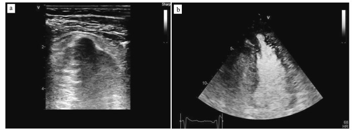
2.4 声学超声造影图像图 1和图 2为观察组行声学超声造影的典型图像。
图 1
 | 图 1 某患者,女,37岁,超声造影诊断为非对称性肥厚性心肌病合并左心室致密化不全a.左心造影后左室乳头肌水平短轴;b.左心造影胸骨旁前四腔心切面 |
图 2
 | 图 2 某患者,男,65岁,超声造影诊断为左室心尖部憩室a.左心造影前左室心尖部;b.左心造影后左室心尖部 |
3 讨论目前临床上测量左心室室壁厚度的常用手段为二维超声心动图,但由于左心室尖部位于声场的近场,容易受到肺气干扰、近场伪像等影响,导致图像质量不清晰,难以清晰界定结构异常,无法准确测量部分节段左心室室壁厚度[7, 8]。左心声学造影解决了二维超声心动图检测图像质量不佳的问题,能够准确勾画出心室边界,测量出左心室室壁的准确厚度,对诊断心尖部结构和功能的异常具有积极作用[9, 10]。
本次研究的数据显示,两组患者的左心室室壁厚度,在A2CH切面舒张期全节段及收缩期3、6、15、18节段、A3CH切面舒张期4、10、13、16节段及收缩期4、10、16节段、A4CH切面舒张期5、14、17节段及收缩期5、17节段,比较差异具有统计学意义(P<0.05),说明两种检测方法在A2CH切面检测结果差异最大,在A4CH切面检测结果差异最小。且二维超声心动图检测对舒张期室间隔厚度测量的准确度低,收缩期准确度较高,因此收缩期使用二维超声心动图检测,舒张期使用左心声学造影进行检测,能够得到相对准确的检查结果。分析原因为收缩期心尖收缩后与近场距离较远,能够更准确地测量左心室室壁,因此收缩期使用二维超声心动图进行检测[11],与国外研究结果基本一致[12]。观察两种检测方法的结果发现,二维超声心动图检测结果均小于左心声学造影检测结果,因此在临床检测中,使用二维超声心动图需要考虑结果偏小的因素,左心声学造影的检测结果更为可信[13]。且两种方法在前侧壁中段室壁厚度以及前后间隔基底段及中段的测量结果非常接近,因此均准确可信。而二维超声心动图在前壁心尖段、下壁中段及心尖段以及后侧壁基底段及中段测量结果不够准确,因此这些节段使用左心声学造影检测更准确[14, 15]。但在临床中,在由于造影剂价格昂贵且检测时间长,左心声学造影暂时无法广泛使用[16],且本次研究纳入样本量较少,需要扩大样本量进一步研究。
综上所述,二维超声心动图对左心室室壁厚度的检测,在舒张期1、2、7、8、11节段以及收缩期1、2、7、8、9、11、12、13、14节段的测量结果准确可靠,而左心声学造影检测能提供更为准确的左心室厚度指标,对提高临床对左心室室壁厚度检测的准确度具有重要意义。
参考文献
| [1] | 毛静远, 赵志强, 王贤良, 等. 中医药治疗心血管疾病研究述评[J]. 中医杂志, 2019, 60(21): 1801-1805. Mao J Y, Zhao Z Q, Wang X L, et al. Study review on traditional chinese medicine in treatment of cardiovascular disease[J]. Journal of Traditional Chinese Medicine, 2019, 60(21): 1801-1805. |
| [2] | 王佳音, 李利平. 21世纪心血管疾病研究展望[J]. 养生保健指南, 2019(40): 213. Wang J Y, Li L P. Research prospects of cardiovascular diseases in the 21st century[J]. Health Care Guide, 2019(40): 213. |
| [3] | Sobchak C, Akhtari S, Harvey P, et al. The value of carotid ultrasound in cardiovascular risk stratification in patients with psoriatic disease[J]. The Journal of Rheumatology, 2018, 45(7): 997. |
| [4] | Antoni S T, Lehmann S, Neidhardt M, et al. Model checking for trigger loss detection during Dopplerultrasound-guided fetal cardiovascular MRI[J]. International Journal of Computer Assisted Radiology And Surgery, 2018, 13(11): 1755-1766. DOI:10.1007/s11548-018-1832-5 |
| [5] | Kidoh M, Nakaura T, Nakamura S, et al. Novel contrast-injection protocol for coronary computed tomographic angiography: contrast-injection protocol customized according to the patient's time-attenuation response[J]. Heart and Vessels, 2014, 29(2): 149-155. DOI:10.1007/s00380-013-0338-x |
| [6] | 宋艳萍.左心声学造影的成像原理及临床应用[D].石家庄: 河北医科大学, 2018. Song Y P. Imaging principle and clinical application of left heart contrast angiography[D]. Shijiazhuang: Hebei Medical University, 2018. http://cdmd.cnki.com.cn/Article/CDMD-10089-1018859223.htm |
| [7] | Kondo M, Nagao M, Yonezawa M, et al. Improvement of automated right ventricular segmentation using dual-bolus contrast media injection with 256-slice coronary CT angiography[J]. Academic Radiology, 2014, 21(5): 648-653. DOI:10.1016/j.acra.2014.01.022 |
| [8] | Cao J X, Wang Y M, Lu J G, et al. Radiation and contrast agent doses reductions by using 80-kV tube voltage in coronary computed tomographic angiography: a comparative study[J]. European Journal of Radiology, 2014, 83(2): 309-314. |
| [9] | Shelton S E, Lindsey B D, Dayton P A, et al. First-in-human study of acoustic angioraphy in the breast and peripheral vasculature[J]. Ultrasound in Medicine and Biology, 2017, 43(12): 2939-2946. DOI:10.1016/j.ultrasmedbio.2017.08.1881 |
| [10] | Lindsey B D, Shelton S E, Martin K H, et al. High resolution ultrasound superharmonic perfusion imaging: in vivo feasibility and quantification of dynamic contrast-enhanced angiography[J]. Annals of Biomedical Engineering, 2017, 45(4): 939-948. DOI:10.1007/s10439-016-1753-9 |
| [11] | 田锦润, 丁云川, 王庆慧, 等. 左心声学造影的临床应用进展[J]. 临床超声医学杂志, 2017, 19(12): 838-840. Tian J R, Ding Y C, Wang Q H, et al. Progress in clinical application of left heart contrast angiography[J]. Clinical Ultrasound Medical Journal, 2017, 19(12): 838-840. DOI:10.3969/j.issn.1008-6978.2017.12.017 |
| [12] | Ma J, Martin K H, Dayton P A, et al. A preliminary engineering design of intravascular dual-frequency transducers for contrast-enhanced acoustic angiography and molecular imaging[J]. IEEE Transactions on Ultrasonics, Ferroelectrics, and Frequency Control, 2014, 61(5): 870-880. DOI:10.1109/TUFFC.2014.6805699 |
| [13] | 陈桑, 李慧忠. 超声心动图及右心声学造影诊断右房憩室1例[J]. 中国超声医学杂志, 2017, 33(8): 766. Chen S, Li H Z. Diagnosis of right atrium by echocardiography and right heart contrast echocardiography[J]. Chinese Journal of Ultrasound Medicine, 2017, 33(8): 766. DOI:10.3969/j.issn.1002-0101.2017.08.034 |
| [14] | Azzalini L, Abbara S, Ghoshhajra B B, et al. Ultra-low contrast computed tomographic angiography (CTA) with 20-mL total dose for transcatheter aortic valve implantation (TAVI) planning[J]. Journal of Computer Assisted Tomography, 2014, 38(1): 105-109. DOI:10.1097/RCT.0b013e3182a14358 |
| [15] | 申斌, 耿召华, 陈瑜, 等. 左心声学造影与二维超声心动图测量左心室厚度的对照研究[J]. 第三军医大学学报, 2018, 40(15): 1413-1418. Shen B, Geng Z H, Chen Y, et al. A comparative study of left heart contrast echocardiography and two-dimensional echocardiography in measuring left ventricular thickness[J]. Journal of Third Military Medical University, 2018, 40(15): 1413-1418. |
| [16] | Radu B, Liliana C, Olimpia C, et al. Associated gastroduodenal artery aneurysm aortic aneurysm: the diagnostic contribution of contrast-enhanced ultrasound in correlation with computed tomography angiography[J]. Journal of Medical Ultrasonics, 2014, 41(2): 217-221. DOI:10.1007/s10396-013-0507-7 |
